


 English (pdf)
 Article in xml format
 Article references
 Send this article by e-mail
 Cited by SciELO
  Similars in
SciELO
Similars in
SciELO 
 Permalink
Introduction
The success of endodontic treatment depends on the control of endodontic infection of the root canal system (1). The ideal filling technique should fill the root canal three-dimensionally, including areas with irregularities and isthmus (2), in order to favor the prognosis (3). However, incomplete obturation is observed in endodontic treatments (4), increasing the chance of persistence of apical periodontitis (5). No root canal obturation technique is capable of completely filling the root canal system (2, 6,7,8).
Lateral compaction is considered a simple, low-cost filling method and promotes sealing (9), with less overextension of root canal filling material (3). However, cold gutta-percha may not allow adaptation to the irregularities of root canals, allowing the presence of voids between the guttapercha cones and root canal (10, 11). To overcome these disadvantages, thermoplastic root canal obturation methods are proposed (1, 12, 13).
Thermoplastic obturation techniques, with vertical compaction of heated gutta-percha and subsequent injection of thermoplastic gutta-percha, promote a homogeneous mass of obturation material capable of filling irregularities (2, 13, 14). This technique has been used in different devices, such as: System B (2), Obtura II (9), and others (13,14,15,16). However, thermoplastic filling technique used in curved canals depends on the enlargement of the root canal and apical penetration of the plugger (2). Thermo Pack II system (Easy Dental Equipment, Brazil) has a thermoplastic plugger with a diameter #50 which performs heating and compaction of the apical gutta-percha. The Thermo Pack II system has a thermal injector with gutta-percha in the alpha phase that is injected with a flexible needle.
Micro-computed tomography (micro-CT) is used to analyze root canal filling techniques (9, 17, 18). However, most micro-CT studies for compari- sons between obturation techniques used straight root canals (2, 7, 9). Although some studies have evaluated mesial roots of mandibular molars, most studies performed 20μm average resolution scans (13,14,15).
The aim of this study was to compare the percentage of filling and voids in mesial canals of mandibular molars filled by a continuous wave of condensation system or lateral compaction, using micro-CT analysis with the resolution of 9μm. The null hypothesis was that there would be no significant difference in the percentage of voids and filling using continuous wave of condensation system or lateral compaction.
Materials and methods
After approval from the Institutional Ethics Committee (Registration number 6473611.40000.5416), mesio-buccal and mesio-lingual root canals from mandibular molars were selected. Digital radiography system (Kodak RVG 6100, Kodak Dental Systems, NY, USA) and microcomputed tomography were used to confirm the inclusion criteria and to perform a homogeneous distribution of the samples. The initial scanning was performed at 35µm, using SkyScan 1176 (Skyscan 1176, Bruker-MicroCT, Kontich, Belgium) with the following parameters: copper and aluminum filter, exposure time of 87ms, frame average 3, rotation of 180°, 80kV, and 300uA.
Total of 12 first and second human mandibular molars with two mesial root canals, presenting type IV configuration according to the classification of Vertucci (19) were selected. Complete apical formation, absence of root fractures, angle of curvature between 20° and 40° and radius of curvature smaller than 10mm, were observed in accordance with Schneider method (20). The size of the teeth was standardized at 18mm with a tolerance of ± 2mm of discrepancy. The root canals selected were stored in a 0.1% Thymol solution at 5°C.
Root canal preparation
After the specimens were washed in water for 48 hours, access to the canals was obtained with a high speed bur (n.2, KG Sorensen, São Paulo, Brazil), and the root canals were irrigated with 2.5% sodium hypochlorite, a size 10 K-file (Dentsply Sirona, Ballaigues, Switzerland) was used to explore the mesial root canals until its tip was visible through the apical foramen. The working length (WL) was established 1mm short of the apical foramen. The roots were embedded in condensation silicone (Oranwash, Zhermack SpA, Badia Polesine, Italy) to simulate the periodontal ligament. Afterwards, a single, experienced operator performed the root canal preparations.
Root canal preparation
The ProDesign Logic (PDL) instruments, size 25, .01 taper, were activated by an electric motor (VDW.SILVER, VDW GmbH, Munich, Germany) in rotary motion at 350 rpm and 1 N cm-1 of torque in accordance with the manufacturer’s instruc- tions, with in-and-out movements up to the WL. Then, a PDL instrument, size 25, .06 taper, was used at 600 rpm and 3 N cm-1 of torque (manufacturer’s instructions for curved root canals), with movements in the apical direction up to the WL. The root canals were enlarged with using ProDesign Logic instrument size 35, .01 taper, and ProDesign Logic instrument size 35, .05 taper as described above. Each root canal was irrigated with 10 ml of 2.5% sodium hypochlorite. Final irrigation was performed with 2.5ml EDTA under agitation for 3 minutes, and afterwards, irrigation with 5 ml of 0.5% sodium hypochlorite.
Filling by the continuous wave of condensation technique
Gutta-percha cones size 35, .05 taper (Tanari industry Ltda., São Paulo, Brazil) were used. The gutta-percha cones were selected according to the tip size and taper measured in the profilometer (Profile Projector Nikon Model 6C-2). After radiographic evaluation of adaptation of the gutta-percha cone selected, the AH Plus sealer (Dentsply DeTrey GmbH, Konstanz, Germany) was placed into the root canal by using a Lentulo size 35 instrument (Dentsply Maillefer, Ballaigues, Switzerland) and manual K-file size 35 pre-curved (Dentsply Maillefer, Ballaigues, Switzerland). After this, the gutta-percha cone covered by endodontic sealer was placed in the root canal. The thermoplastic plugger from the Termo Pack II System (Easy Dental Equipments, Brazil) was used for plasticization, cutting and compaction of the gutta percha within the apical root canal (Downpack) up to 3mm of the working length. After this, the thermal injector of the system was used to inject the warmed gutta- percha that was compacted. Compaction was performed with Nickel and Titanium condensers (Easy Dental Equipments, Brazil). A radiograph was taken to verify the quality of the obturation.
Filling by the lateral compaction technique
Gutta-percha cones size 35, .02 taper (Tanari Indústria Ltda., São Paulo, Brazil), were also selected after being measured in a Profilometer (Profile Projector Nikon Model 6C-2). The filling sealer AH Plus (Dentsply, DeTrey GmbH, Konstanz, Germany) was placed to the root canal by using a Lentulo size 35 (Dentsply Maillefer, Ballaigues, Switzerland) instrument and pre-curved manual K-file size 35 (Dentsply Maillefer, Ballaigues, Switzerland). After this, the gutta-percha cone covered by endodontic sealer was placed in the root canal. The finger spreader B (Dentsply Maillefer, Ballaigues, Switzerland) was used for inserting the auxiliary XF cones (Tanari industry Ltda., São Paulo, Brazil) into the apical third. Afterwards, the finger spreader C (Dentsply Maillefer, Ballaigues, Switzerland) was used for inserting the auxiliary FF gutta-percha cones (Tanari industry Ltda., São Paulo, Brazil) into the middle and coronal thirds. Lateral compaction was performed on a precision balance to control the force applied, which did not exceed 1.5 kg. A radiograph was taken to verify the quality of the obturation.
After filling the root canals, coronal sealing was performed with restorative material Coltosol (Vigodent, Rio de Janeiro, Brazil), and the roots were kept in an oven at a temperature of 37°C, in 95% humidity, for three days, for sealer setting.
Micro-ct analysis
The roots were scanned using micro-CT (SkyScan 1176; Bruker microCT, Kontich, Belgium) after preparation, and after the filling of the root canals. The roots were positioned in a standardization device, allowing the specimens to remain in the same position at all times during scanning procedures. The scanning parameters were: copper and aluminum filter, 90 kV power, 278 mA energy, evolution cycle 0,5, rotation 180° and frame average 3 with 9µm voxel size. The post-preparation and post-obturation images obtained were reconstructed using the NRecon software (NRecon v.1.6.10.4, Bruker Micro-CT), Volumetric analysis was performed in all canals after the preparation, through the software CTAn (CTAn v.1.14.4, Bruker Micro-CT) with the use of specific task lists.
The reconstructed images, obtained before and after obturation, were superimposed by means of geometric alignment in the Data Viewer software (Data Viewer v.1.5.1, Bruker Micro-CT). Quantitative analyses were then made using the CTAn software by applying task lists, and arithmetic and logic operations between the superimposed sections. Were quantified root canal volume, filling material volume (gutta-percha and sealer), and voids volume. The gray scale range required to recognize each object under study was determined in a density histogram by using an adaptive threshold method. The percentages of filling material were taken as a basis on the post-preparation volume of the canal and on the filling volume: (percentage of filling = (volume of filling x 100) / volume of canal). The volume of voids was calculated by subtracting the filling material volume from the post-obturation root canal volume: (volume of voids = volume of canal - volume of filling). The percentage volume of voids was calculated by using the following formula: (Percentage of voids = (volume of voids X 100)/volume of canal). Representative images were performed by means of models obtained by using CTVol software (CTVol v. 2.0, Bruker Micro-CT).
Evaluation was performed at all root canal extension and by thirds (Coronal, Middle and Apical). The value of approximately 9mm was determined for the total length analysis and approximately 3 mm for each third. In the software (CTAn), the value of bottom was corresponding to the working length, and to this value was added 9mm, as the value of top, corresponding to the furcation level.
Statistical analysis
The Graph Pad Prism 7.00 (GraphPad Software, Inc., California, USA) software was used for data analysis. Data obtained for canal volume values after preparation, percentage of filling material and percentage of voids between the groups were submitted to Student’s t-test. ANOVA/Tukey tests were used for comparison of filling material and voids between thirds. The significance level was set at 5%.
Results
The volume of the root canals prepared was similar for the two obturation techniques (p>0.05) (Table 1). Canals obturated by the lateral compaction technique presented a higher percentage of voids than the canals obturated by continuous wave (p<0.05) (Table 2 and Figure 1). A higher percentage of filling material was obtained for the canals filled by the continuous wave of condensation technique (p<0.05) (Table 2).
Table 1 Mean percentage (± standard deviation) of volume (mm³) - total and by thirds of canals after preparation.
| - | Continuous wave of condensation | Lateral compaction |
|---|---|---|
| Total | 3.858 ± 0.562 | 3.573 ± 0.501 |
| Coronal | 2.086 ± 0.306 | 1.951 ± 0.256 |
| Middle | 1.121 ± 0.195 | 1.079 ± 0.148 |
| Apical | 0.596 ± 0.105 | 0.544 ± 0.089 |
*There was no statistical difference between the groups (p>0.05).
Table 2 Mean percentage (± standard deviation) of voids and filling material in the filling after use of lateral compaction and continuous wave of condensation.
| - | - | Continuous Wave of Condensation | Lateral Compaction |
|---|---|---|---|
| % voids | Total | 7.77 ± 2.23ᵇ | 18.21 ± 4.49ª |
| - | Coronal | 10.06 ± 4.09ᵇᴬ | 17.07 ± 4.43ªᴬ |
| - | Middle | 5.87 ± 2.60ᵇᴮ | 20.60 ± 7.77ªᴬ |
| - | Apical | 2.92 ± 1.12ᵇᴄ | 18.43 ± 4.43ªᴬ |
| % filling material | Total | 92.22 ± 2.32ᵇ | 81.79 ± 4.49ª |
| - | Coronal | 89.94 ± 4.09ᵇᴬ | 82.93 ± 4.43ªᴬ |
| - | Middle | 94.12 ± 2.60ᵇᴮ | 74.84 ± 7.77ªᴬ |
| - | Apical | 96.27 ± 1.12ᵇᴄ | 81.57 ± 4.43ªᴬ |
*Different superscript lowercase letters in same line indicate statistical difference between the groups (p<0.05). Superscript uppercase letters in the same column indicate statistical difference among the thirds ( p<0.05).
Discussion
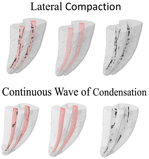
Our results showed that no obturation technique was able to completely fill the root canals, in agreement with previous studies (7, 8, 12, 15, 18, 21, 22). A higher percentage of filling material was observed for the continuous wave of condensation Termo Pack II System, in comparison with lateral compaction. Therefore, the null hypothesis was rejected. The micro-CT images showed gutta-percha cones covered by sealer in the root canals filled by lateral compaction, and a uniform mass of filling material for Termo Pack II (Figure 2), as observed by Ho et al. (14). Cold gutta percha has been shown to be incapable of filling artificial irregularities in mandibular incisors (10). A previous study (14) comparing three techniques of lateral compaction (manual, mechanical, and ultrasonic), presented that all the methods caused void areas in the root filling. The authors attribu- ted the voids to the spreader, that creates a larger space than required for the accessory cones.
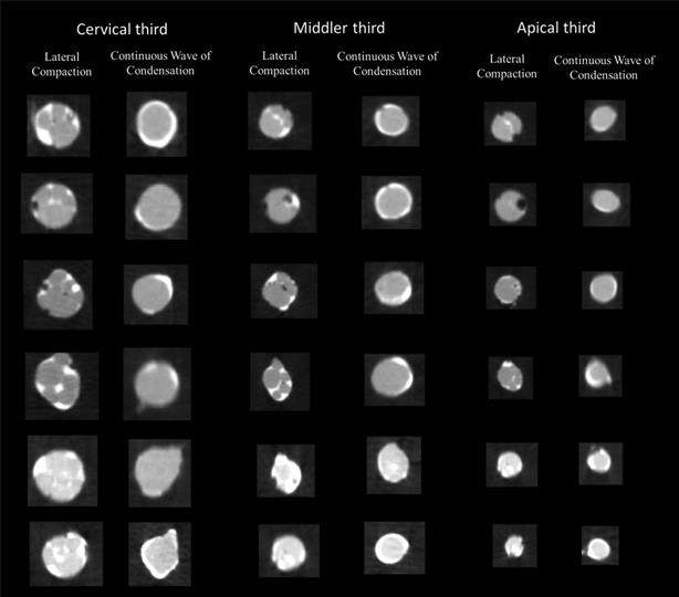
Figure 2 Representative micro-CT cross-sections from the coronal, middle and apical thirds of six different mesial root canals of mandibu- lar molars filled by continuous wave of condensation and six different mesial root canals of mandibular molars filled by lateral compaction, showing the presence of voids (black spots within the filling materials), sealer (in white) and gutta-percha (in grey).
Endodontic sealers must fill irregularities and areas that are difficult to access (23). AH Plus has sealing capacity (18) and promotes adaptation in the root canal walls (24), and is compatible with thermal compaction techniques (25). Proper filling was observed for continuous wave of condensation evaluated by micro-CT in mesial root canals from mandibular molars (13, 14) and mandibular premolars (7, 9, 10). However, the comparison by micro-CT of the filling volume in oval canals (mandibular premolars) after lateral compaction techniques has demonstrated that the finger spreaders may leave spaces in the gutta-percha, which are occupied by sealer (AH Plus), or remain empty (7). The continuous wave of condensation technique allows plasticization of the gutta percha throughout the entire extension of the canal promoting a homogeneous mass capable of filling irregularities of the root canal (7, 15), consequently decreasing the layer of sealer and formation of voids. Considering that gutta-percha is a dimensionally stable material (7, 26) and sealers are subject to solubilization, a higher proportion of gutta percha in the apical region in relation to sealer, may favor the prognosis in the long term (11, 27).
The apical third is considered critical for endodontic treatment success (28) and control of re-infection (29). Our results showed that the root canals obturated with the Termo Pack II System obtained a lower percentage of voids in the apical region, than the root canals obturated by the lateral compaction technique. These results corroborated previous results that compared the quality of filling in the apical regions in mesial roots of mandibular molars, prepared up to the ProTaper Next X4 instrument, and obturated by continuous wave of condensation or lateral compaction (13).
Moreover, the continuous wave of condensation Easy Termo-Pack II presented a lower percentage of voids in the apical third than in the other thirds. Other studies that have evaluated continuous wave of condensation systems obtained better results in the coronal third (7, 15). According to Iglecias et al. (15), this occurred because in the continuous wave of condensation technique the coronal third received the force of compaction more directly. However, this study used mesial root canals of mandibular molars prepared with Reciproc R25 and observed a percentage of 4.93±1.9 of voids in the apical third. In the present study, the root canals were prepared up to ProDesign Logic 35/.05, with a lower percentage of voids in the apical third (2.923±1.124). Considering that the quality of endodontic obturations may be influenced by the enlargement of root canals (7), the greatest enlargement may have contributed for inserted up to 3mm of the working length in the continuous wave of condensation technique. Besides that, the gutta-percha used for thermocompaction may affect the results (30). The warmed gutta-percha may undergo volumetric changes with heating (3, 21). Thus, different compositions of gutta-percha and temperatures of thermoplastic techniques may influence filling.
The homogeneous filling of the root canal system is just one of the many factors that contribute to the success of an endodontic treatment. Overall, within the limitations of the current study, this is the first research to evaluate the Termo Pack II as a continuous condensation wave technique. Moreover, a high resolution was performed in order to allow the detection of small voids in the filling root canals. Further studies are needed to investigate this system.
Conclusion
In conclusion, none of the techniques used was able of completely filling the root canals. The continuous wave of condensation Termo Pack II System produced a higher percentage of filling material and lower incidence of voids when compared with the lateral compaction technique.
Author Contribution Statement
Conceptualization and design: J.C.P and M.T-F.
Literature review: J.C.P
Methodology and validation: J.C.P, M. M. B. P-J., J.M.G-T and M.T-F.
Formal analysis: J.C.P and M.T-F.
Investigation and data collection: J.C.P and M.T-F.
Resources: J.C.P and M.T-F.
Data analysis and interpretation: J.C.P, M.M.B. P-J., J.M.G-T and M.T-F.
Writing-original draft preparation: J.C.P, J.F.R-C and M.T-F.
Writing-review & editing: J.C.P, M.M.B. P-J., J.M.G-T, J.F.R-C., and M.T-F.
Supervision: M.T-F.
Project administration and funding acquisition: M.T-F.

