


 English (pdf)
 Article in xml format
 Article references
 Send this article by e-mail
 Cited by SciELO
  Similars in
SciELO
Similars in
SciELO  uBio
uBio 
 Permalink
Introduction
Four genera of the so-called mojarras, Family Gerreidae occur in Mexico, namely: Eugerres (Jordan & Evermann, 1927), Diapterus (Ranzani, 1842), Eucinostomus (Baird & Girard, 1855), and Gerres (Quoy & Gairmard, 1824). According to González-Acosta and Rodiles-Hernández (2013) the Eugerres genus is represented by three species in the Eastern Pacific: E. axillaris (Günther, 1864), E. brevimannus (Gúnther, 1864) and E. lineatus (Humboldt, 1821), and by three in the Western Atlantic: E. awlae (Schultz, 1949), E. brasilianus (Cuvier, 1830) and E. plumieri (Cuvier, 1830).
The “Mexican mojarra”, Eugerres mexicanus (Steindachner, 1863), called it as Diapterus mexicanus (Miller, Minckley, & Norris, 2005) is a good representative of the ichthyofauna of the Usumacinta fluvial system. While González-Acosta and Rodiles-Hernández (2013) reported Eugerres castroaguirreias a new species, Martínez-Guevara et al. (2015), synonymized as E. mexicanus by means of morphological and molecular analyses. The Mexican mojarra is distributed in Southeastern Mexico and Northern Guatemala (Miller et al., 2005).
Anatomical studies of the Gerreidae family are relatively scarce, among them are those of: Green (1971), Andreata and Barbieri (1981), Cyrus and Blaber (1982), Kobelkowsky (2004), Parmentier, Mann and Mann (2011), Hernández et al. (2012), González-Acosta, Rubio-Rodríguez and Ruiz-Campos (2014), and Terán-Martínez (2015).
The splanchnology (study of organs in vertebrates) implies anatomical knowledge of the coelomic cavities, which in fish are the pericardial and the visceral. While the pericardial cavity contains the heart, the visceral cavity includes the lung or its homologous, the gas bladder, the gonads, the spleen, and the digestive tube with its annexed glands. Additionally, this morphological discipline includes the study of the kidney, even though it is extra peritoneal. The anatomical knowledge of the organs of the teleosts provides the basis for the study of several biological aspects of the species, such as feeding, reproduction, osmoregulation, excretion and, in part, the gas exchange and buoyancy. In order to provide with anatomical information of structures that support aspects of the biology of E. mexicanus, here, we describe and illustrate the boundaries of the visceral cavity and the morphology of the organs contained in it.
Materials and methods
We used 10 male and female adult specimens of the Mexican mojarraE. mexicanus, collected in the state of Chiapas (ECO-SC-048, ECO-SC-398). The specimens were fixed in 10 % formaldehyde and stored in 70 % ethyl alcohol.
In order to topographically locate the organs within the visceral cavity, the coelomic cavity was exposed and described through the following dissection technique: the skin was removed from the left side of the body, between the opening of the operculum and the level of the anal and urogenital openings (Fig. 1). Next, the myotomes were removed from that side, until the pleural (ventral) ribs, the epipleural (dorsal) ribs, and the precaudal vertebrae were evident. The opercular and subopercular bones were removed and the branchial filaments were detached. The muscles associated with the scapular and pelvic girdles were removed. The infracarinalismedius muscle was left in place. The pleural and epipleural ribs were then disarticulated, with the exception of the first two epipleural ribs.
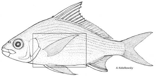
Fig. 1 Lateral view of the Mexican mojarra Eugerres mexicanus. The squared area marks the boundary of the visceral cavity.
The postemporal bone was separated from the neurocranium, the Baudelot ligament was sectioned, and, both cleithra were separated by their symphysis, in order to remove the scapular girdle. Then, we determined the location of the organs within the visceral cavity and described their topographic relationships.
The mesonephric kidney was analyzed insitu, removing from its anterior portion the cranial and spinal nerves, to which contacts, as well as the anterior cardinal vein. The archinephric ducts were identified, which were sectioned in a point of their free trajectory. The ventral aspect of this organ was observed after the removal of the gas bladder.
The mature ovaries and testes were analyzed insitu from their lateral and ventral views, while their dorsal aspect was evident after sectioning the corresponding mesenteries that hang them from the gas bladder. After the gonads were separated from the visceral cavity, they were sectioned transversally to analyze their interior.
The gas bladder was examined insitu, describing its components, its anatomical relationships, and its relative volume. It was separated from its contact with the vertebrae and with the floor of the visceral cavity.
The digestive tube and its glands were observed insitu from their lateral view; they were then separated from the pharynx by sectioning the contact of the esophagus with the pharyngeal bones; likewise, they were separated from the floor of the visceral cavity by cutting around the anus. The location and shape of the spleen was described.
The illustrations were made using a drawing tube (cameralucida) attached to a Leica Wild M3Z stereomicroscope. The terminology for the endoskeleton followed the criteria of Gregory (1959) and Kobelkowsky (2004), and that of the musculature followed Winterbottom (1974) criterion.
Results
Visceral cavity: The visceral cavity of E. mexicanus is oval shaped, both from its lateral aspect (Fig. 2A) and transverse section (Fig. 3); its length equals the cephalic length. It is delimited by the scapular and pelvic girdles and by the appendicular musculature, the precaudal vertebrae, the first pterygiophore of the anal fin, the infracarinalismedius muscles, the pleural ribs, and the hypaxialismuscles.
The largest volume of the visceral cavity is occupied by the gas bladder. The digestive tube, the liver, and the gonads (Fig. 2A, Fig. 2B, Fig. 3A, Fig. 3B) are within the lower portion of the cavity. The ovaries and testes are located between the digestive tube and the gas bladder (Fig. 2A, Fig. 2B, Fig. 3B). The spleen is situated among some anterior intestinal loops (Fig. 3A, Fig. 6D). The kidney is placed, with an extraperitoneal character, dorsal to the gas bladder and ventral to the precaudal vertebrae (Fig. 2A, Fig. 2B, Fig. 3A, 3B), and it is extended towards the base of the neurocranium (Fig. 2A).
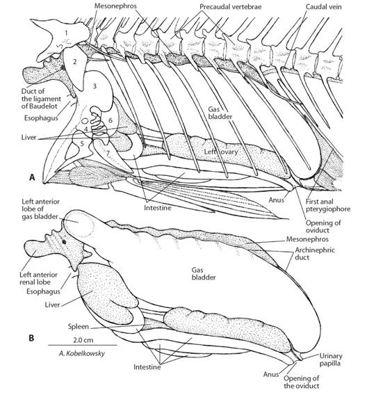
Fig. 2 Visceral cavity of Eugerres mexicanus. A. Left lateral view of the visceral cavity and its boundaries. B. Left lateral view of the organs of the visceral cavity.
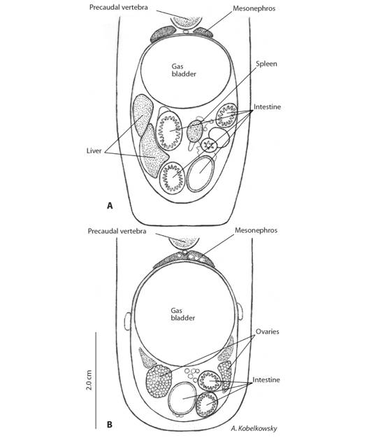
Fig. 3 Visceral cavity of Eugerres mexicanus. A. Transverse section at the level of the liver and spleen. B. Transverse section at the level of the ovaries.
Urogenital system: Kidneys. The kidneys or mesonephros are elongated and fuse with each other from the level of the ninth vertebra to the back (Fig. 4B). The beginning of its left and right portions (Fig. 2A) is located at the level of the basioccipital and exoccipital bones, these portions are called anterior renal lobes (Fig. 2A, Fig. 2B, Fig. 4A, Fig. 4B). These lobes are connected with the lateral ends of the sinus venosus of the heart and each one is crossed by the Baudelot ligament (Fig. 2A, Fig. 4A), which holds the corresponding scapular girdle.
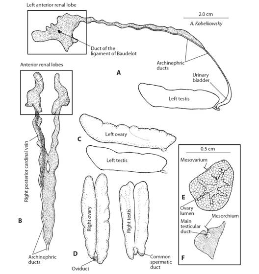
Fig. 4 Urogenital system of Eugerres mexicanus. A. Left lateral view of the mesonephros. B. Ventral view of the mesonephros. C. Left lateral view of the gonads. D. Dorsal view of the gonads. E. Transverse section of the left ovary. F. Transverse section of the left testicle.
On the ventral surface of the kidney, and parallel to the right archinephric duct, the right posterior cardinal vein is observed (Fig. 4B), and reaches the anterior left renal lobe. On both edges of the posterior portion of the kidney, the archinephric ducts are observed (Fig. 4A, Fig. 4B), they fuse together at the posterior end of the organ forming the common urinary duct. This duct descends attached to the first anal pterygiophore; however, it penetrates the back of the gas bladder where it connects to the urinary bladder. In this way, both the urinary bladder and the distal portion of the common urinary duct are located within the gas bladder (Fig. 5E). In males, the common sperm duct joins the common urinary duct forming the urogenital duct, whereas in females it remains independent.
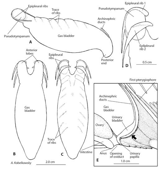
Fig. 5 Gas bladder of Eugerres mexicanus. A. Left lateral view. B. Ventral view. C. Dorsal view. D. Dorsal view of the left anterior lobe. E. Sagittal section of the posterior portion of the gas bladder. The arrow points to the floor of the visceral cavity.
Gonads. Both the ovaries and the testes are paired organs, which reach similar volumes when they mature. They are located ventrally to the gas bladder (Fig. 2A, Fig. 2B) from which they are suspended by lengthwise mesenteries (Fig. 4E, Fig. 4F). Both in the ovaries as in the testes, their left and right portions are in contact with each other (Fig. 4D).
The ovaries are elongated and their lateral edges are lobed (Fig. 2A, Fig. 2B, Fig. 4C, Fig. 4D); from their dorsal surface the mesovarium emerges (Fig. 4E), and joins to the ventral surface of the gas bladder. Both ovaries are fused in its most posterior portion, where the unique oviduct is formed as a continuation of the ovarian tunic. The female genital opening is located anteriorly to the urinary orifice. In transverse section, the ovigerous lamellae are recognized starting from the entire internal surface and maintaining the lumen (Fig. 4E). The testes are elongated (Fig. 4C, Fig. 4D), with smooth edges, and from their dorsal surface the mesorchia, that attach to the ventral surface of the gas bladder, emerge. In transverse section, each testis presents a triangular shape, with numerous fine grooves, corresponding to groups of seminiferous lobules, which converge in the main testicular duct. Dorsal to this duct, the testicular vessels, and the mesorchium can be observed (Fig. 4F). Both main testicular ducts joined together to form the common spermatic duct, which in turn joins to the common urinary duct. The urogenital opening is located anterior to the anal fin.
Gas bladder: The gas bladder of E. mexicanus (Fig. 2A, Fig. 2B, Fig. 3A, Fig. 3B) is bulky, ovoid, elongated, with two anterior lobes (Fig. 5A, Fig. 5B, Fig. 5C) which form an anterior notch, and with the posterior end of tubular aspect which is fixed to the floor of the visceral cavity (Fig. 5E). It is anchored to the first vertebral centrum and to the pleural ribs (Fig. 2A). The anterior lobes are separated from each other by the retractoresdorsalis muscles, which originate in the centrum of the first vertebrae and insert into the pharyngobranchial bones of the branchial arches. Each anterior lobe of the gas bladder is attached to the first two dorsal or epipleural ribs (Fig. 5D), in that way delimiting a small oval area of thinner walls named “pseudotympanum”. Each pseudotympanum is close to the postemporal and supracleithrum bones. The posterior end of the gas bladder is directly fixed to the floor of the visceral cavity and it houses the urinary bladder and part of the archinephric ducts (Fig. 5E).
Digestive tube and liver: As a whole, the digestive tube of E. mexicanus shows an anterior portion in an oblique position and formed by several intestinal loops, and a posterior portion of longitudinal orientation formed by three intestinal loops (Fig. 6). The digestive tube is regionalized into esophagus and intestine, and lacks stomach and pyloric caeca.
The esophagus is narrow (Fig. 6B, Fig. 6D) and starts immediately behind the upper and lower pharyngeal bones, it passes between the anterior renal lobes and the two portions of the sinus venosus of the heart it is of medium length and curves down until it reaches the intestine. The stomach is not defined, in such way; the esophagus is directly connected with the intestine; at that point the entrance of the common bile duct is recognized. The most anterior portion of the intestine is the duodenum (Fig. 6B, Fig. 6D), which shows, in its beginning, a slight bulge. The intestine forms two forward facing loops and other two facing back; the first loop makes extensive contact with the liver. From the second anterior loop, the intestine continues straight to the back to form the rectum, which opens to the outside through the anus (Fig. 2A), and it is anterior to the anal fin. The length of the intestine is about 1.3 of the standard length of the fish.
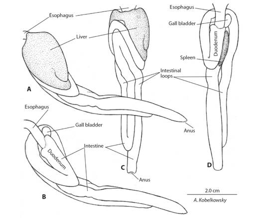
Fig. 6 Digestive tube and liver of E. mexicanus. a) Left lateral view. b) Left lateral view after removing the liver. c) Ventral view. d) Dorsal view after removing the liver.
The liver is relatively small (Fig. 2B, Fig. 6A, Fig. 6C). It surrounds the esophagus ventrally, and its largest volume is located on the left side with two small lobes in its posterior end (Fig. 3A, Fig. 6A, Fig. 6C), whereas the right portion constitutes the right hepatic lobe. The gallbladder is oval-shaped and is located on the inner surface of the right hepatic lobe (Fig. 6B, Fig. 6D). The common bile duct is short and it is connected to the digestive tube at the junction of the esophagus with the intestine.
Spleen: The spleen is a dark red surface lanceolate organ located on the right side of the duodenum (Fig. 3A, Fig. 6D).
Discussion
The morphology of the visceral cavity in the Mexican mojarraE. mexicanus, and the distribution of its organs, shows an organization that is within the general pattern of the Teleostei. The shape of the visceral cavity in the teleosts is related to the shape of the body. In that way, teleosts with a notable lateral flattening, such as the Clupeiformes and the Pleuronectiformes, show a compressed shape; those with a cylindrical body, such as the Anguilliformes, Beloniformes, and Syngnathidae, present a cylindrical cavity; and in those, whose bodies are dorso-ventrally flattened, like the Ogcocephalidae, this coelomic cavity is depressed.
The gross anatomy of the kidney of E. mexicanus is similar to that of other Perciformes, and in general the shape of the kidney in teleosts is determined by its contact with a considerable number of adjacent structures (Kobelkowsky, 2013), which is also observed in the present study. However, the special anatomical condition consisting in the entrance of the archinephric ducts to the gas bladder is to be noted, whereas in most teleosts the excretory system and the gas bladder are completely independent from each other.
Among the organs of the visceral cavity, the one that shows the highest number of special characters is the gas bladder. It develops the anterior lobes reinforced by the epipleural ribs and that carry the pseudotympana, it attaches its posterior end to the floor of the visceral cavity, and it includes the urinary bladder and part of the archinephric ducts in its interior.
The presence of the anterior lobes of the gas bladder of E. mexicanus, and its proximity with the postemporal and supracleithrum bones, is similar to that found in the genus Chaetodon (Chaetodontidae) (Webb, Smith, & Ketten, 2006) and in Eucinostomusargenteus (Gerreidae) by (Parmentier et al., 2011). Possibly, this area of the anterior lobes corresponds to the structures that Parmentier et al. (2011) referred to as “pseudotympanum” in E. argenteus, and that probably participates in a hearing mechanism. However, the pseudotympana of E. argenteus are directly coupled to foramina of the otic capsules, whereas those of E. mexicanus are located close to the skin and to the postemporal and supracleithrum bones.
The role of the gas bladder in the transmission of sound is relatively frequent in teleosts, such as the Clupeidae (Whitehead & Blaxter, 1989), the Sciaenidae (Sasaki, 1989), and mainly in the superorder Ostariophysi, in which the gas bladder is part of the Weber apparatus (Alexander, 1962), which transmits the sound to the otic capsules. Unlike E. mexicanus, the posterior end of the gas bladder of E. axilaris, E. lineatus, and E. plumieri does not reach the floor of the visceral cavity.
The most notable characters of the digestive tube of E. mexicanus are the absence of stomach and of pyloric caeca. According to Wilson and Castro (2010) the agastric character corresponds to around 20-27 % of the teleostean fishes, and the absence of pyloric caeca is common in numerous groups of fishes. Stomachless condition is also reported in wrasses (Labridae) and parrotfishes (Scaridae) (Helfman, Collette, Facey, & Bowen, 2009) and in silversides (Atherinopsidae) (Kobelkowsky & Figueroa Lucero, 2018). The intestinal length/length ratio of E. mexicanus indicates an omnivorous feeding habit.
The outstanding aspect of the gonads of E. mexicanus is the remarkable volume of the mature testes, in contrast with many teleosts. The morphology of the ovaries of E. mexicanus is similar to that described Bairdiellachrysoura (Sciaenidae) (Kobelkowsky, 2012). While the spleen in E. mexicanus is located between two intestinal loops, in teleosts generally it has a diversity of locations, as it is functionally not related to other viscera.
Ethical statement: authors declare that they all agree with this publication and made significant contributions; that there is no conflict of interest of any kind; and that we followed all pertinent ethical and legal procedures and requirements. All financial sources are fully and clearly stated in the acknowledgements section. A signed document has been filed in the journal archives.

