


 English (pdf)
 Article in xml format
 Article references
 Send this article by e-mail
 Cited by SciELO
  Similars in
SciELO
Similars in
SciELO 
 Permalink
Introduction
Mucoepidermoid carcinoma (MC) is a malignant epithelial neoplasm of the salivary glands, as described in 1945 by Stewart, Foote, and Becke. It consists of epidermoid, intermediate, columnar, and clear cells in a cystic to solid histological pattern, differentiated into three severity degrees, and has a 3:2 predilection for the female gender (1,2).
The term epigenetics was coined in 1942 by Conrad H. Waddington for changes in gene expression without a change in gene sequence (3,4). A significant relationship is observed in the expression regulation or inactivation of tumor suppressor genes (TSG) and neoplasm progression and proliferation (5) DNA methylation occurs in normal cells for the specific inactivation of certain genes; this is the most frequent epigenetic modification (6,7,8).
DNA methylation analysis has become a powerful biomarker for early changes in malignant lesion pathogenesis. In malignant neoplasms of the lung, prostate, head and neck, bladder, colon, and rectum, clinical variables such as specific histological subtypes and grades of severity are considered important predictors of prognosis (5,9). The methylation state could be considered a new associated factor; however, it is important to determine whether there is any association with TSG (9).
Rb (retinoblastoma gene) is one of the most representative TSGs. It plays a regulation role in three processes: cell cycle control, apoptosis, and differentiation (10,11). Hypermethylation of the Rb promoter region has been reported in hepatoce- llular carcinoma and gastric cancer (12,13) This shows that this feature produces modification in the cell cycle and proliferation of both neoplasms (11,14).
The CDKN2A/p16 encodes for cell cycle inhibitory proteins, which normally block cell growth and proliferation by binding to cyclin D-cyclin dependent kinase complexes (15) This binding inhibits kinase activity, arresting the cell cycle in the G1 phase. Mutations or deletions of this gene are fundamental features in carcinogenesis because their presence promotes the progress of neoplasms (15,16).
Methylguanine-DNA methyltransferase (MGMT) is involved in cellular defense against mutagene- sis and toxicity from alkylating agents. Promoter methylation has been associated with colorectal, lung, and kidney carcinomas, lymphomas, and other neoplasms (17,18,19). Another tumor suppressor gene whose promoter methylation has been detected in premalignant and malignant endometrial lesions is hMLH-1 (20,21,22)This gene participates in the DNA mismatch repair system when forming the heterodimer with mismatch repair endonuclease PMS2 to form MutL alpha, a component of the post-replicative DNA mismatch repair system (23) Considering that epigenetic changes are important features proven in many different neoplasms, it is crucial to know what happens with salivary gland neoplasms, particularly with MC (23).
Our aim was to identify the methylation state of RB, P16, MGMT, and hMLH genes in the three severity grades of MC.
Materials and methods
Population
An analytical observational pilot study was conducted with five samples (two low, two moderate, and one high severity cases) using formol fixed and paraffined embedded MC obtai- ned from the archive of histopathology diagnosis of the Oral Pathology and Medicine Department (ISO-9001:2015 certified CMX C SGC 157 2017).
An informed consent form was obtained for each sample in accordance with the integral privacy notice for patients of the Dentistry School, National Autonomous University of Mexico, affirming that the obtained data could be employed to disseminate scientific, technological, and professional knowledge (24). Clinical and demographic variables, such as age, sex, anatomic zone, and grade of severity, were collected from each data file. The histopathological diagnosis and severity grade were reevaluated and confirmed by oral pathologists according to the WHO 2017 Classification of tumors of the salivary glands. Briefly, the low degree shows a cyst histological pattern with a rich population of mucous cells and is generally well circumscribed. Intermediate degree lesions more commonly show a solid histological pattern and are less circumscribed with a diversity of appearance, including mucin extravasation. High-grade severity displays one or more of the following features: nuclear anaplasia, necrosis, an increased mitotic rate, and perineural, lymphovascular, or bony invasion, but the diagnosis of high severity requires at least focal intracellular mucin positivity (25). Pathologists CMRM and ERLH standardized their diagnostic criteria according to the criteria mentioned above. Only the five samples selected met all clinical, demographic, and histological criteria for inclusion.
DNA Extraction
A 50-µm section was obtained from each sample. Genomic DNA extraction was performed using the ReliaPrep FFPE gDNA Miniprep System (A2352, Promega, Madison, WI, USA), following the manufacturer's instructions. The sections were incubated at 80°C, and lysis buffer was added, followed by proteinase K and incubated at 56°C for one hour, and finally at 80°C for 4 hours. The samples were stored overnight at 4°C. RNase was then added, incubating for 5 minutes at room temperature, and subsequently buffer BL and 95% ethanol were added, and they were centrifuged and collected using collection tubes. The DNA collected was washed twice in washing solution and centrifuged to recover the DNA by elution buffer and final centrifugation. Quantification and evaluation of the DNA quality was carried out with the Nanodrop2000 system (Thermo Fisher Scien- tific), and samples that met the quality criteria (260/280>1.8) were considered.
Bisulfite reaction for DNA: 1μg of DNA was added in 25μl of distilled water, subsequently adding 2.75μl of 2M NaOH solution, incubated for 10min at 37C°, then 15μl of hydroquinone were added together with 260μl of sodium bisulfite for 16 hours incubation at 54 C°. DNA was extracted and 300µl of SV lysis solution and 95% ethanol were added. Finally, 5.5μl of 3M NaOH solution was added and incubated for 5min at room temperature to posteriorly add: 1μl of 10mg/ml glycogen, 6μl of 3M sodium acetate at pH 5.2 and 150μl of absolute ethanol for the DNA precipitation at -20C°. After 12 hours, the DNA was resuspended in 50μl of 10mM Tris-Cl at pH 8. The quantification and evaluation of the quality of the DNA were carried as above mentioned by Nanodrop200 system.
PCR analysis: We used 40 ng of converted DNA using the PCR Master Mix System (M7502, Promega, Madison, WI, USA). For the design of the primers we employ MethPrimer software(26) Taking the sequences for each gene form the GenBank. The primers designed were specifically to localize CpG methylation in promoter region. Oligonucleotide RT-PCR primers, were purchased from Eurofins Mwg/Operon (Louisville, KY, USA). Amplifications were performed by using a Maxygen II (Axygen, NY, USA). The temperature adjust for denaturalization and extension was to 95C° respectively, and the Tm for extension was different for each primers (Table 1). Posterior at 40 cycles a final extension at 72C° was performed for to be stored at 4C° until their resolution.
Table 1 Sequence of primers used.
| Primers for MS-PCR | - | - | - |
|---|---|---|---|
| - | Unmethylated sequence | Methylated sequence | °C Tm |
| RB1 NC_000013 | up: 5'-GGGAGTTTTGTGGATGTGAT-3' ds:5'-ACATCAAAACACACCCCA -3' | up: 5'-GGGAGTTTCGCGGACGTGAC -3' ds:5'-ACGTCGAAACACGCCCCG -3' | 54 |
| p16 NC_000009 | up: 5'-TTATTAGAGGGTGGGGTGGATTGT -3' ds: 5'-CAACCCCAAACCACAACCATAA-3 | up: 5'-TTATTAGAGGGTGGGGCGGATCGC -3' ds:5'-GACCCCGAACCGCGACCGTAA -3' | 56 |
| MGMT NC_000010.11 | up: 5'- TTTGTGTTTTGATGTTTGTAGGTTTTTGT -3' ds: 5'- AACTCCACACTCTTCCAAAAACAAAACA -3' | up: 5'- TTTCGACGTTCGTAGGTTTTCGC -3' ds: 5'- GCACTCTTCCGAAAACGAAACG -3 | 58 |
| hMLH-1 NC_000003 | up: 5'- TTTTGATGTAGATGTTTTATTAGGGTTGT -3' ds: 5'- ACCACCTCATCATAACTACCCACA -3' | up: 5'- ACGTAGACGTTTTATTAGGGTCGC -3' ds: 5'- CCTCATCGTAACTACCCGCG -3' | 52 |
Up: Upstream Ds: Downstream.
Amplification reaction products were resol- ved by using 2.0% agarose/TAE gels (Amresco, Solon, OH, USA) that were electrophoresed at 100 mV and visualized by ethidium bromide staining in a gel documentation system (Axygen). Product expression was determined through optical density by using GelQuantNet software 1.8.2. (Bioche- mLabSolutions, UCSF, CA, USA).
Briefly, a TIF image obtained for each gel was open in the software. A standardized box for select for each band was used for determinate the raw optical density in methylated and non-methylated genes.
Statistical analysis
The mean and standard deviation were obtained from the optical densities of the genes methylated and non-methylated in all grades of severity, and for the age of the study population. We performed non-parametrical Kruskall-Wallis tests to determine differences between methyla- tion in relation to severity grade. A p-value ≤0.05 was considered significant (IBM, SPSS, version 22, IBM SPSS, IL, USA).
Results
Our population has a mean age of 52.6±18.6 years old, the gender of all population was female and representing 2 low grade, 2 intermediate grade and 1 high grade of severity.
The histopathological features observed were according to the reported for MC. Briefly, in our 5 samples we observed that they are composed mostly of squamous, mucosecretory and intermediate cells, as well as, with occasionally presence of clear cells, with microcystic to cystic growth patterns.
Specifically in low grade MC, we identify mucosecretory cells with clear and granular cytoplasm, as well as intermediate cells in a cystic histological growth pattern (Figure 1. A-B). In the intermediate severity grade, we observe abundant presence of intermediate cells; as well as the presence of mucosecretory and squamous cells that were arranged in both a cystic pattern and a solid pattern (Figure 1. C-D). In the high grade, we find an abundant presence of clear cells, which are septa- ted by highly vascularized fibroconjunctive tissue, interspersed with mucosecretory and epidermoid cells in a solid pattern (Figure 1. E-F).
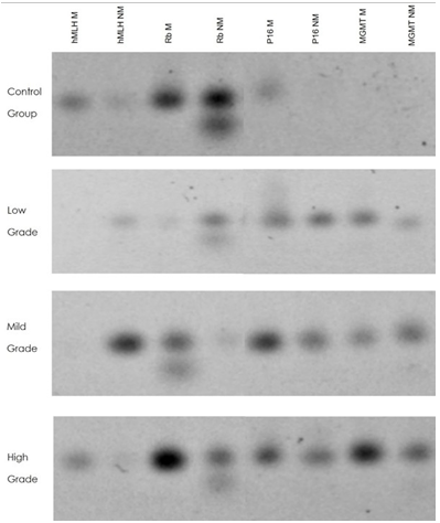
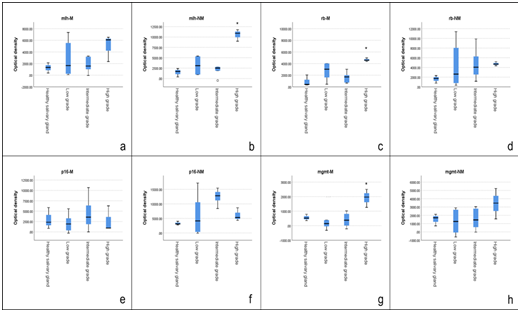
In the comparison of the presence of methylation and non- methylated genes (Figure 2) in the three histopathological degrees of the MC with respect to the control group, a statistically significant difference was observed for Rb-M in the high grade of severity with a P=0.03, as well as for MGMT-M when made the comparison of the high grade of severity, statistical significance was found with a P=0.05 In other way, statistical significance was observed for hMLH-1-NM with a P=0.04 in the high grade of severity.
Finally, we observed in hMLH-1-M and P16, that non exist a trend or statistical significance in their methylation state in any of their grades of severity (Figure 3).
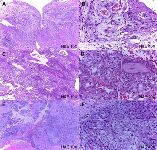
Figure 1 Histopathological photomicrographs whit hematoxylin and eosin of mucoepidermoid carcinomas. A) & B) low-grade mucoepider- moid carcinoma; C) & D) intermediate-grade mucoepidermoid carcinoma, and E) & F) high-grade or clear cell mucoepidermoid carcinoma. A), C), & E) 10 X magnification and B), D) & F) 40X magnification.
Discussion
MC is a malignant neoplasm of the salivary glands, that can develop from any salivary gland structure. This is an aggressive behavior tumor and the search of specific pathogenic pathway, has come to consider epigenetic mechanisms as possible candidates to improve knowledge about biological behavior and prognosis (1,2)
The role of Rb in the normal cell cycle as well as its involvement in the progression of malignant lesions has been a historically important research topic. The main change observed for this gene has been mutations (27,28,29). However, Zhang et al. reported that patients with hepatocellular carcinoma analyzed by MS-PCR to identify the methylation mark of the Rb, P14 and INK4 genes, Rb methylation was observed in 27.3% of the cases (23). Our results suggest a trend in methylation in relation to high grade of severity for MC. However, the unique relation between Rb and salivary gland tumor is reported for De Souza et al. that showed deregulation of Rb signaling pathway in pleomor- phic adenoma, recurrent pleomorphic adenoma and carcinoma ex pleomorphic adenoma using the immunohistochemical technique (11,30). Our results are the first time that showing a relationship between Rb, methylation and a salivary gland tumor, particularly, MC.
Michaildidi et al. in 2015 reported the expression and promotion of the methylation status of hMLH-1, MGMT, APC and CDH1, in colorectal carcinoma, indicating that 25% and 66% of cases presented increased methylation for hMLH-1and MGMT, respectively, mainly in lesions in early stages (22,23,31). In our results for MGMT gen, we observe an inverse pattern, where a higher state of methylation is observed in advanced or more severe lesions, which suggests that methylation in the pathogenesis of MC could be a late pheno- menon. In our results, p16 it did not show a trend or statistical significance, different from what was previously has been reported that inactivation by hypermethylation ocurrs mainly in early stages of carcinogenesis (Guo, X. et al.)(13,20).
However, something that must highlight is that this is the first time that this group of genes has been analyzed in MC. Therefore, we could consider that this pilot study shows us that epigenetics, particularly hypermethylation, is an area of knowledge that raises new issues to be resolved. This study could be granting more knowledge about pathogenesis of MC, even with a Little number of samples employed. Among the future achievements that could obtain by continuing with the study of the methylation of MC is to collect representative sample through multicenter studies and establish analyzes with more precise and sophisticated techniques such as pyrosequencing or microarrays. With these approaches we could correlate with a better inference if methylation, clinical features and their correlation with others important variables could be representative keys factors in MC carcinogenesis.
Conclusions
Based on the results observed, we can conclude that there is methylation in someone of the gens studies. Nevertheless, we observed a trend towards methylation for the Rb, hMLH- 1and MGMT genes in relation to the degree of severity. If we consider the basic functions of these genes, we could consider that high-grade mucoepidermoid carcinomas have alterations in cell cycle regulation, mismatch correction, and defects caused by alkylating mutagens. There- fore, these three characteristics could be involved in the heterogeneity of tumors and the intrinsic difficulty in treating them. For this reason, it is necessary to further investigate this neoplasm with more abundant populations and perhaps with a multicenter approach. Since showing a reduced population and a single case for the high-grade form of mucoepidermoid carcinoma, it is difficult to establish a trend.
Conflict of interest
The authors declare that there is no conflicto of interest for the publication of this manuscript.
Author contribution statement
Conceptualization and design: L.A.M.M., C.M.R.M. and L.F.J.A.
Literature review: L.A.M.M. and C.M.R.M.
Methodology and validation: L.A.M.M., L.F.J.A. and D.A.T.R.
Formal analysis: L.A.M.M. and L.F.J.A.
Investigation and data collection: L.A.M.M., D.A.T.R. and L.F.J.A.
Resources: L.F.J.A. and E.R.L.H.
Data analysis and interpretation: L.A.M.M., L.F.J.A., and C.M.R.M.
Writing-original draft preparation: L.A.M.M., L.F.J.A. and C.M.R.M.
Writing-review & editing: L.A.M.M., L.F.J.A. and C.M.R.M.
Supervision: C.M.R.M. and E.R.L.H.
Project administration: C.M.R.M. and E.R.L.H.
Funding acquisition: L.F.J.A. and E.R.L.H.

