


 English (pdf)
 Article in xml format
 Article references
 Send this article by e-mail
 Cited by SciELO
  Similars in
SciELO
Similars in
SciELO  uBio
uBio 
 Permalink
The purpose of this study is to implement RED Perspex dosimeters as a high-dose dosimetry system for a self-shielded irradiation system for routine control of irradiated products. The materials involved, the control during irradiation process and its intended purpose are the factors that define the application of high dose ionizing radiation procedures (International Atomic Energy Agency (IAEA), 2013). High-dose applications encompass a range of fields. In the context of food processing, doses typically range from 2kGy to 10kGy or even higher doses (Aquino et al., 2017; Eichholz 2003; Farkas & Mohácsi-Farkas, 2011). In addition, the sterilization of several materials, such as medical products require dose reaching up to 25kGy (International Organization for Standardization (ISO), 2006). Tissue irradiation, primarily for medical purposes, falls within the range of 10kGy to 25kGy (IAEA, 2005) and for cultural heritage preservation irradiation dose lie within the same ranges (IAEA, 2017). Literature provides extensive references on irradiation processes (Aquino et al., 2017; Eichholz 2003; Farkas & Mohácsi-Farkas, 2011; IAEA, 2005, 2013; ISO, 2006), including quality assurance and calibration of dosimetry systems in industrial irradiation plants (ISO, 2013a, 2017, 2020; Sharpe & Miller, 2009).
Dosimetry systems are categorized into several classes: primary standards, reference standards, routine (or working) standards, and transfer standards (Eichholz 2003; ISO, 2020). Within the routine or working dosimeters, there are RED Perspex dosimeters, which are manufactured from polymethylmethacrylate (PMMA). These dosimeters undergo changes in their optical properties when exposed to ionizing radiation. These dosimeters find applications in facilities equipped with both radioactive sources and ionizing radiation generators. They exhibit a relatively low sensitivity to radiation, making them suitable for use within a range spanning from 5kGy to 50kGy (ISO, 2019). In this study, we used the RED Perspex type 4034 dosimeters (batch PS) of a nominal thickness of 3±0.5mm (Harwell Dosimeters, 2022). These were manufactured by Harwell Dosimeters in the UK. We used a self-shielded gamma irradiator of the Isotop brand: Ob-Servo Ignis model by Izotop in Hungary, equipped with 24 Co-60 sources for irradiation of the dosimeters. The dosimeters were exposed to 5, 10, 15, 20, 25, 30, 35, and 40kGy, following the recommendations of Sharpe and Miller (2009) and ISO (2013a). The uncertainty associated with dose determination was 1,95%. We employed a cylindrical phantom made of polymethylmethacrylate (PMMA) located at the center of the irradiation chamber to irradiate the dosimeters. The phantom had 1,26cm external radius, 0,86cm internal radius, and 6,82cm in height. The phantom had the capacity to hold 2 dosimeters, so we conducted two separate irradiation rounds, utilizing a total of 4 dosimeters for each dose (ISO, 2013a), as shown in Fig. 1. The dosimeters were read using a Thermo UV-Visible Evolution 220 spectrophotometer (Thermo Fisher Scientific, Massachusetts, USA) in absorption mode at a wavelength (λ) of 640nm (Harwell Dosimeters, 2022), with a single measurement taken for each dosimeter. For this study, we utilized the specific absorbance quantity as provided in ISO
(2019).
To construct the calibration curve, the specific absorbance (A𝜆) was plotted as a function of the dose and applying a polynomial fit. The fitted curve accounted for variations in dosimeter readings at the same dose (Sharpe & Miller, 2009). The normality of the residuals was verified by the Anderson-Darling method (Montgomery, 2012) and its homoscedasticity with the Breusch-Pagan- Godfrey test (Gujarati & Porter, 2009).
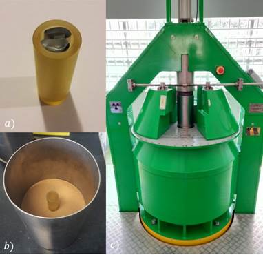
Fig. 1 (a) dosimeters position inside the PMMA phantom. (b) phantom position inside the irradiation chamber and (c) chamber placed inside the irradiation equipment
We examined several sources of dose uncertainty within the calibration process, a summary of this information is provided in Table 1.
Table 1 Uncertainty in the calibration process of the RED PMMA PERSPEX dosimeters
| Source | Type | Probability distribution |
| Dose determination | B | Normal |
| Dosimeter-Dosimeter dispersion | A | Normal |
| Calibration graph | A | Normal |
| Temperature effect | B | Normal |
| Spectrophotometer | A | Normal |
Duarte-Ladeira L. et al. (2015) reported a value of 0,01% for source decay uncertainty; for this reason, that uncertainty was not included in the uncertainty budget.
We studied the uncertainties in terms of dose. With the calibration curve, A𝜆 values were transformed into their corresponding dose. The uncertainties of calibration curve and dosimeters dispersion were determined through residual analysis (Sharpe & Miller, 2009). In the dosimeter-dosimeter dispersion, reproducibility was also included.
Regarding the influence of the spectrophotometer, two aspects were examined. The first aspect to consider was the repeatability, it was assessed in measuring the absorption by taking five measurements of absorption for the same dosimeter. The second aspect was the spectral resolution of the spectrophotometer that was 0,1nm and measured the variation in absorption resulting from this change. Both uncertainties were estimated as type A uncertainty (Joint Committee for Guides in Metrology, 2008).
The selected fitted method of the calibration curve was a third-degree polynomial Aλ = (−1.46x10⁻⁶)D3 + (1.83x10⁻⁶)D2 + (9.34x10⁻³)D + 3.94x10⁻² (Fig. 2). We confirmed
homoscedasticity and normality with p value = 0,80.

We calculated the uncertainties of the calibration curve and dosimeter-dosimeter dispersion, with results of 0,22% and 1,39%, respectively, for a total uncertainty of these two components of 1,41%. The dose uncertainty of the dosimeters as a function of the irradiated dose with the results mentioned above are showed in the Fig. 3. These uncertainties have a coverage factor k = 1.
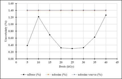
Fig. 3 Black line shows the uncertainty of the dose of the dosimeters as a function of the used dose. The orange line is the uncertainty due the dosimeter-dosimeter dispersion and the blue line is the contribution of the uncertainty due at the calibration curve and dispersion of the dosimeters.
The spectrophotometer’s uncertainties in measuring repeatability of absorption for the same dosimeter was found to be 0,03% and its contribution to dose uncertainty was 0,04%. Consequently, these uncertainties were not included in the overall dose uncertainty budget. Regarding the influence of temperature, Whittaker and Watts (1998) established that temperatures impact is insignificant at 30°C, and this was the maximum temperature to which the dosimeters were exposed while positioned inside the irradiator. Hence, the sources of uncertainty in dose as determined through dosimeter calibration were reduced to the influence of the calibration curve, dosimeters dispersion, and dose determination. A summary of this information is provided in table 2.
Table 2 Uncertainty budget of the calibration process of the RED PMMA PERSPEX dosimeters
| Source | - | Probability distribution | Sensitivity coefficient | Contribution Type | |
| Dosimeter calibration | Uncertainty | - | - | - | |
| - | - | - | - | A | B |
| Dose determination | 1,95 | N | 1 | - | 1,95 |
| Uncertainty from calibration curve | 0,22 | N | 1 | 0,22 | - |
| Dosimeter-dosimeter dispersion | 1,39 | N | 1 | 1,39 | - |
| Combined uncertainty (𝑘 = 1) | - | - | - | 2,40 | |
| Expanded uncertainty (𝑘 = 2) | - | - | - | 4,80 | |
Accurate determination of dose levels in irradiated material is of great importance to ensure quality in the irradiation process (IAEA, 2013; ISO, 2013b, 2020). The objective of this work was to implement the RED Perspex dosimeters as a high dose dosimetric system for a self-shielded irradiator in the routine dosimetric control of irradiated products. This process was validated by comparing the results obtained with standards used in industrial irradiation plants.
According to Sharpe and Miller (2009), the uncertainty resulting from the calibration curve and dosimeter-to-dosimeter dispersion yielded an average uncertainty across the dose range. This average uncertainty may result in an underestimation of uncertainty at the boundaries and an overestimation in the center of the dose range. The authors noted in their work that the effect at the boundaries of the dose range is attributed to the increase in signal-to-noise ratio at low doses. Additionally, the saturation of dosimeters at higher dose also contributes to an underestimation of uncertainty. The results obtained demonstrated this effect at the boundaries of the dose range used.
ISO (2019) indicates the estimate of expanded uncertainty achievable with measurements conducted using a routine dosimetry system, such as PMMA, typically is found within a range of approximately ±6% for k = 2. This range corresponds to approximately to 95% confidence level within a normal distribution of data. The results were obtained within this range and therefore deemed acceptable. It's important to note that these results are valid specifically for the irradiation system and dosimetry system utilized. It's crucial to recognize that calibration processes must consider the interdependence between irradiation and environmental conditions, as discussed by Whittaker and Watts (1998), Sharpe and Miller (2009) and ISO (2013a). It remains for a future study to evaluate the uncertainty of the calibration process of the AMBER Perspex dosimeters, under the same conditions carried out in this work.
The successful calibration of RED Perspex dosimeters for a self-shielded irradiation system yielded an uncertainty value in the calibration process within acceptable range indicated by ISO (2019) standard. This validation highlights to guarantee the quality and safety of irradiated product.
Ethical, conflict of interest and financial statements
The authors declare that they have fully complied with all pertinent ethical and legal requirements, both during the study and in the production of the manuscript; and that there are no conflicts of interest of any kind. This research received funding from Technological Institute of Costa Rica Research Vice Rectory under grant 1510101/1510150 from project “Pre-Clinical Treatment and Validation of Bone Tissue for Therapeutic Use”. They fully agree with the final edited version of the article. A signed document regarding all of this has been filed in the journal archives.
The statement of each author’s contribution to the manuscript is as follows: Conceptualization, WVS; Funding acquisition, WVS; Investigation, WVS; Methodology, WVS; Writing - original draft, WVS; Writing - review & editing, LRR. All authors have read and agreed to the published version of the manuscript.

