




 Inglês (pdf)
 Artigo em XML
 Referências do artigo

 Tradução automática
 Enviar este artigo por email
 Citado por SciELO
 Similares em
SciELO

 Permalink


Introduction
The diagnostic process in dentistry involves conducting a patient anamnesis and thorough clinical examination to identify the patient’s diagnostic needs (1). Based on this information, the dentist decides whether complementary radiographic examinations are necessary. The interpretation of these examinations, along with the clinical evaluation, enables the clinician to establish a diagnosis, develop a treatment plan, assess the effectiveness of the treatment, and follow-up (2).
Conventional radiographic examinations provide two-dimensional images, which can include both intraoral and extraoral radiographs. In contrast, cone-beam computed tomography (CBCT) offers three-dimensional imaging, specifically designed for dental applications. It reveals the anatomical complexity of the maxillofacial bone structures, addressing the limitations of two-dimensional images, such as the overlap of structures, distortion, and magnification (2,3,4).
Both two-dimensional and three-dimensional radiographic dental examinations use low and very low doses of ionizing radiation, so the biological effects related to patient exposure are associated with sublethal damage to DNA, mainly radiation-induced cancer (5,6,7). These effects are called stochastic (5, 6) because of their random proba-bility of occurring without a dose threshold (5). Therefore, there is no safe dose; even minimal doses of radiation carry the probability of inducing cancer (5, 6) and the probability increases as the received dose increases.
Given the inherent risk associated with exposure to ionizing radiation, the justification for requesting and performing a radiographic examination is essential. The potential benefits to the patient must outweigh any potential risks. Additionally, the chosen examination should be the most appropriate for the patient's clinical needs to ensure that the benefits surpass the risks involved (7).
The process of justifying a radiographic acquisition is a collaborative effort between the maxillo-facial radiologist and the referring dentist. The specialized training of the maxillofacial radiologist in imaging and health technologies enables them to make evidence-based decisions (5,6). Before performing or requesting a radiographic examination, the clinician should review the patient’s history, clinical information, and any previous radiographic examinations. It is also essential for the clinician to ensure that the requested study is appropriate for addressing the clinical need.
The maxillofacial radiologist is responsible for determining whether the request is justified. If it is not, the radiologist should suggest an alternative examination. They must be aware of the available alternatives and choose the most suitable one based on the diagnostic efficacy of each examination, which refers to how well each exam serves the patient’s benefit (6,8). Generally, the examination with the highest efficacy is recommended, as it provides the greatest benefit to the patient (9).
The advantages of three-dimensional examinations suggest they could replace two-dimensional examinations. However, this substitution should only occur if the three-dimensional examination proves to be more effective than the two-dimensional option for the specific needs of the patient. Currently, there is limited evidence regarding the diagnostic efficacy of CBCT for various indications in dentistry (8,10,11,12). Additionally, many clinicians receive training solely from equipment manufactu-rers and suppliers. This training often focuses only on their specific equipment and may lack evidence supporting its effectiveness in diagnostics and, consequently, its benefits for patients (8).
Due to the variety of radiographic examinations, clinical guidelines recommend their use based on evidence and diagnostic effectiveness, ensuring informed and responsible decision-making (6, 8). These recommendations help clinicians select appropriate examinations and allow radiologists to suggest tailored options (13).
Routine X-rays for new patients are not supported by evidence (14), and they are unneces-sary if there are no signs of disease or pathology since the last X-ray (1). Therefore, this review provides guidelines for prescribing radiographic examinations and serves as a consultation document for clinicians. Recommended imaging techniques are based on diagnostic efficacy and their respective advantages and disadvantages (15). These recom-mendations should be tailored to each patient’s specific needs.
Main characteristics of image formation by x-ray techniques
Different X-ray techniques applied to the same anatomical region can produce images showing the same structures. However, the projection phenomena in two-dimensional images and the reconstruction processes in three-dimensional images affect the quality and performance of the images obtained. Therefore, it is crucial to unders-tand the key aspects of image formation in the X-ray techniques used in dentistry. This knowledge will help select the technique that provides the most comprehensive information based on diagnostic needs.
Intraoral radiographic techniques
Intraoral radiographic techniques consist, mainly, of periapical and interproximal or bitewing radiographs. Periapical radiographs are useful for observing the entire tooth, from the apex to the crown, as well as the alveolar bone and the alveo-lar cortex. These radiographs can be obtained using either parallel or bisecting technique. When using parallel technique, periapical radiographs provide an accurate evaluation of the proximal coronal region. However, since the central X-ray beam does not strike this area perpendicularly, the effectiveness of this technique for evaluating enamel on the proximal surfaces and the dentin-enamel junction at the occlusal level is diminished.
Bitewing radiographs are useful for examining dental crowns, roots, and the supporting structures up to the middle third of the root. This technique enables a more accurate assessment of the interproximal surfaces and the distance between the cementoenamel junction and the crestal bone, thanks to the perpendicular angle of the X-ray beam directed at the dental crowns and alveolar crests. It allows for the detection of inter-proximal and secondary carious lesions, as well as the assessment of restorations. Additionally, it can reveal some occlusal carious lesions.
Both techniques provide high spatial resolution and contrast and can be performed in a dental office at a relatively low cost (14,15 16).
To achieve optimal performance with intraoral radiographic techniques, it is preferable to use image receptor-holders. These devices help maintain the parallelism between the longitudinal axis of the teeth and the image receptor, resulting in less distortion of structures in the images. Additionally, they enhance the reproducibility of the technique, which is essential for monitoring changes in structures over time. This also reduces the likelihood of retakes due to technical errors, ultimately leading to a lower radiation dose for patients (14,17).
In contrast, the bisecting-angle and adhesive techniques are less advisable. The bisecting-angle technique tends to produce more distortion errors, such as shortening and elongation, as well as issues with the centralization of the localizing cylinder. The adhesive bitewing technique complicates both vertical and horizontal angulation of the image receptor, often leading to increased overlap of proximal surfaces and excessive vertical angulation, which can impair diagnostic accuracy.
Extraoral radiographic techniques
The most common extraoral radiographic techniques in dentistry are panoramic radiography and lateral cephalometric teleradiography.
Panoramic radiography provides an overall view of the teeth, alveolar bone, and surrounding structures in the maxillofacial area. However, it has lower spatial resolution compared to periapical radiographs. In addition to the general limitations of two-dimensional imaging -such as overlap-ping structures, distortion, and magnification-panoramic radiography has a particular drawback: vertical distortion (16). This occurs because of the negative angulation of the X-ray beam during the imaging process, which affects the vertical relationships of the structures in the image and can obscure lesions on the crowns of the teeth. Along with horizontal distortion, positioning errors and artifacts further compromise the reliability of structural measurements (16).
While panoramic radiography has specific applications that are discussed in this article, it is crucial to note that using it routinely for evaluating new patients or at regular intervals is not justified.
Lateral cephalometric or profile teleradiography is a technique that allows for the visualization of the bones of the face and skull, as well as the profile of facial soft tissue. This standardized method is particularly useful for patients with skeletal discrepancies, as it helps evaluate the skeletal pattern (17).
Cone beam computed tomography (CBCT)
In CBCT, the X-ray tube produces a cone- or pyramid-shaped beam, generating three-dimensional images of the dentoalveolar or maxillofacial region. This technique addresses the limitations of two-dimensional images, although it has a variable spatial resolution compared to periapical radiographs (18).
The size of the tomographic volume is known as the field of view or FOV, which is measured in diameter and height and varies from small, to medium, or large, depending on each equipment. The resolution of CBCT examinations is influenced by several factors, one of which is the size of the voxel, the smallest unit of the tomographic image. Smaller voxel sizes lead to higher-resolution images. Additionally, the acquisition parameters, such as tube voltage and current, can be adjusted based on the patient's characteristics and the specific diagnostic requirements of the image (3). Therefore, a CBCT study should be customized for each individual case.
CBCT has specific indications that will be discussed later in this article. However, current evidence does not support its use for certain diagnoses because of the artifacts inherent to the technique. These artifacts can reduce diagnos-tic quality and complicate the evaluation of the coronal area of the teeth.
Criteria for the recommendations in this article
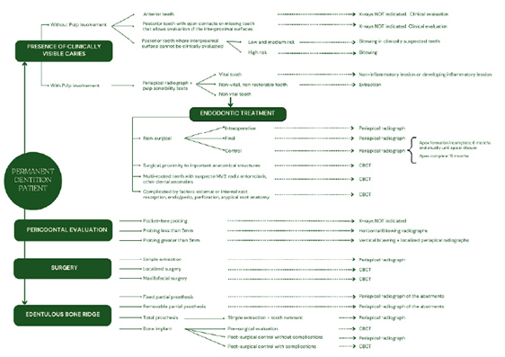
This article aims to justify the use of radio-graphic images based on specific needs. However, it's important to remember that the diagnostic approach should be comprehensive. After conducting a clinical examination, any necessary radio-graphs should only be defined if the required information cannot be obtained through clinical assessment. Once the radiographs are obtained, they should be thoroughly analyzed. Figure 1 presents a flowchart outlining the process for selecting radiographic images in dentistry.
The patient's record should include the clinical examination, symptoms and signs, the reason for the radiographic examination, the techni-que used, the date and number of radiographs taken, and the written radiological interpretation. Relevant information for specific needs should also be documented. Diagnostic errors occur when an imaging study is not thoroughly assessed, when findings are not reported, or are reported inaccurately (19, 20).
Diagnostic errors can lead to adverse outco-mes for both patient and clinician, such as late diagnosis of serious diseases, unnecessary or harmful treatments, patient mortality (21,22,23), malpractice lawsuits, and damage to the clinician's reputation (24). Factors associated with these errors include a lack of clinical history, nondiagnostic radiographic images obtained with poor technique, poor knowledge update on the part of the interpreter, observation and lighting conditions (20,25).
The dentist must provide the maxillofacial radiologist with the patient's complete clinical history and be prepared to supply previous images or additional information if required for interpreting the images (13, 15, 20). When the dentist takes and interprets the images, he or she is responsible for the entire process, which must be documented in the patient's record.
Radiographic diagnosis of caries lesions
The request for radiographs to diagnose caries is based on evaluating caries risk. This evaluation also determines the interval for taking radiographs, which is personalized for each patient.
The date of the risk assessment, the risk level (low, moderate, high), and the date of the next assessment must be documented in the patient's record (26). The intervals between subsequent radiographic examinations must be reassessed periodically, along with the evaluation of caries risk (15).
The use of bitewing radiographs with adhesive bitewings or periapical radiographs is not recommended due to previously mentioned limitations. Panoramic radiographs and CBCT are likewise not indicated (6, 15) (Table 1).
Diagnosis in endodontics
In endodontics, it is essential to use diagnostic tests such as palpation, mobility, percussion, periodontal evaluation, occlusal examination, and sensitivity (27).
The sensitivity tests for the diagnostic stage (27), the preoperative evaluation, the working length, the assessment of the final treatment, the presence of material extrusion, the type of restorative material, among others, must be recorded in the patient's record (15, 27).
Paralleling technique is the recommended technique for evaluating teeth in endodontics. The bisecting-angle technique and panoramic radiography are not recommended for the reasons previously described regarding posible vertical distortion in the obtained images (28). Routine use of CBCT is not indicated since its use has been found to lead to overdiagnosis and overtreatment (29, 30). Never-theless, there are specific cases in which CBCT must be used (Table 2).
Radiographic diagnosis in periodontal evaluation
The goal of diagnosing the periodontal disease is to identify its stage and severity (15, 31, 32), considering the depth of the probe, the loss of insertion, the extent of bone loss, the presence of bone defects, and furcation lesions (32). There is no evidence to establish the periodic acquisition radiographs for periodontal evaluation (15).
Panoramic radiographs, the bisecting technique, and the adhesive bitewing technique are not recommended due to due to their inherent technical limitations (15, 32).
Although panoramic radiographs and the complete set of periapical radiographs have been used for periodontal evaluation, it is currently recommended to avoid this practice due to the limitations of panoramic radiographs and the fact that it involves a higher dose of radiation to the patient (15, 32) (Table 3).
Radiographic diagnosis in surgery
Surgical treatment requires appropriate diagnostic tests, which include radiographic imaging (34,35,36,37). Minor procedures are detailed below. Major surgeries are not considered, as they are performed by specialists in a hospital setting (Table 4).
Radiographic diagnosis of implants
X-rays are crucial for assessing the quantity and quality of bone, as well as their relation to the position and design of prosthetic rehabilitation (6, 15, 34).
While CBCT is indicated for treatment planning, the entire acquired tomographic volume must be evaluated and its findings documented in the patient's record (Table 5).
Radiographic diagnosis of edentulous bone ridge
The request for radiographic evaluation is based on the clinical signs or symptoms and the treatment to be performed. In the absence of clini-cal signs and symptoms, taking x-rays is not justified (15, 26) (Table 6).
Radiographic diagnosis of temporomandibular disorders (TMD)
The initial evaluation of TMD is based on the clinical examination of the TMJ region and the masticatory system (38). The request for a radio-graphic examination is based on the result of the clinical evaluation, when the evaluation of the bone component is required (15, 38) (Table 7).
Radiographic diagnosis of the development of the dentition
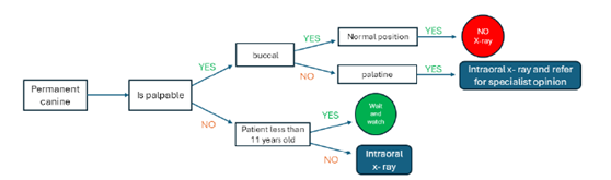
Figure 2, Figure 3, Figure 4 detail speci-fic procedures for developing dentition, and do not include orthodontic treatments performed by specialists (15, 17) (Table 8).
Table 1 Recommendations for radiographic examinations according to the risk level of dental caries.
| Risk of caries | Interproximal surface | Initial radiographic examination | Frequency of radiographic examinations |
|---|---|---|---|
| High, Medium and Low | Clinically visible | Not indicated | -- |
| High | - | Bitewing radiograph | 6 months |
| Medium | -- | Bitewing radiograph | 12 months |
| Low-primary dentition | Non-Clinically visible | Bitewing- only in clinically suspected teeth | 12- 18 months |
| Low- permanent dentition | - | 24 months |
Table 2 Recommendations for radiographic examinations in endodontics.
| Treatment stage | Radiographic examination |
|---|---|
| Diagnosis | - |
| Teeth with endodontic treatment or with extensive restorations | Paralleling technique-periapical radiography |
| Teeth with overlapping roots or nearby anatomical structures | Localization or sliding technique, using a second periapical radiograph with modification of the horizontal angulation |
| Preoperative | - |
| Evaluation of the root anatomy and the configuration of the root canal | Paralleling technique-periapical radiography |
| Nonspecific signs and symptoms | CBCT |
| Teeth with complex morphology and accessory canals (premolar, molar, and mandibular anterior teeth) | CBCT |
| Dental anomalies | CBCT |
| Intraoperative | - |
| Confirm the previously determined working length using an electronic apex locator | Paralleling technique-periapical radiography |
| Postoperative | - |
| Radiographic confirmation of the completion of treatment. Base radiograph to compare with subsequent treatment reviews and evaluate the healing process. | Paralleling technique-periapical radiography |
| Complications | - |
| Vertical fracture | Paralleling technique-periapical radiography CBCT, if periapical fails to demonstrate fracture |
| Control | - |
| There is no consensus on the radiographic interval for a control, but it is consi-dered reasonable to evaluate healing changes in the periradicular tissues 12 months after the end of treatment (15,27) | Paralleling technique-periapical radiography |
| Decrease in the size of the lesion and evidence of healing | Paralleling technique-periapical radiography-no further controls are necessary |
| No changes or evidence of healing are observed | Paralleling technique-periapical radiography-control 12 months later |
| Reassessment of treatment: no healing | CBCT |
| Non-surgical retreatment, with possible complications | CBCT |
Table 3 Recommendations for radiographic examinations according to periodontal evaluation.
| Clinical evaluation - probing depth | Radiographic examination |
|---|---|
| Less than 5 mm | Horizontal bitewing |
| 5 mm or more | Vertical bitewing |
| Paralleling technique-periapical radiography that complements specific teeth | |
| Suspected endo-periodontal lesion | Paralleling technique-periapical radiography |
| Evaluate bone defects to determine treatment or prognosis of teeth (15,31,33) | CBCT |
| Evaluate furcation lesions to determine treatment or prognosis of teeth (15,31,33) | CBCT |
Table 4 Recommendations for radiographic examinations in tooth extraction and surgery.
| Extraction | - |
|---|---|
| Single, erupted tooth | Paralleling technique-periapical radiography |
| Third molars | Panoramic |
| Surgery | - |
| Third molars: suspicion of external root resorption in the adjacent tooth (35,36) | CBCT |
| Third molars: Signs of proximity to the mandibular canal. | CBCT |
| Unerupted tooth | CBCT |
| Trauma | - |
| Dentoalveolar | Paralleling technique-periapical radiography |
| Alveolar fracture | CBCT |
Table 5 Recommendations for radiographic examinations in implantology.
| Treatment stage | Radiographic examination |
|---|---|
| Diagnosis | - |
| Post-extraction alveolar socket | Paralleling technique-periapical radiography |
| Evaluation of dental implant site | CBCT |
| Planning | - |
| Planning implant placement | Panoramic |
| Planning prosthodontic rehabilitation | CBCT |
| Intraoperative | - |
| Immediately after placement, when there is doubt about the position in relation to anatomical structures or when there are complications during surgery. | Paralleling technique-periapical radiography |
| Post surgery | Paralleling technique-periapical radiography |
| After the prosthodontic rehabilitation phase | Paralleling technique-periapical radiography |
| Baseline radiograph for comparison over time. | - |
| Control | - |
| To evaluate marginal bone levels and osseointegration. | - |
| Single implant | Paralleling technique-periapical radiography at 12 months Paralleling technique-periapical radiography every year for 5 years |
| Multiple implants | Paralleling technique-periapical radiography for each implant or a panoramic. Check-up at 12 months Check-up every year for 5 years |
Table 6 Recommendations for radiographic examinations in preprosthetic evaluation.
| Edentulous area | Clinical examination | Rehabilitation | Radiographic examination |
|---|---|---|---|
| Total | Absence of signs and symptoms | Total prosthesis | Not indicated |
| Total | Clinical suspicion of small area | Total prosthesis | Paralleling technique-peria-pical radiography |
| Total | Clinical suspicion of larger area | Total prosthesis | Panoramic |
| Total | Absence or presence of signs and symptoms | Implants | CBCT |
| Partial | Evaluation of abutments | Removable partial prosthesis | Paralleling technique-peria-pical radiography |
| Partial | Evaluation of abutments | Fixed partial prosthesis | Paralleling technique-peria-pical radiography |
Table 7 Recommendations for radiographic examinations in temporomandibular disorders.
| Clinical examination | Radiographic examination |
|---|---|
| Absence of signs or symptoms | Not indicated |
| Joint sound (“click”), in the absence of other signs or symptoms | Not indicated |
| Indicated to detect joint and soft tissue changes in the TMJ. | Magnetic resonance imaging (MRI) |
| Indicated to detect bone changes associated with the TMJ | CT or CBCT. |
Table 8 Recommendations for radiographic examinations according to the development of the dentition.
| Dentition | Radiographic examination |
|---|---|
| Temporary | Not indicated |
| Mixed | |
| Removable appliances without indication for extractions | Not indicated |
| Removable appliances with indication for extractions | Paralleling technique-periapical radiography |
| Extraction of temporary or permanent tooth | Paralleling technique-periapical radiography |

Figure 2. Radiographic image selection in early and late mixed dentition. Adaptedfrom Horner K. (15) y Isaacson K. (17).
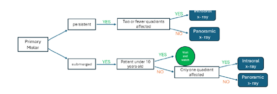
Figure 3 Radiographic image selection for a persistent second primary molar in late mixed dentition. Adapted from Horner K. (15) y Isaacson K. (17).
Conclusions
Radiographic images are an essential diagnostic tool that should be employed only after a compre-hensive history and clinical evaluation. Their use must be justified, as each type of intraoral and extrao-ral radiographic study-whether two-dimensional or three-dimensional-serves specific diagnostic purposes. It is crucial to carefully consider the limitations of each radiographic technique, as well as the effects of projection phenomena and reconstruction processes on the resulting images.
Given the rapid advancements in oral radiology, it is imperative to continuously update the criteria for imaging indications in dentistry based on the latest scientific evidence. This proactive approach not only enhances diagnostic accuracy but also prioritizes the well-being of patients and supports clinicians in their practice.
Author contribution statement: Conceptualization and design: L.B.R, A.C.R.I. and D.C.S.; Literature review: L.B.R., A.C.R.I. and D.C.S.; Formal analysis: L.B.R., A.C.R.I. and D.C.S.; Writing-original draft preparation: L.B.R., A.C.R.I. and D.C.S.; Writing-review & editing: L.B.R., A.C.R.I. and D.C.S.; Supervision: L.B.R.

