Introducción
Uno de los principales efectos del cambio climático en América Latina y El Caribe es la alteración en la distribución de las precipitaciones anuales, que puede expresarse mediante temporadas de lluvias irregulares. Proyecciones realizadas sugieren que los impactos del cambio climático afectarán la disponibilidad de agua, lo que compromete la producción de alimentos (Intergovernmental Panel on Climate Change, 2015). Los sistemas de pequeña escala son los más vulnerables, ya que son sistemas productivos que se caracterizan por depender de las precipitaciones y de la capacidad de almacenamiento de humedad en el suelo (Lemi & Hailu, 2019). Para mitigar los posibles efectos adversos del cambio climático, es necesario facilitar la adopción de alternativas de adaptación y mitigación al cambio climático, para así garantizar la sostenibilidad de los sistemas agroalimentarios.
En los últimos años, se han desarrollado múltiples técnicas y equipos para la estimación del contenido de humedad en el suelo, donde los más relevantes han sido los sensores de humedad de suelo (Lekshmi et al., 2014; Usuga & Pauwels, 2008). Independiente de su importancia en el manejo de los cultivos, el acceso a tecnologías de agricultura de precisión como sensores de humedad de suelo es un desafío para pequeños y medianos productores. Un costo elevado, complejidad de uso y la dependencia de conectividad a internet son algunas de las razones que limitan la adopción de nuevas tecnologías en agricultura (Escobar, 2016).
El proceso de adopción de innovaciones agrícolas es liderado por los grandes productores, quienes, son líderes en sus respectivos rubros, adoptan las tecnologías más temprano y por lo tanto, se benefician primero de estas (Avendaño-Ruiz et al., 2017). La razón principal por la que existe esta brecha de adopción entre pequeños y grandes productores es por falta de acceso a recursos financieros que permitan invertir en nuevas tecnologías. Por lo tanto, los pequeños productores se vuelven adoptantes tardíos, con mucha dependencia de programas gubernamentales, de extensión y organismos internacionales. El desarrollo de tecnologías adaptadas a las necesidades de sistemas productivos de pequeña escala, como los sensores de humedad de suelo, pueden reducir la brecha de adopción existente entre pequeños y grandes productores (Avendaño-Ruiz et al., 2017).
Con el propósito de reducir la brecha de adopción de tecnologías de agricultura de precisión y digital, el proyecto de Digitalización de la Agricultura de Pequeña Escala, ejecutado por la Universidad Zamorano, la Alianza Bioversity-Centro Internacional de Agricultura Tropical (CIAT) y la empresa Visualiti, trabajó en el diseño de una solución tecnológica que contribuya a la democratización de la era digital en la agricultura, con un dispositivo que permita medir y registrar humedad de suelo, robusto, de bajo costo y alta usabilidad (medida en la cual un producto puede ser usado por usuarios específicos para conseguir objetivos específicos con efectividad, eficiencia y satisfacción en un contexto de uso especificado), orientado a productores de pequeña y mediana escala.
En la actualidad, los productores de pequeña y mediana escala, que realizan agricultura familiar o de subsistencia y productores con menos de 10 ha, son los menos beneficiados de la revolución digital en la agricultura (Trendov et al., 2019). Con una solución tecnológica que les permita a los productores medir la humedad de suelo, estos pueden tomar decisiones informadas sobre el cultivo, adaptarse a diversas condiciones ambientales y utilizar recursos naturales, como el agua, de manera eficiente. El proyecto ha logrado evidenciar la necesidad y demanda de la tecnología mediante los reconocimientos que ha recibido el mismo, al ser un producto inclusivo en comparación a los dispositivos disponibles en el mercado.
La precisión de medición de humedad del suelo de los sensores está condicionada por el principio de la tecnología que utiliza el sensor y de las características del suelo, como textura, temperatura, densidad aparente y salinidad (Mittelbach et al., 2012). Múltiples autores han realizado validaciones de sensores de humedad de suelo, donde evalúan su funcionamiento del mismo en diferentes condiciones de tipo de suelo, salinidad, temperatura, contenido de materia orgánica, y densidad aparente, entre otros (Adeyemi et al., 2016; Datta et al., 2018; Gonzalez Ortiz, 2020). Luego, la precisión de las mediciones puede ser optimizada con la utilización de curvas de calibración específicas a los tipos de suelo (Usuga & Pauwels, 2008).
La evaluación del funcionamiento de los dispositivos y sensores puede realizarse en condiciones controladas de laboratorio o en condiciones de campo, mientras que las calibraciones de fábrica de los sensores de humedad de suelo se realizan en condiciones de laboratorio, que suelen ser limitadas y no reflejar las condiciones de campo (Feng & Sui, 2020). Es necesario calibrar los sensores de humedad en campo, en las condiciones en que se utilizarán, y con métodos de referencia confiables (Robinson, 2009). Algunos fabricantes afirman que los sensores no necesitan de calibración, pero esto sería cierto si los sensores son utilizados en condiciones ideales (International Atomic Energy Agency (IAEA), 2008).
Este estudio contribuye a la literatura mediante la evaluación de tres prototipos para medir y registrar humedad de suelo diseñados para agricultores de pequeña escala, en agricultura familiar y de subsistencia. A diferencia de la literatura existente, los dispositivos evaluados son productos mínimos viables con una estructura básica para medición de la humedad del suelo, con el objetivo de reducir su costo y aumentar así su probabilidad de adopción. El objetivo del estudio fue evaluar tres prototipos de dispositivos para agricultura de pequeña escala de bajo costo para medir humedad de suelo en diferentes texturas de suelo, así como determinar las respectivas ecuaciones de calibración y los efectos de conductividad eléctrica y temperatura en la medida de humedad.
Materiales y métodos
Área de estudio
Los prototipos se sometieron a pruebas de campo en parcelas seleccionadas por conveniencia que cumplieran con los criterios de: (1) fácil acceso, (2) diversidad de texturas de suelo, y (3) diversidad de cultivos, estableciéndose una parcela en Colombia y cuatro en Honduras.
La parcela de prueba en Colombia se localizó en un cultivo de café (Cofffea arabica), ubicada en Cerrillos Popayán, Valle del Cauca, Colombia. En Honduras, tres parcelas estaban localizadas en el lote La Vega, y una parcela en un lote de una finca agroecológica. Ambos lotes son parte del campus de la Universidad Zamorano, en el Valle del Yegüare, departamento de Francisco Morazán, Honduras. Las parcelas correspondientes a La Vega se establecieron en un cultivo de frijol (Phaseolus vulgaris) y el lote de la finca agroecológica en un cultivo de maíz (Zea mays). Las características de suelo de cada parcela se pueden observar en el Cuadro 1.
Cuadro 1 Características fisicoquímicas de los suelos de las parcelas de prueba, a 30 cm de profundidad, Laboratorio de Suelos Zamorano, Honduras, enero de 2022.
| Parcela de prueba | Textura | g/100g de suelo | pH | M.O. | ||
| Arena | Limo | Arcilla | ||||
| Finca agroecológica* | Franco arenoso | 64 | 20 | 16 | 5,83 | 2,65 |
| La Vega* | Franco | 32 | 46 | 22 | 5,81 | 2,47 |
| La Vega* | Franco | 40 | 40 | 20 | 5,69 | 2,05 |
| La Vega* | Franco | 26 | 50 | 24 | 5,83 | 2,57 |
| Cerrillos Popayán** | Franco arcilloso | 36 | 33 | 31 | 5,22 | 3,10 |
M.O.: materia orgánica. * Honduras. ** Colombia. / M.O.: organic matter. * Honduras. ** Colombia.
Descripción de los dispositivos
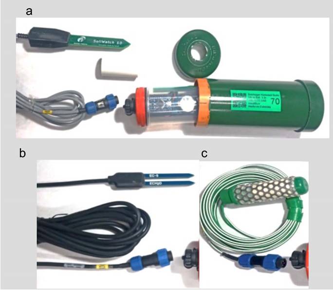
Se evaluaron tres prototipos de sensores de medición y registro de humedad de suelo, denominados prototipo A (SW 10), B (EC5) y C (SS200). Los prototipos fueron diseñados para cumplir con las características de bajo costo, de alta usabilidad y robustez, estos son muy similares en el diseño del registrador de datos y con tres sondas comerciales diferentes. Al momento de las pruebas, los dispositivos contaban con una autonomía teórica de seis meses de operación continua sin necesidad de recarga o cambio de batería. Uno de los prototipos desarrollados en el proyecto se puede observar en la Figura 1. El desarrollo de los prototipos se dio entre los meses de agosto y septiembre de 2021.

Figura 1 Imágenes de los sensores de medición desarrollados y registro de humedad de suelo, se observan el sistema de almacenamiento y batería (capsula verde) y las tres diferentes sondas. a) Sonda del modelo A. b) Sonda del modelo B. c) Sonda del modelo C. Imagen de empresa desarrolladora de tecnológica agrícola Visualiti S.A, Palmira, Colombia, septiembre de 2021.
Los dispositivos contaban con tres componentes principales: 1) la sonda de medición, 2) contenedor interior, con la batería y dispositivo registrador, y 3) la carcasa externa de protección. La carcasa de protección externa, fabricada a partir de tubo cloruro de polivinilo (PVC), les brinda robustez a los dispositivos, al protegerlos contra factores ambientales como humedad, temperatura, radiación ultravioleta, y daño mecánico. Una de las tapas era removible, para facilitar la instalación y desinstalación del contenedor interno y el sensor. El contenedor interior encapsulaba una tarjeta electrónica de procesamiento, una memoria microSD para el registro de datos y una batería de alimentación de cara para el dispositivo. La tarjeta central de procesamiento y registro de datos fue desarrollada por la empresa Visualiti SAS de Colombia, y se diferenciaban en el sensor o sonda de medición. Las sondas comerciales utilizadas fueron Soil Watch 10 (SW 10) de Pinotech, ECH2O EC-5 de Meter Group, y Watermark 200SS de Irrrometer, en los prototipos A, B y C, respectivamente. Las primeras dos sondas son de principio de medición de reflectometría en el Dominio de la Frecuencia (FDR, siglas en ingles) y la última de matriz granular. Las especificaciones técnicas de los dispositivos son de acceso abierto y están disponibles en la página del proyecto (https://www.fontagro.org/new/proyectos/digitalization-small-scale/es). Las características de medición y electrónicas de los prototipos evaluados se muestran en el Cuadro 2.
Cuadro 2 Características de especificaciones de medición de los sensores y registro de humedad de suelo brindadas por fabricante comercial de la sonda, y especificaciones electrónicas de los modelos desarrollados por el fabricante Visualiti proveedor del proyecto, Palmira, Colombia, septiembre de 2021.
| Características de medición | Modelo A | Modelo B | Modelo C |
| Sonda/sensor | SoilWatch-10 | ECH2O-EC5 | SS 200 |
| Fabricante comercial | Pinotech | Meter Group | Irrometer |
| Principio de funcionamiento | FDR | FDR | Matriz Granular |
| Unidad de medida | WCV - % | WCV - m3/m3 | kPa |
| Resolución | 0,1 % | 0,001 m3/m3 | 1 kPa |
| Rango de medición | 0 - 100 % | 0 - 1 m3/m3 | 0 - 200 kPa |
| Características electrónicas | |||
| Amperaje | 24 mA | 20 mA | 100 mA |
| Voltaje de trabajo | 3,1 - 5 v | 2,5 - 3,6 v | 3-5 v |
| Tiempo de respuesta | 200 ms | 100 ms | 60 ms |
| Tipo de salida | Voltaje | Voltaje | Resistencia |
FDR: Reflectometría de dominio de tiempo. / FDR: Frequency domain reflectometry.
Descripción de los dispositivos control o de referencia
Los sensores Teros 12® y Teros 21® (Meter Group, Inc.) fueron identificados como los sensores de referencia contra los cuales se compararon los prototipos desarrollados. Estos sensores son de alta confiabilidad, aprobados por instituciones de prestigio, desarrolladores de equipos tecnológicos, universidades y empresas agrícolas (Meter Group, n.d.a; Meter Group, n.d.b). Ambos sensores fueron acoplados a un dispositivo de carga, almacenamiento y administración de energía diseñado por Visualiti y utilizado a escala comercial.
El sensor FDR Teros 12® brinda estimaciones de contenido volumétrico del agua (m3/m3), conductividad eléctrica (dS/m) y temperatura en el suelo (°C). El sensor Teros 21® mide el potencial hídrico del suelo, compuesto por un sensor de humedad y cerámica porosa. El dispositivo mide el contenido de humedad en la cerámica y lo convierte a potencial hídrico. Este brinda estimaciones del potencial matricial en kpa y la temperatura en el suelo (°C). Los datos obtenidos de los sensores de referencia fueron utilizados para la precisión y exactitud de los prototipos y como valores de referencia para la estimación de las ecuaciones de calibración. Para los prototipos A y B el control utilizado fue el sensor Teros 12®, mientras que para el modelo C se utilizó el sensor Teros 21®.
Establecimiento del ensayo e instalación de los sensores
La instalación de los dispositivos en la parcela experimental de Colombia inició en el mes de diciembre del año 2021 y finalizó en febrero del año 2022. En Honduras, los dispositivos fueron instalados entre enero y febrero del año 2022 para las parcelas de frijol y abril en las parcelas de maíz. Cada una de las parcelas de prueba consistió en cinco dispositivos, que correspondían a los tres prototipos y sus respectivos controles. La profundidad de instalación de los sensores fue a la profundidad promedio esperada de las raíces del cultivo. En el caso del cultivo de frijol se instaló a una profundidad de 15 cm, para cultivo de maíz 30 cm en café a 25 cm.
La instalación de los dispositivos se realizó con un barreno manual donde se preparó un orificio de instalación de 10 cm de diámetro. Se procuró evitar la perturbación del suelo en el punto de instalación del sensor, se realizó un orificio pequeño que permite volver a empaquetar (regreso del suelo según los horizontes) de manera correcta. Se realizó una verificación de la correcta instalación y registro de humedad a las 24 h posterior a la instalación.
Recolección de datos
Se recolectaron datos se realizó durante 60 días en el cultivo de frijol, 90 días en maíz y 90 días en café. A través de los dispositivos de control, se recolectó los datos de conductividad eléctrica (CE) y temperatura del suelo. La frecuencia de registro de todas las variables fue cada 60 min.
Análisis de datos
El análisis de datos constó de tres etapas: 1) estadística descriptiva, 2) evaluación de la exactitud y precisión de las mediciones, y 3) análisis de regresión para la estimación de las curvas de calibración.
Para evaluar la exactitud de las mediciones, se realizaron pruebas t-estudiante emparejadas, de las medidas de los prototipos y su respectivo control a las mismas horas. La precisión fue evaluada mediante un modelo de regresión lineal simple con la medida de humedad del prototipo como variable independiente y la medición del control como variable dependiente, donde el coeficiente de determinación del modelo es la medida de precisión. La estimación de las curvas de calibración se realizó mediante un modelo de regresión lineal múltiple, según se muestra en la ecuación 1.

Donde, β0 es el intercepto, β1 es el coeficiente de ajuste a la medida del prototipo, β2 es el coeficiente de ajuste para la conductividad eléctrica, β3 es el coeficiente de ajuste para la temperatura, %VWC es el contenido volumétrico de agua (%), CE conductividad eléctrica del suelo. La inclusión de las variables de CE y T° se realizó para los datos recolectados en la parcela La Vega, para los modelos A y B, correspondientes a los sensores FDR.
Es importante destacar que se tuvieron todas las consideraciones posibles para igualar la condición en la que se instalaron los sensores, sin embargo, por la variabilidad en los suelos era natural esperar diferencias en las medidas, ya sea por leves diferencias en las condiciones de suelo o por el alto número de observaciones, que tienden a reducir el error estándar y aumentar la probabilidad de encontrar diferencias estadísticas significativas, pero no prácticas. En todos los modelos se contó con más de 800 observaciones, a excepción del prototipo B en La Vega 40 % arena.
Ranking y selección de la mejor alternativa
Para evaluar de forma integral los dispositivos y definir la mejor alternativa para el proyecto, se hizo un ranking de los prototipos según su exactitud, precisión y costo, se calculó un puntaje para cada prototipo donde se sumó sus posiciones en los rankings. La mejor alternativa se identificó por tener el puntaje más bajo.
Resultados
Se encontraron diferencias estadísticas significativas entre las mediciones de humedad de los prototipos y sus controles (Cuadro 3), se rechazó la hipótesis nula de una diferencia media igual a cero. Las medidas del sensor C (SS200) se presentan separadas por tener diferente unidad de medición (Cuadro 4).
Cuadro 3 Estadística descriptiva de la diferencia de la medición de humedad de suelo expresado en volumen de agua por volumen de suelo (%) de los prototipos A y B (sensores de medición y registro de humedad de suelo). Parcelas La Vega, Honduras y Cerrillos, Popayán, Colombia, entre los meses de noviembre 2021 y febrero 2022.
| Parcela | Prototipo | N | Diferencia media* | DE σ | Valor p | Frecuencia porcentual |
| Cerrillos Popayan | A (SW 10) | 1342 | -6,18 | 1,41 | <0,0001 | 100 |
| B (EC5) | 1342 | 11,14 | 1,42 | <0,0001 | 100 | |
| La Vega | A (SW 10) | 810 | -1,24 | 1,80 | <0,0001 | 100 |
| Arena 40% | B (EC5) | 621 | 1,84 | 1,09 | <0,0001 | 77 |
| La Vega | A (SW 10) | 775 | -1,39 | 0,89 | <0.0001 | 100 |
| Arena 32% | B (EC5) | 775 | 6,90 | 1,40 | <0,0001 | 100 |
| La Vega | A (SW 10) | 810 | -4,22 | 1,10 | <0,0001 | 100 |
| Arena 26% | B (EC5) | 810 | 9,08 | 0,72 | <0,0001 | 100 |
| Finca agroecológica | A (SW 10) | 1153 | -13,71 | 0,94 | <0,0001 | 100 |
| B (EC5) | 1153 | 5,47 | 0,54 | <0,0001 | 100 |
*Diferencia media y desviación estándar expresados en puntos porcentuales donde se utilizó el valor de prototipo menos valor de referencia. N: número de observaciones; DE: desviación estándar. / *Mean difference and standard deviation expressed in percentage points using prototype value minus reference value. N: number of observations; SD: standard deviation.
Cuadro 4 Estadística descriptiva de la diferencia de la medición de humedad de suelo expresado en volumen de agua por volumen de suelo (%) del prototipo C (sensor de medición y registro de humedad de suelo). Parcelas La Vega, Honduras y Cerrillos, Popayán, Colombia, entre los meses de noviembre 2021 y febrero 2022.
| Parcela | Prototipo | N | Diferencia media | DE σ | Valor p | Frecuencia porcentual |
| La Vega: arena 32% | C (SS200) | 775 | -59,47 | 6,17 | <0,0001 | 100 |
| La Vega: arena 40% | C (SS200) | 810 | -174,30 | 10,32 | <0,0001 | 100 |
| La Vega: arena 26% | C (SS200) | 810 | -20,73 | 17,13 | <0,0001 | 100 |
| Finca agroecológica | C (SS200) | 1003 | -1,94 | 14,18 | <0,0001 | 87 |
*Diferencia media y desviación estándar expresados en puntos porcentuales donde se utilizó el valor de prototipo menos valor de referencia. N: número de observaciones; DE: desviación estándar. / *Mean difference and standard deviation expressed in percentage points using prototype value minus reference value. N: number of observations; SD: standard deviation.
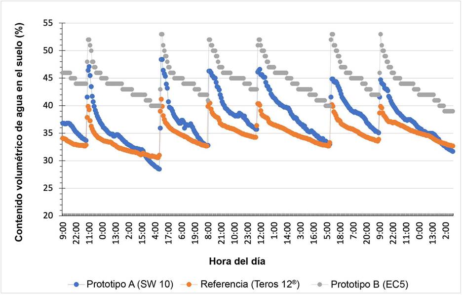
En la diferencia de las medias se puede observar que el prototipo A (SW 10), para todas las parcelas evaluadas, subestimó el contenido de humedad de suelo real del suelo (diferencia media negativa). Mientras que el prototipo EC5 sobrestimó el contenido de humedad del suelo con respecto al sensor de referencia (diferencia media positiva). La Figura 2 muestra un ejemplo de las mediciones de los prototipos A (línea azul) y B (línea gris) con respecto al control (línea naranja), en un suelo franco en Honduras.

Figura 2 Ejemplificación de comportamiento de sensores de medición y registro de humedad de suelo de los prototipos A (SW 10) y B (EC5) y sensor de referencia (Teros 12®), a través del tiempo, Parcela de La Vega 40 %, Honduras, enero de 2022.
Para el prototipo C se observó el mismo comportamiento, donde se observó la diferencia de las medias estadísticas diferente de cero. En cuanto a las mediciones del prototipo C se observó valores negativos en el valor de la diferencia media, por lo que el prototipo subestimó las mediciones de humedad con respecto al sensor de referencia (Cuadro 4).
Comportamiento según tipo de suelo
En los suelos con mayor contenido de arena, correspondientes a la parcela de la finca agroecológica y Cerrillos Popayán, fue donde los dispositivos mostraron menor precisión para los tres prototipos evaluados. En los suelos arenosos, los coeficientes de determinación de los modelos fueron menores a 0,70. En contraste, en los suelos con menor contenido de arena y mayor arcilla, tuvieron mejor ajuste de los modelos. Se observó que los tres prototipos evaluados presentaron una mejor precisión o ajuste a la regresión lineal en el suelo con 32 % de arena, donde todos obtuvieron un ajuste superior a 0,95 (R2=0,95). El segundo mejor ajuste de los modelos se observó en el suelo con 26 % de arena, con valores de los coeficientes superiores a 0,92 (R2=0,92) (Cuadro 5).
Cuadro 5 Resultados de los análisis de regresión simple entre prototipo y sensor de medición y registro de humedad de suelo, de referencia para cada parcela de prueba, Parcelas La Vega, Honduras y Cerrillos, Popayán, Colombia, entre los meses de noviembre 2021 y febrero 2022.
| Parcela %A-%Ar | Prototipo | R | R 2 | RMSE | Modelo de regresión y = β0 + β1 x1 + ε |
| Cerrillos Popayán | A- SW 10 | 0,78 | 0,60 | 0,53 | %VWC = 14,36 + 0,81 (% VWC SW10) |
| 36 - 31 | B- EC5 | 0,78 | 0,62 | 0,53 | %VWC = -1,43 + 0,84 (% VWC EC5) |
| La Vega: | A- SW 10 | 0,90 | 0,80 | 1,80 | %VWC = 1,68 + 0,98 (% VWC SW10) |
| 40 - 20 | B- EC5 | 0,95 | 0,90 | 1,08 | %VWC = 3,35 + 0,64 (% VWC EC5) |
| C - SS200 | 0,95 | 0,90 | 84,68 | Kpa = 95,25 + 7,78 (kpa SS200) | |
| La Vega: | A- SW 10 | 0,99 | 0,98 | 0,90 | %VWC = 12,84 + 0,59 (% VWC SW10) |
| 32 - 22 | B- EC5 | 0,97 | 0,95 | 1,40 | %VWC = 4,54 + 0,68 (% VWC EC5) |
| C - SS200 | 0,95 | 0,90 | 28,08 | Kpa = 78 + 5,19 (kpa SS200) | |
| La Vega: | A- SW 10 | 0,93 | 0,86 | 1,10 | %VWC = 17,03 + 0,40 (% VWC SW10) |
| 26 - 24 | B- EC5 | 0,97 | 0,94 | 0,72 | %VWC = 15,91 + 0,38 (% VWC EC5) |
| C - SS200 | 0,94 | 0,89 | 17,07 | Kpa = 27,54 + 3,23 (kpa SS200) | |
| Finca Agroecológica 64 - 16 | A- SW 10 | 0,84 | 0,70 | 0,94 | %VWC = 17,74 + 0,76 (% VWC SW10) |
| B- EC5 | 0,95 | 0,90 | 0,54 | %VWC = 14,77 + 0,44 (% VWC EC5) | |
| C - SS200 | 0,80 | 0,65 | 14,18 | Kpa = 11,34 + 2,07 (% kpa SS200) |
%A: porcentaje de arena; %Ar: porcentaje de arcilla; R: coeficiente de Pearson; R 2 : coeficiente de determinación; %VWC: % contenido volumétrico de agua. / %A: percentage of sand; %Ar: percentage of clay; R: Pearson coefficient; R 2 : coefficient of determination; %VWC: % volumetric water content.
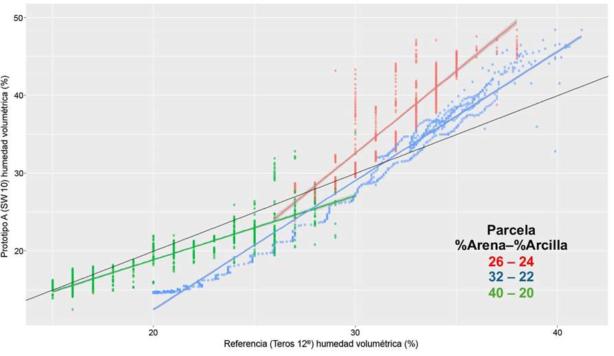
El tipo de suelo afectó la medición del prototipo con la sonda SW10 (Figura 3). La sonda SW10 subestimó el contenido de humedad en suelos con mayor contenido de arena, en los suelos con menos arena y más arcilla, con respecto a la sonda de referencia (Figura 3).
Comparación entre prototipos
Al comparar los tres prototipos se determinó que en cuatro de los cinco sitios evaluados, el prototipo B presentó un mejor ajuste al modelo de regresión, con valores del coeficiente de determinación (R2) superiores a 0,9, a excepción de la parcela Cerrillos Popayán donde se obtuvo un valor de 0,62. En la parcela La Vega (arena 32 %), el prototipo A fue el que presentó un mejor ajuste al modelo de regresión, con un valor de R2 de 0,98. Por lo tanto, según el criterio de precisión, el mejor prototipo corresponde al modelo B. Los ajustes del prototipo A fueron similares a los del prototipo C.
La inclusión de las variables independientes de conductividad eléctrica y temperatura del suelo a los modelos de regresión resultaron en diferencias estadísticas significativas con respecto a los modelos simples. En todos los casos, los modelos múltiples tuvieron mejor ajuste, es decir, un coeficiente de determinación mayor, en comparación con los modelos de regresión lineal simple (Cuadro 6). Por lo tanto, la inclusión de las variables de conductividad eléctrica y temperatura contribuyen a la estimación de mejores curvas de calibración de los dispositivos.
Cuadro 6 Cambio en el ajuste del modelo de regresión con la inclusión de las variables de conductividad eléctrica y temperatura en los modelos A y B (sensores de medición y registro de humedad de suelo). Parcelas de La Vega, Zamorano, Honduras, enero de 2022.
| Parcela %A-%Ar | Prototipo o Modelo | R2 ajustado | R2 ajustado inclusión CE y T | Cambio del R2 | Valor p |
| La Vega: | A (SW 10) | 0,80 | 0,91 | 0,11 | < ,001 |
| 40 - 20 | B (EC5) | 0,90 | 0,93 | 0,03 | < ,001 |
| La Vega: | A (SW 10) | 0,98 | 0,99 | 0,01 | < ,001 |
| 32 - 22 | B (EC5) | 0,95 | 0,98 | 0,03 | < ,001 |
| La Vega: | A (SW 10) | 0,86 | 0,93 | 0,07 | < ,001 |
| 26 - 24 | B (EC5) | 0,94 | 0,96 | 0,02 | < ,001 |
%A: porcentaje de arena; %Ar: porcentaje de arcilla; R: coeficiente de Pearson; R 2: coeficiente de determinación; %VWC: % contenido volumétrico de agua; CE: conductividad eléctrica del suelo. / %A: percentage of sand; %Ar: percentage of clay; R: Pearson coefficient; R 2: coefficient of determination; %VWC: % volumetric water content; CE: electrical conductivity of the soil.
El mayor cambio en el coeficiente de determinación se puede observar en el prototipo A, que presentó un aumento en el coeficiente de determinación de 11 puntos porcentuales en el suelo con mayor cantidad de arena. El sensor que presentó el mejor ajuste al incluir las variables de conductividad y temperatura fue el modelo A.
Curvas de calibración
En cuanto a las curvas de calibración de los modelos A y B, se puede observar que, para ajustar el porcentaje de humedad del suelo al valor de referencia, esta debe reducirse entre 0,19 y 0,52 puntos porcentuales por cada grado adicional de temperatura del suelo (Cuadro 7). Se observó un comportamiento similar en el caso de la conductividad eléctrica, ya que en casi todas las ecuaciones el coeficiente de conductividad eléctrica fue negativo en la curva de calibración. Valores más altos de conductividad eléctrica resultaron en sobrestimación de la medición del contenido de humedad de suelo para ambos prototipos modelos (A y B). Los coeficientes muestran que por cada dS/m la lectura se debe ajustar entre 8 y 55 puntos porcentuales. Los valores de conductividad en los suelos evaluados se ubicaron entre 0,10 y 0,45 dS/m.
Cuadro 7 Curvas de calibración con la inclusión de las variables conductividad eléctrica y temperatura para los modelos A y B (sensores de medición y registro de humedad de suelo). Parcelas de La Vega, Zamorano, Honduras, enero de 2022.
| Parcela | Modelo | Curva de calibración |
| La Vega: arena 40% | A- SW 10 | %VWC = 23,84 + 0,20 (% VWC SW10) + 28,78 (CE) - 0,52 (Temp. C) |
| B- EC5 | %VWC = 11,68 + 0,76 (% VWC EC5) - 8,66 (CE) - 0,47 (Temp. C) | |
| La Vega: arena 32% | A- SW 10 | %VWC = 21,84+ 0,58 (% VWC SW10) - 19,77 (CE) - 0,34 (Temp. C) |
| B- EC5 | %VWC = 15,84 + 0,73 (% VWC EC5) - 55,63 (CE) - 0,44 (Temp. C) | |
| La Vega: arena 26% | A- SW 10 | %VWC = 32,45 + 0,37 (% VWC SW10) - 32,04 (CE) - 0,47 (Temp. C) |
| B- EC5 | %VWC = 22,91 + 0,37 (% VWC EC5) - 18,51 (CE) - 0,19 (Temp. C) |
%VWC: % contenido volumétrico de agua; CE: conductividad eléctrica del suelo. / %VWC: % volumetric water content; CE: electrical conductivity of the soil.
Ranking y selección de la mejor alternativa
El prototipo B con la sonda EC5 fue el más preciso, mientras que el prototipo A fue más exacto y de menor costo, mientras que el prototipo C no fue el mejor en ninguno de los criterios (Cuadro 8). La mejor alternativa fue el prototipo A con la sonda SW-10.
Cuadro 8 Resultados de mejor desempeño de prototipos desarrollados (sensores de medición y registro de humedad de suelo) según las pruebas realizadas y selección final, análisis de Parcelas La Vega y Finca agroecológica, Honduras y Cerrillos, Popayán, Colombia, entre los meses de noviembre 2021 y febrero 2022.
| Descripción de selección o prueba | Modelo de dispositivo | ||
| A (SW-10) | B (EC5) | C (SS200) | |
| Resultado de análisis de medida de precisión (1: mejor desempeño) | 2 | 1 | 3 |
| Resultado de análisis de medida de exactitud (1: mejor desempeño) | 1 | 2 | 3 |
| Resultado análisis económico (1: menor costo) | 1 | 3 | 2 |
| Sumatoria | 4 | 6 | 8 |
| Puntaje | 1,33 | 2 | 2,67 |
Puntaje: valores cercanos a 1, presentan un mejor desempeño. / Score: values close to 1 present a better performance.
Discusión
Los resultados de la sección anterior sugieren que el sensor más preciso fue el prototipo B, mientras que el más exacto fue el prototipo A. La precisión de los sensores medida a través de los coeficientes de determinación (R2) en los modelos de calibración fueron menores en los suelos con un mayor porcentaje de arena. Esto concuerda con los hallazgos de Gonzalez Ortiz (2020) donde se observó que sensores FDR de bajo costo presentaron un mayor ajuste (R2) en suelos Arcillosos y Francos, con respecto a los suelos de texturas gruesas (arenosos). En contraste, las mediciones realizadas por los sensores en los suelos arcillosos y francos fueron los menos exactos, al tener menor diferencias con respecto al control. Si se prefiere precisión a exactitud la mejor alternativa sería el prototipo B.
Los modelos con los mejores coeficientes de determinación se encontraron en los suelos francos-arcillosos, con valores entre 0,86 y 0,98, mientras que en los suelos con mayor contenido de arena tuvieron un valor de 0,65 y 0,9. Estos resultados son consistentes con González-Teruel et al. (2019), quienes en una evaluación de sensores de humedad de suelo capacitivos de bajo costo obtuvieron coeficientes de determinación de 0,8 y 0,98, para suelos arenosos y francos, respectivamente. Las ecuaciones de calibración determinadas por González-Teruel et al. (2019) fueron funciones de segundo y tercer orden, mientras que las del presente se utilizó regresión simple y múltiple (primer orden).
Los resultados encontrados sugieren que, en los suelos con mayor contenido de arcilla los sensores de capacitancia fueron más precisos, pero otros estudios afirman que la precisión disminuye a medida que incrementa el contenido de arcilla (Datta et al., 2018; Sugita et al., 2016). En suelos con altos niveles de arcillas y sales se presentó un mayor error en la predicción que en sitios de menor contenido de arcilla al evaluar cinco sensores de humedad electromagnéticos y capacitivos (Datta et al., 2018). A diferencia del presente estudio, los suelos arcillosos del estudio de Datta et al. (2018), tenían un alto contenido de sales expresado en CE. Este contraste de resultados sugiere que la textura del suelo por sí misma, no es determinante en la precisión de los sensores, y que otros factores como la salinidad son de igual importancia.
Las curvas de calibración donde se incluyó CE como variable explicativa sobreestiman el contenido volumétrico de agua a medida que aumenta el valor de la CE. Diversos estudios han destacado el impacto que tiene la salinidad en los sensores electromagnéticos en estimar el contenido volumétrico del agua (Dalton, 1992; Wyseure et al., 2005). Por ejemplo, Dalton (1992) determinó que, en suelos salinos la constante dieléctrica estimada por los sensores TDR aumenta, y por lo tanto, sobrestima el contenido de humedad. Esto sugiere que de forma general y sin importar el tipo de sensor utilizado, en suelos más salinos las mediciones serán más imprecisas.
Las parcelas donde se evaluó los prototipos presentaron valores de conductividad eléctrica en un rango entre 0,1 y 0,5 dS/m, lo que los cataloga como suelos no salinos. Según Dalton (1992) la sobrestimación ocurre cuando los valores de CE sobrepasan 8 dS/m, mientras que Wyseure et al. (2005) determinaron que el error era aceptable en niveles de CE menores a los 2 dS/m, y que la sobreestimación de la lectura de contenido volumétrico inferiores pueden despreciarse. Sin embargo, la salinidad medida como CE es una variable que debe ser incluida en las curvas de calibración, como se demostró en este estudio. La inclusión de la CE como variable explicativa incrementó hasta en 11 puntos porcentuales la capacidad del modelo de explicar la variación en humedad del suelo (R2).
Las curvas de calibración sugieren que al incrementar la temperatura del suelo los modelos sobrestiman el contenido volumétrico de agua. Cada 1 °C adicional de temperatura en el suelo, aumentó la estimación del contenido volumétrico de humedad entre 0,19 % y 0,52 %. Esto es consistente con el estudio de Oates et al. (2017), donde indican que el aumento en la temperatura del suelo incrementa la capacitancia medida por el sensor y por ende, sobrestima la humedad. Esto difiere con lo encontrado en el estudio de Zhu et al. (2019), donde aseguran en su estudio de evaluación de sensores FDR en suelos de textura fina y gruesa, que los sensores exhibieron sensibilidad sustancial a los incrementos de temperatura del suelo, al subestimar en 0,034 % (m3/m3) el contenido de humedad volumétrico por cada 1 °C adicional. Por ende, la influencia de cambios en la temperatura del suelo en la precisión de sensores difiere entre las clases texturales y tipo de sonda. Se sugiere que los sensores deben incluir detector de temperatura para realizar una corrección de la influencia de este factor (González-Teruel et al., 2019).
La inclusión la CE y temperatura del suelo en el desarrollo de las curvas de calibración incrementó la capacidad predictiva de los modelos, con aumentos entre un 2 % y 11 % del R2 para todos los sensores de las Parcelas de la Vega. Por lo que, se sugiere que las curvas de calibración de sensores de humedad de suelo sean realizadas en las condiciones donde operara el dispositivo y no utilizar las calibraciones proporcionadas por los fabricantes. En un estudio se evaluó el ajuste de ecuaciones de calibración desarrolladas por las empresas que fabrican sensores de humedad en condiciones de campo y se determinó que cada sensor necesita de una ecuación de calibración única que contemple tipo de suelo (Varble & Chávez, 2011).
Conclusión
El prototipo más exacto fue el prototipo A, mientras que el más preciso fue el prototipo B. El prototipo A fue la mejor alternativa al considerar precisión, exactitud y costo. Las curvas de calibración estimadas demostraron que los prototipos sobrestimaron las medidas del contenido de humedad volumétrico al presentarse valores mayores de conductividad eléctrica y temperatura, y que, por lo tanto, su estimación es necesaria al instalar los dispositivos.


