




 Inglês (pdf)
 Artigo em XML
 Referências do artigo

 Tradução automática
 Enviar este artigo por email
 Citado por SciELO
 Similares em
SciELO
 uBio

 Permalink


Introduction
Pseudoplatystoma magdaleniatum (family Pimelodidae) described by Buitrago-Suárez & Burr (2007), commonly known as striped catfish, surubí or pintadillo, is a potadromous fish endemic of the Magdalena River basin and the largest within the ichthyofauna of this fluvial system, reaching up to 1.4 m in length and 70 kg in weight. It inhabits the main rivers of the basin, as well as its floodplains. It is characterized by its elongated dark gray body and white belly with dark transverse stripes, large head, and small eyes located in a dorsal position (Buitrago-Suárez & Burr, 2007), the upper jaw is longer than the lower jaw and the teeth are small (Lasso et al., 2011). It is a carnivorous species that is located among the higher trophic positions within the ecosystems of the basin, feeding mainly from fish and shrimp, which represent approximately 78 % of its stomach content (Cortés-Millán, 2003).
P. magdaleniatum is a carnivorous catfish, primarily feeding on smaller fish and crustaceans, which places it at the top of the river’s trophic chain (Ayala, 2022). This species typically inhabits the main branches and floodplains of the Magdalena River basin, with seasonal migrations related to reproduction and feeding patterns (Mojica et al., 2012). Therefore, understanding the gastrointestinal morphology of P. magdaleniatum in relation to its feeding habits and habitat is crucial for comprehending its ecological role and for developing effective conservation strategies.
P. magdaleniatum is one of the species with the highest commercial value in the Magdalena River basin; however, this fish is subjected to high fishing pressure, which has led to a decline in natural populations and a reduction in catch by approximately 90 %, which is why this species is currently listed as critically endangered in the red book of Colombian freshwater fish (Mojica et al., 2012). Different conservation strategies have been proposed for this species, such as the establishment of a legal minimum capture size, which is currently 80 cm (Lasso et al., 2011), as well as closed seasons and semen cryopreservation programs (Herrera-Cruz et al., 2019). In Colombia, other strategies have been proposed aimed at the recovery and conservation of fishing resources through fish stocking programs, taking into account technical recommendations, such as the stocking time and site, the guarantee of genetic purity, the diversity of fish in the body of water and the survival of the individuals destined for repopulation programs (Atencio, 2001), as well as optimal environmental conditions for adaptation; however, these processes imply an appropriate management of the reproducers through factors such as density and feeding (Atencio, 2001; Esmaeili et al., 2024).
The study of the structure and organization of the gastrointestinal tract in fish is of great interest because, in general, there is a correlation between the structure of a fish’s digestive system and its dietary habits (Hassan, 2013); thus, the basic understanding of this system allows optimizing procedures required in the management of broodstock in fish stocking programs and consequently, the conservation of species of great ecological, reproductive, nutritional, economic and cultural value such as P. magdaleniatum. Therefore, the objective of this study was the morphological, histological, and histochemical characterization of the gastrointestinal tract of the P. magdaleniatum, an endemic fish species of the Magdalena River basin.
Material and methods
Animal ethics: All procedures related to animal handling were performed in accordance with the Guide for the Care and Use of Laboratory Animals (Albus, 2012), the meeting minute 134 of the committee, and a permit granted by the National Aquaculture and Fisheries Authority of Colombia-AUNAP, under the Resolution 0955 (May 27th, 2020).
Animals and husbandry: A total of 22 adult specimens (males and females) of P. magdaleniatum were used, which were captured from the natural environment in the Magdalena River basin, between the years 2018-2020 and kept in the facilities of the Piscícola San Silvestre Fish Farm (PSS). Prior to the experimental procedures, the individuals were transferred to land ponds at a density of one fish per m2 and were subjected to a 24 h quarantine period in filtered water and two baths with salt (20 ppm for 30 seconds).
Body indexes: For the morphometric characterization, from the 22 fish initially collected, 15 specimens were selected for detailed analysis based on their condition and size range to ensure a representative sample. The remaining 7 specimens were excluded due to damage during collection or to avoid bias from extreme size variations of which total (TL), standard (SL), intestinal (IL) and gastrointestinal (GIT) length (cm) measurements were recorded; body weight (g) (BW), eviscerated (EW), gastrointestinal content (esophagus, stomach, intestine-GIC) and individual weight of the following organs: esophagus, stomach, intestine, liver, gonads. For these measurements, an ichthyometer and an Ohaus® digital scale were used.
Measurements of all individuals were made with the same instrument. Photographic records were made with an EOS Rebel T3i camera with a Canon EF-S 18-55 mm f/4-5.6 IS STM lens for the description of the anatomical characteristics.
From these measurements the following morphometric relationships were calculated:
Condition factor (K) = (Body Weight (BW)) / (Total length3(TL3) ×100
Intestinal Coefficient (CO) = (Intestinal Lenght (IL)) / (Total Lenght (TL))
Zihler’ s Index (Z.I) = (IL) / (10× ∛ (TW))
Gonadosomatic Index (GSI) = (Gonads Weight (GW)) / (TW)×100
Hepatosomatic Index (HSI) = (Liver Weight (LW)) / (TW) × 100
Carcass Yield (CY) = (Dressed Carcass Weight (DCW)) / (TW)
Histology and histochemistry analysis: To characterize the microscopic morphostructure of the gastrointestinal tract, the organs of the gastrointestinal tract (esophagus, stomach, and intestine) were removed, and fixed in 10 % buffered formalin solution in a ratio of 1 : 10 for 24 hours. The organs were also injected with this solution for their subsequent preservation.
Tissues were dehydrated in ethanol at serial concentrations and subsequently cleaned with toluene or equivalent, followed by imbibition in hot paraffin and cold molding. Sections with a thickness of 5 to 6 μm were made with a rotary microtome.
The sections obtained were stained with hematoxylin and eosin (H&E), according to standard procedures, and photographs were taken with an Olympus CX 23 optical microscope and Basler ACA5472-17UC digital camera for characterization of the structure of the digestive system (Gosavi et al., 2019).
For the detection of neutral mucins, the PAS (Periodic Acid-Schiff) technique was used, and for acidic mucins, AB staining (Alcian Blue) was performed with pH 1.0 and pH 2.5 (AB pH1.0 and AB pH 2.5, respectively). The sequential technique AB pH 2.5 + PAS was carried out to detect the association between neutral and acidic mucins. For the specific detection of mucins of epithelial origin and acidic mucins from the gastrointestinal tract, staining with mucicarmine was used. Masson’s trichrome staining was used to detect collagen fibers in connective tissue.
Transmission Electron Microscopy (TEM): A tissue fragment is fixed in 2.5 % buffered glutaraldehyde in PBS. Samples were post-fixed in 1 % osmium tetroxide and 3 % uranyl acetate, progressively dehydrated, infiltrated in 1 : 1 plastic resin mixed with acetone, and embedded in SPURR resin (Electron Microscopy Sciences, Fort Washington, PA, USA). Plastic blocks were cut with a Sorvall MT2-B ultramicrotome. Semithin sections (1 µm) were stained with toluidine blue and evaluated to identify appropriate areas for ultrathin sections. These areas were diamond cut to a thickness of 80-100 nm (interference color yellow-gold) and placed on 200 mesh copper grids. Subsequently, the sections were contrasted with uranyl acetate and lead citrate and were examined and photographed with a JEOL 1400 Plus transmission electron microscope from the Hospital Universitario Fundación Santa Fe de Bogotá, Department of Pathology.
Statistical analysis: A completely randomized experimental classification design was applied. Descriptive statistics were used to characterize the sample regarding morphological and zootechnical parameters, body parameters and indices were expressed as mean ± STD (standard deviation). The histochemical results were interpreted based on the staining intensity of the tissue for each staining technique. The level of intensity was determined by qualitative inspection and the staining intensity results were described by means of a table of crosses.
Results
The gastrointestinal tract of P. magdaleniatum was characterized by presenting the oropharyngeal region followed by a short sac-shaped esophagus connected to the stomach followed by a short, slightly coiled intestine that lacks pyloric caeca (Fig. 1).
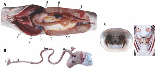
Fig. 1 A. Internal body organization of P. magdaleniatum showing branchial arch, liver, esophagus, stomach, intestine, gallbladder, swim bladder, and gonads. B. Gastrointestinal tract: esophagus (es), stomach cardial region (ca), stomach fundic region (fu), stomach pyloric region (pyl), anterior intestine (ai), middle intestine (im), posterior intestine (ip). C. Oral cavity with structures similar to dental plaques observed in the background.
The data for body measurements and digestive organs of P. magdaleniatum is presented in Table 1. On average, the 15 individuals analyzed weighed 1690.33 ± 827.6 g. The total and standard body heights were 65.8 ± 6.1 cm and 55.3 ± 5.7 cm, respectively (Table 1).
Table 1
Records of body data of 15 individuals of striped catfish (P. magdaleniatum).
Measurement
Mean
SD
Minimum
Maximum
Weight (gr)
1690.3
827.6
1075.0
4426.0
Eviscerated (gr)
1586.9
805.0
978.0
4220.0
TL (cm)
65.8
6.1
59.0
83.0
SL (cm)
55.3
5.7
50.0
72.0
GIT (cm)
26.6
9.5
13.8
48.7
GIC (gr)
53.4
40.1
26.6
192.6
Esophagus (gr)
8.2
3.1
4.5
14.2
Stomach (gr)
12.9
6.2
6.3
32.2
Intestine (gr)
7.6
2.8
5.5
16.5
Intestine (cm)
32.4
6.2
24.5
53.0
Liver (gr)
8.7
4.5
5.1
22,7
Gonads (gr)
4.7
4.6
1.2
16.3
Abbreviations: TL= Total length, SL = Standard length, GIT = Gastrointestinal length, GIC = Gastrointestinal content, SD = Standard deviation.
The body indices for the 15 specimens analyzed are presented in Table 2. The average value of the condition factor (K) was 0.56 ± 0.07. Additionally, the intestinal coefficient (IC) measured 0.49 ± 0.08, while the Zihler index (ZI) averaged 0.64 ± 0.17. The hepatosomatic index (HSI) had an average value of 0.51 ± 0.12, the carcass yield (CY) was 0.94 ± 0.10, and the gonadosomatic index (GSI) averaged 0.3 ± 0.24.
Table 2
Body index of 15 individuals of striped catfish (P. magdaleniatum).
Measurement
Mean
SD (±)
Minimum
Maximum
0,56
0,07
0,47
0,77
CO
0,49
0,08
0,40
0,75
ZI
0,64
0,17
0,23
0,93
HSI
0,51
0,07
0,38
0,66
CY
0,94
0,10
0,63
1,12
GSI
0,30
0,24
0,10
0,94
Abbreviations: K = Condition factor, CO = Intestinal coefficient, ZI = Zihler’ s Index, HIS = Hepatosomatic index, CY = Carcass yield, GSI = Gonadosomatic index, SD = Standard deviation.
Histology and histochemistry analysis
Esophagus: Histologically, the esophagus exhibits a mucosal lining characterized by a non-keratinized stratified squamous epithelium throughout its entire length. Abundant mucus-secreting cells are present along the length of the organ, as well as below the epithelium. Additionally, round and oval cells with eosinophilic cytoplasm were observed, each containing a centrally located basophilic round nucleus; some of these cells appeared binucleated (Fig. 2A), potentially corresponding to club cells. Beneath the basal membrane of the epithelium, a thick layer of loose connective tissue was observed. This connective tissue layer is likely the submucosa propria, as no muscular layer was observed within the mucosa, resulting in an absence of separation between the lamina propria and the submucosal layer.
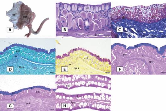
Fig. 2 A. Macroscopic image of the portion of the digestive tract of P. magdaleniatum made up of the esophagus (esf), stomach (est) and intestine (int). From image. B. histological organization and distribution of esophageal mucins. Stratified squamous epithelium made up of goblet cells (cc) and club cells (ccl) is observed on the basal lamina (up arrow) and submucosal lamina propria (lp-s). The presence of neutral (image d - sign +) and acid (images e, f and g - sign *) mucins were detected in the goblet cells. Staining: b) Hematoxylin and Eosin 100X. C. Masson’s trichrome 40X D. PAS 40X. E. Mucicarmin 40X. F. Alcian Blue (AB) pH 2.5 40X. G. Alcian Blue (AB) pH 1.0. H. PAS/AB pH 2.5.
Moving towards the outer layers, we observed a layer of striated skeletal muscle, which corresponds to the muscular tunic. In certain sections, two layers became apparent: an inner circular layer and an outer longitudinal layer, with the latter appearing thicker. External to the tunica muscularis, a thin layer of loose connective tissue was observed, which corresponds to the tunica adventitia (Fig. 2B). Within the initial third of the esophageal mucosa, the presence of corpuscles or taste buds was noted. Employing combined staining techniques, we observed numerous goblet cells with an oval morphology that covered the entire length of the esophagus.
In the cranial third, we exclusively observed labeling for sulfated and carboxylated acid mucins (Fig. 2E, Fig. 2F). In the middle portion, labeling was observed for both sulfated and carboxylated acidic mucins, along with neutral mucins, with the labeling of acidic mucins being predominant (Fig. 2H). Finally, in the distal third, which is closest to the stomach, we exclusively observed labeling for neutral mucins (Fig. 2D).
Stomach: The stomach exhibited a sac-like structure divided into three regions: the cardiac, fundic, and pyloric regions, each of which, presenting mucosa with folds. Histologically, this organ consisted of four layers (mucosa, submucosa, muscular, and serosa). The mucosal layer was lined by a simple columnar epithelium composed of tall, slender cells with oval nuclei primarily located toward the basal region. Beneath the epithelium was the lamina propria (LP), and within it, there were abundant gastric glands that formed clusters. These glands were either divided or surrounded by connective tissue septa and primarily comprised oxynticopeptic cells, which were most prominently observed in the cardiac and fundic regions.
In the pyloric region, the glands predominantly presented cylindrical cells. Beyond the lamina propria, the mucosal layer presented a slender layer of smooth muscle known as the muscularis mucosae. Below the mucosal layer, there was a substantial layer of connective tissue, corresponding to the submucosal layer, comprised of TCL (type connective tissue layer). External to this, the muscularis layer was observed, consisting of two layers of smooth muscle: an inner circular layer and an outer longitudinal layer. Externally, a thin layer of TCL, termed the tunica serosa, was present. The distribution of mucins in all stomach regions exclusively corresponded to neutral mucins in both the epithelial and glandular regions (Fig. 3).
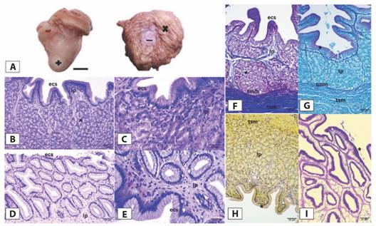
Fig. 3 A. Macroscopic image of the stomach of P. magdaleniatum, external view of the stomach (+), internal view and folds inside the stomach (x), opening to the intestine (-). From image. B. Histological organization and distribution of stomach mucins. The tunica mucosa of the stomach is made up of a simple columnar epithelium (ecs) and the lamina propria (lp) (images b, c, d and e). The presence of gastric glands (up arrow) is observed in this region. On the tunica mucosa, the tunica muscularis mucosa (tmm) and the tunica submucosa (tsm) made up of connective tissue (image f) can be seen. The presence of neutral mucins is detected (image g - sign *); however, acid mucins are not detected (image g - sign *). B.-E. Hematoxylin and eosin 100X. F. Masson’s trichrome 40X. G. PAS 40X. H. Mucicarmin 40X. I. PAS/AB pH 2.5.
Intestine: The intestine of P. magdaleniatum was divided into three regions: anterior (Fig. 4), middle (Fig. 5) and posterior-rectal (Fig. 6). The three regions presented a mucosal tunic covered by a simple columnar epithelium composed mainly of absorptive cells (enterocytes) in the anterior and middle portions and a smaller number of mucus cells (goblet cells). In the posterior portion, goblet cells are more abundant, and the number of enterocytes is reduced. Additionally, the mucosa was characterized by the presence of long folds that are projected towards the lumen and its branches, forming an intricate pattern of distribution. Beneath the epithelium, we observed a well-visible layer of TCL corresponding to the lamina propria. External to the mucosal layer, we observed the muscularis layer, consisting of two layers of smooth muscle: an inner circular layer and an outer longitudinal layer. Beyond this layer, there was a thin layer of loose connective tissue known as the tunica serosa. Histochemical analysis revealed the presence of acid sulfated, carboxylated, and epithelial mucins, with a predominant presence. Additionally, neutral mucins were also detected. The labeling was more pronounced in the apical portion of the folds, as the number of mucus-secreting cells was higher in this region compared to the middle and basal sections.
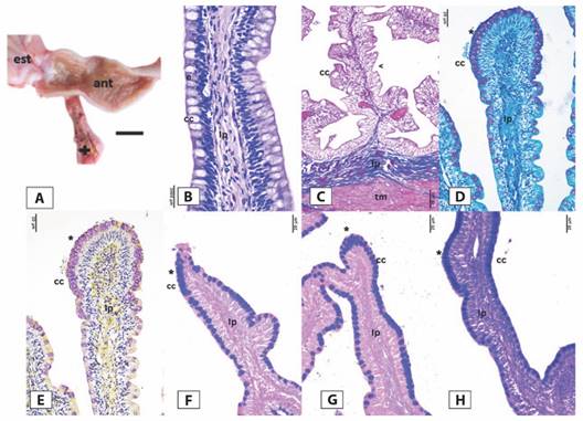
Fig. 4 A. Macroscopic image of the anterior region of the intestine (ant) of P. magdaleniatum. Part of the stomach (est) is also observed, as well as an extension of the intestine (+). From image. B. Histological organization and distribution of mucins from the anterior region of the intestine of P. magdaleniatum. The simple columnar epithelium made up of enterocytes (e) and goblet cells (cc) is observed (image b). Followed by the epithelium, the lamina propria (lp) and the muscular tunic (tm) are observed, as well as the folds of the intestine that branch off (arrow to the left - image). C. The presence of neutral mucins is detected in the cc (image d), as well as carboxylated and sulfated acidic mucins (images E., F. and G.). Stains: B. Hematoxylin and eosin 100X. C. Masson’s trichromic 40X. D. PAS 40X. E. Mucicarmin 40X. F. Alcian Blue (AB) pH 2.5 40X. G. Alcian Blue (AB) pH 1.0. H. PAS/AB pH 2.5.
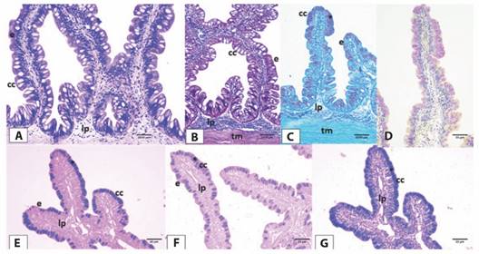
Fig. 5 A. Histological organization and distribution of mucins from the middle region of the intestine of P. magdaleniatum. The simple columnar epithelium made up of goblet cells (cc) and enterocytes (e) is observed. B. The presence of connective tissue corresponding to the lamina propria (lp) and the muscular tunic (tm) is observed. C. The presence of neutral mucins in the goblet cells is detected. D.-G. As well as the presence of carboxylated and sulfated acidic mucins. Staining: A. Hematoxylin and eosin 100X. B. Masson’s trichrome 40X. C. PAS 40X. D. Mucicarmin 40X. E. AB pH 2.5 40X. F. AB pH 1.0 40X. H. PAS/AB pH 2.5.
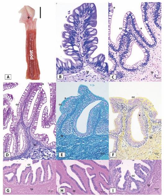
Fig. 6 A. Macroscopic image of the posterior region of the intestine (post) and rectum (+) of P. magdaleniatum. B. Histological organization and distribution of mucins in the posterior region of the intestine. A.-B.The simple columnar epithelium of the posterior region of the intestine is observed, made up of goblet cells (cc) and enterocytes. C. Followed by loose connective tissue that forms the lamina propria (lp). D. The presence of neutral mucins. E.-H. as well as carboxylated and sulfated acidic mucins is detected. Stains: B.-C. Hematoxylin and eosin 100X. D. Masson’s trichrome 40X. E. PAS 40X. F. Mucicarmin 40X. G. AB pH 2.5. H. AB pH 1.0. I. PAS/AB pH 2.5.
The presence of neutral mucins was detected throughout the digestive tract of P. magdaleniatum, observing a higher intensity of staining in the stomach. Acidic mucins were detected in the esophagus, except in the caudal portion and in the entire stomach. They were also observed in all three regions of the intestine.
In the organ epithelium of the GIT of striped catfish, goblet cells are identified in the tunica mucosa of the esophageal epithelium, some projecting into the lumen (Fig. 7A). In the stomach, the epithelium is observed with a simple organization and with the presence of some more electron-dense nuclei compared to the cytoplasm; likewise, the presence of goblet cells is not detected (Fig. 7B). Concerning the intestine, we observed the presence of enterocytes in the mucosal layer of the epithelium. These enterocytes featured microvilli projecting toward the lumen from the apical region of the cell, effectively increasing the surface area for absorption and secretion in the intestine. Additionally, goblet cells were also evident, characterized by the presence of mucin granules within them. These granules varied in electron density, with some being more electron-dense than others. Furthermore, these goblet cells projected their contents toward the lumen of the organ (Fig. 7C, Fig. 7D, Fig.7E).
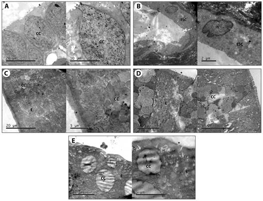
Fig. 7 TEM micrographs of the epithelia of the gastrointestinal tract of P. magdaleniatum. A. Esophagus with the presence of goblet cells (cc). B. Stomach, with simple columnar epithelium (ESC). The presence of goblet cells is not detected. C.-E. Anterior, mid and posterior intestines, respectively. In all three regions, the presence of goblet cells, enterocytes. E. And microvilli (asterisk *) projecting towards the lumen of the intestine is observed.
Discussion
The relationship between body length and weight is commonly employed in fish studies as it enables the establishment of potential growth and nutrition patterns (Bagenal & Tesch, 1978). The condition factor (K) is used as a health indicator in studies on fish biology (Muller-Gomiero et al., 2008). This parameter is influenced by variables such as the fish’s age, the sampling season, sex, individual’s stage of sexual maturity, the amount of stomach and intestinal content, as well as fat reserves and muscle mass (Barnham & Baxter, 1998).
This factor allows comparisons to be made between fish populations subjected to different conditions of climate, temperature, feeding, densities, and water quality (de Oliveira-Felizardo et al., 2011). High condition factor values indicate that animals have experienced favorable conditions, whereas low values suggest less favorable conditions (Radkhah & Eagderi, 2015). The catfishes analyzed were captured in their natural environment in the Magdalena River basin at different times and their condition factor K at the time of sampling was 0.56 ± 0.07.
According to Nandita & Ujjania (2017), K factor values equal to or greater than 1.0 are adequate since they would indicate a good level of feeding and appropriate environmental conditions. On the other side, Bagenal & Tesch (1978) recommended K value ranges for freshwater fish between 2.9 and 4.8. Although the K factor calculated for the specimens analyzed in this study represents a single measurement taken at one point in time, the range of values obtained (0.44 and 0.77) may be related to variability in the reproductive cycle of the specimens, which has been reported in captive fish populations (Herrera-Cruz et al., 2019).
On the other hand, intestinal length is a morphometric parameter commonly associated with diet and eating habits in vertebrates (Kramer & Bryant, 1995). In teleost fish, there is a well-documented close relationship between intestinal morphology and diet. Herbivorous fish tend to exhibit elongated, thin, loop-organized intestines, whereas fish with carnivorous diets typically have larger intestines, less folding, and shorter lengths relative to their body size (Day et al., 2014). Based on various studies, it has been possible to categorize fish according to their feeding habits (Al-Hussaini, 1949; Kapoor et al., 1976; Ward-Campbell et al., 2005). Taking these references into account, along with the results of the intestinal coefficient (0.49 ± 0.08) and Zihler index (0.64 ± 0.17) obtained for the analyzed catfish in this study, it can be concluded that they fall within the range associated with carnivorous fish habits (0.2 to 2.5).
The hepatosomatic (HSI) and gonadosomatic (GSI) indices serve as indicators of reproductive activity in fish due to the mobilization of energy reserves from the hepatopancreas to the gonads (Revathi et al., 2012). According to Hisao (1985), teleost fish have an average HSI between 1-2 with a maximum of 3. The average HSI of P. magdaleniatum in this study was 0.51, suggesting that the analyzed catfish had low liver reserves at the time of sampling, possibly as a result of the 24 h quarantine period. Additionally, the average GSI was 0.3, which may be related to the low HSI. Mitu (2017) suggests that during the spawning period, the HSI tends to increase in correlation with the GSI. This, in turn, would indicate that variations in the HSI are linked to the storage of energy reserves for reproduction. However, it is important to note that this effect could not be conclusively determined in the catfish analyzed since these indices were assessed at a single time point. Hence, it is recommended to consider this aspect in future studies.
Based on the histological analysis of the esophagus in P. magdaleniatum, it exhibited a stratified epithelium within the mucosal layer, featuring goblet cells and taste corpuscles. This histological observation aligns with descriptions in catfish, such as Glyptosternum maculatum, whose esophageal epithelium is also stratified in nature and characterized by an abundance of goblet cells and the presence of taste buds in the anterior region, as reported by (Xiong et al., 2011). These authors suggest that the presence of taste buds on the lips and in the esophagus would indicate that the food is selected before and during the ingestion process, so that the presence of striated skeletal muscle in the esophagus of P. magdaleniatum would be involved in the generation of voluntary movements allowing the fish to regurgitate unwanted food.
In the esophagus of P. magdaleniatum, the presence of Club cells or alarm cells was also observed. Faccioli et al. (2014) report the presence of Club cells in the esophagus of Hemisorubim platyrhynchos (family Pimelodidae), with a negative reaction to mucin production, indicating a possible defense function against epithelial damage when food is ingested. According to (Chivers et al., 2007), club cells exhibit holocrine secretion within epithelial wounds of the esophagus, suggesting their role as a defense mechanism for the underlying tissues during prey ingestion. The organization of the stratified epithelium is associated with the esophagus’s protective function against abrasion. Additionally, the presence of mucosal cells aids in lubrication during swallowing, facilitating the ingestion process (Wilson & Castro, 2010).
On the other hand, while in P. magdaleniatum there is a separation between the epithelium and the lamina propia-submucosa by means of the basal lamina and absence of the muscularis mucosae, in G. maculatum the muscularis tunica extends directly into the lamina propia and submucosa (Xiong et al., 2011). In the case of the esophagus of Trachelyopterus striatulus (Siluriformes, Auchenipteriadae), the submucosa tunica presented abundant connective tissue, similar to P. magdaleniatum. The absence of the muscularis mucosa might be countervailed by the presence of abundant connective tissue, which plays an important role in the protection and strengthening of the esophagous in carnivorous fish, similar to the walking catfish, Claris batrachus (Raji & Norouzi, 2010). The muscular tunica presented circular and longitudinal muscle fibers, like T. striatulus (dos Santos et al., 2015).
In fish, the stomach is involved in food storing and hydrochloric acid production (Purushothaman et al., 2016). In a study conducted on the morphology and histology of the gastrointestinal tract of the catfish Pimelodus maculatus (Pimelodidae), similar findings to those observed in P. magdaleniatum were documented. These similarities included the histological organization of the three regions of the stomach and the presence of a simple columnar epithelium in the mucosa. Additionally, the study identified glands composed of oxynticopeptic cells within the lamina propria, followed by the muscular layer of the mucosa, which consists of smooth muscle, and, finally, by the serosa (Santos et al., 2007). In H. platyrhynchos, gastric glands made up of oxynticopeptic cells related to the production of hydrochloric acid were observed (Faccioli et al., 2014), so these cells are associated with a high energy demand due to ionic exchange (Naguib et al., 2011). The presence of abundant gastric glands is a characteristic of carnivorous fish, since they are essential in the digestion of protein-rich foods (Purushothaman et al., 2016). In catfish such as Pachypterus khavalchor, a reduction in the number of gastric glands is observed towards the pyloric region of the stomach, which could indicate that this part is more involved with food storage than with digestion (Gosavi et al., 2019).
Similar results were found in P. khavalchor (Siluriformes: Horabagridae), where the examination revealed the presence of four layers (mucosa, submucosa, muscularis, and serosa) in the gastrointestinal tract. The transition from a stratified epithelium in the esophagus to a simple columnar epithelium in the stomach was observed, as well as the absence of mucous cells. Notably, the lamina propria and gastric glands were observed in detail. Like in P. magdaleniatum, the separation between the tunica mucosa and submucosa was evident due to a thin layer of smooth muscle known as the lamina muscularis mucosae. Additionally, both circular and longitudinal muscle layers were recognized within the muscularis layer, and the outermost layer, known as the serosa, was also identified (Gosavi et al., 2019). The presence of thick muscular and serous layers in the stomach is associated with the fragmentation and unwinding of the food bolus, which favors the secretion of digestive enzymes (Faccioli et al., 2014).
H. platyrhynchos is a pimelodid with distribution in the Amazon and Orinoco basins (Lasso et al., 2011). As in striped catfish, the presence of a simple columnar epithelium made up of enterocytes and goblet cells is reported for the intestine of this species, with greater abundance of the latter in the posterior region of the intestine (Faccioli et al., 2014). The enterocytes and goblet cells present in the epithelium could fulfill a lubrication and absorption function in the intestine, facilitating the transit of food (Purushothaman et al., 2016). Other studies reported that the microvilli present in the apical zone of enterocytes could amplify the area of intestinal absorption. The intestine of the striped catfish presented a high degree of folding and an absence of pyloric caeca, which is consistent with that reported for G. maculatum, whose intestinal mucosa presented a great abundance of folds and goblet cells (Xiong et al., 2011).
In terms of histochemical characterization, neutral mucins were observed throughout the gastrointestinal tract of the striped catfish. However, acidic mucins were detected in the cranial and middle sections of the esophagus and throughout the intestine, but they were not observed in the caudal part of the esophagus or in the stomach. These findings align with those reported for the catfish P. khavalchor (Gosavi et al., 2019), T. striatulus (dos Santos et al., 2015), H. platyrhynchos (Faccioli et al., 2014), Pelteobagrus fulvidraco (Cao & Wang, 2009) and Rhamdia quelen (Hernández et al., 2009). Some authors suggest that the abundance of goblet cells and the presence of acidic mucins in the esophageal epithelium are associated with the ability to direct food more easily due to the lubricating action provided by these proteins, as well as protection against pathogens and mechanical damage (de Oliveira-Ribeiro & Fanta, 2000; Xiong et al., 2011) which, along with taste buds, would allow a high degree of food selection by P. magdaleniatum. Conversely, the presence of neutral mucins would be associated with the transformation of the bolus into chyme, which would indicate pre-gastric digestion (Cao & Wang, 2009).
Similar to the yellow catfish P. fulvidraco (Cao & Wang, 2009), the exclusive presence of neutral mucins in the stomach of striped catfish would be associated with enzymatic cooperation during digestion, as well as the transformation of food into chyme, the absorption and transport of molecules across cell membranes. They also have a buffering effect on the acidity level in the stomach (Petrinec et al., 2005). According to Cao & Wang (2009) the presence of gastric glands, as well as the secretion of neutral mucins in the stomach of yellow catfish provide a great absorption and digestion capacity, as well as a high concentration of collagen fibers that make up the stomach lamina propria-submucosa, which increases stomach elasticity, and favors food storage capacity.
The presence of neutral mucins in the intestine could be related to enzymatic activity in food degradation (Anderson, 1986), while acidic mucins in the intestine would be linked to the protection of the epithelium against degradation by the action of glycosidases (Carrassón et al., 2006). In the intestinal epithelium of striped catfish, the presence of sulfated and carboxylated acid mucins was observed, which are related to the absorption of proteins, ions, among other particles and, in turn, are involved in the protection of the epithelium against the action of pathogens, which facilitates the transit of food (Díaz et al., 2008).
The abundance and distribution of goblet cells observed in the intestine of P. magdaleniatum provide important insights into its digestive adaptations. Similar to other carnivorous fish species, such as the sea bass Dicentrarchus labrax (Carrassón et al., 2006), P. magdaleniatum exhibits a high concentration of goblet cells, particularly in the posterior region of the intestine. This pattern is consistent with the need for increased mucus production to facilitate the transit of protein-rich food and protect the intestinal epithelium from mechanical damage (Sukkhee et al., 2024).
However, the distribution pattern of goblet cells in P. magdaleniatum differs markedly from that observed in omnivorous species like Oreochromis niloticus, which shows a more uniform distribution along the entire length of the intestine (Díaz et al., 2008). This difference likely reflects the distinct digestive requirements of carnivorous and herbivorous diets. In herbivorous fish, the uniform distribution of goblet cells may aid in the continuous lubrication necessary for processing plant material, which often requires longer transit times and more extensive mechanical breakdown (de Matos et al., 2021).
Furthermore, the types of mucins produced by these goblet cells in P. magdaleniatum, with a predominance of acidic mucins in the posterior intestine, align with observations in other carnivorous fish species such as the European catfish Silurus glanis (Kozarić et al., 2008). This prevalence of acidic mucins may serve multiple functions, including protection against bacterial colonization, facilitation of ion transportation, and possibly helping in the final stages of protein digestion (Matheus et al., 2021; Yun-Chieng et al., 2020).
The observed goblet cell distribution and mucin composition in P. magdaleniatum, when compared to fish with different feeding habits, underscore the species’ adaptation to a carnivorous diet (Gonçalves et al., 2024; Nunes et al., 2020). These features likely contribute to efficient protein digestion and absorption, while also providing necessary protective functions in the gastrointestinal tract (Kotzé & Huysseune, 2020). Such adaptations are crucial for P. magdaleniatum’s role as a top predator in its native ecosystem and highlight the intricate relationship between diet, digestive physiology, and ecological niche in teleost fishes.
This study revealed that P. magdaleniatum presents a gastrointestinal tract with histological and ultrastructural features adapted for carnivorous feeding. Our findings include a relatively short intestine lacking a submucosal layer, distinct mucin distribution patterns in the esophagus, stomach, and intestine, abundant gastric glands in the stomach, and the presence of specialized cells such as club cells in the esophagus and enterocytes with prominent microvilli in the intestine. These adaptations provide insights into the digestive physiology of this endangered catfish species, contributing to our understanding of its biology and potentially informing conservation strategies. However, these findings are based on a limited sample size from a single location and period of time, using only histological methods. Further research incorporating larger sample sizes, multiple populations, and advanced techniques such as immunohistochemistry is needed to fully characterize the digestive physiology of this species and its implications for conservation.
Ethical statement: The authors declare that they all agree with this publication and made significant contributions; that there is no conflict of interest of any kind; and that we followed all pertinent ethical and legal procedures and requirements. All financial sources are fully and clearly stated in the acknowledgments section. A signed document has been filed in the journal archives.

