




 English (pdf)
 Article in xml format
 Article references

 Automatic translation
 Send this article by e-mail
 Cited by SciELO
 Similars in
SciELO
 uBio

 Permalink

Cyanobacteria or blue-green algae are photosynthetic prokaryotes widely distributed in almost all habitats, from aquatic marine and freshwater ones to terrestrial environments, being also associated with various marine organisms such as corals and sponges (Glas et al., 2010; Paerl & Paul, 2011).
In the last decades, cyanobacteria have been gaining attention in ecology, biochemistry, physiology and molecular biology, because of their high potential for antibiotics and pharmacologically active compounds production (Cardozo et al., 2007; Al-Wathnani, Ara, Thamaz, Al-Dayel, & Bakir, 2012). The exploitation of natural cyanobacterial products can result in the discovery of new compounds (lipopeptides, amino acids, fatty acids, macrolides) with antiprotozoal, antiviral, antibacterial, antifungal, antitumoral, cytotoxic, and other biological activities (Ehrenreich, Waterbury, & Webb, 2005; Singh, Tiwari, Rai, & Mohapatra, 2011; Costa et al., 2012). Among the toxins produced by cyanobacteria, the microcystins have been the most commonly found in blooms around the world, and they are produced by several cyanobacterial genera such as Microcystis, Anabaena, Nodularia, Oscillatoria, Nostoc, Cylindrospermopsis, Aphanizomenon, Planktothrix, Anabaenopsis, Synechocystis, Lyngbya, and others (Siqueira & Oliveira-Filho, 2005; Bortoli & Pinto, 2015). The toxic strains can be identified by the presence of mcy gene encoding the polyketide synthases and peptide synthetases involved in the biosynthesis of microcystins (Ross, Santiago-Vázquez, & Paul, 2006; Dyble, Fahnenstiel, Litaker, Millie, & Tester, 2008). Among the mcy genes, a region of the mcyB, has been often used as a molecular marker for the detection of microcystin producers (Bittencourt-Oliveira, 2003; Dyble et al., 2008; Bittencourt-Oliveira, Oliveira, & Pinto, 2011).
The importance of secondary metabolites of cyanobacteria and unicellular algae with antimicrobial properties has been extensively revised by Senhorinho, Ross and Scott (2015). Additionally, the antibacterial and antifungal activity of cyanobacteria exometabolites has been previouly reported by Volk and Furkert (2006) and Ramos et al. (2015). In this work, we isolated and identified the cyanobacteria belonging to the genera Synechocystis, Synechococcus and Romeria from seawater and reef benthic invertebrates (sponges, corals and ascidians) from the Brazilian Northeastern coast and we evaluated their capacity for microcystin production and antibacterial activity.
Materials and methods
Isolation and identification of marine cyanobacteria
Cyanobacteria were isolated from samples of seawater, brackish water of the river mouth, and tissue of reef benthic invertebrates: the zoanthid Protopalythoa variabilis, the sponges Cinachyrella sp. and Haliclona sp., the coral Siderastrea stellata, and ascidian (Ascidiacea) collected at the states of Paraíba and Rio Grande do Norte, Northeastern Brazil.
The water and reef organism samples were collected between July 2010 and February 2014 at the Northeast coast of Brazil. The samples of coral Siderastrea stellata, ascidian and sponge Cinachyrella sp. were collected in a coral reef of Cabo Branco Beach (7°08’50” S - 34°47’51” W), João Pessoa, Paraíba State, while the samples of sponge Haliclona sp. were collected in the coral reefs of Carapibus (7°17’ 59.14” S - 34°47’45” W), Conde, Paraiba State. The seawater were obtained from a coral reef of Cabo Branco Beach (7°08’50” S - 34°47’51” W) (João Pessoa), Cabedelo Beach (Cabedelo) and Acau Beach (Acau), Paraiba State. The brackish water samples were collected at Intermares Lagoon (07º02’52” S - 34º51’34” W), João Pessoa, Paraiba State, Mamanguape River estuary (6°47’19” S - 34°59’22” W), Mamanguape, Paraiba State, Bucatú River estuary (7º18’19.85”S - 34º47’47.14” W), Conde, Paraiba State, and Pirangi River estuary, Rio Grande do Norte State.
The 500 mL water samples were collected using sterilized bottles, and the samples of reef organisms were placed in plastic bags containing seawater, and were sealed hermetically. The samples were transported on ice to the Laboratory of Reef Environments and Biotechnology of Microalgae of Federal University of Paraíba, João Pessoa, Paraíba State.
The Siderastrea spp. tissue was extracted using a high-pressure jet of sterile seawater (Waterpik®) according to the protocol of Costa, Sassi, and Gorlach-Lira (2008). The zoanthid, sponges and ascidian samples (5.0 g) were fragmented and macerated with the steril porcelain pestle and mortar with the addition of 2 mL of filtered seawater.
The aliquots of seawater (2 mL) or tissue samples of each reef invertebrate (2 mL), were transferred to 250 mL autoclaved flasks containing Conway medium (Walne, 1970) or BG11 medium (Stanier, Kunisawa, Mandel, & Cohen-Bazire, 1971), in order to grow biomass, and isolate the cyanobacteria. The culture media were made with natural filtered seawater and was sterilized at 121 °C for 30 min. The cultures were incubated for 14 days in climate-controlled growth chamber (MARCONI MA402) at 25°C under 12-hours photoperiod.
The isolation of cyanobacteria was done using capillary micropipettes by collecting one cell of each kind of cyanobacteria from the drop of culture placed on the slide observed under the microscope (LEICA DM1000). The collected cell was incubated in the culture medium and the procedure was repeated until the singlespecies culture was obtained (Lourenço, 2006).
The cyanobacterial strains were incorporated into the Collection of Microalgae of the Laboratory of Reef Environments and Biotechnology of Microalgae of Federal University of Paraíba, João Pessoa, Paraíba State. The strains were kept in liquid media (Conway or BG11 medium) in climate-controlled growth chamber (MARCONI MA402) at 25 °C under 12-hours photoperiod. The cyanobacterial cultures were also preserved on solid Conway or BG11 medium (2.5 % of agar) in Petri plates following a protocol of Syiem and Bhattacharjee (2010).
The morphological characteristics of strains were verified using the optical microscope (Leica DM2500), and the genera/species were identified on the base of the key characteristics described by Bicudo and Menezes (2006) and Franceschini, Prado, and Burliga (2010).
Cultivation of cyanobacterial strains
The cultivation and identification of cyanobacteria were done in the Laboratory of Reef Environments and Biotechnology of Microalgae of the Federal University of Paraiba. The culture of the strains was carried out in bottom flat flasks containing 5 L of filtered seawater with Conway and BG11 medium. The cultures were incubated in acclimatized culture chamber (25 °C) under constant aeration provided by Resun AOC2 minicompressor for 15 days with lighting system of 12-hours photoperiod. Then, these were centrifuged at 3 500 g, 25 °C, 15 min., and the obtained 80 mg (wet biomass) of cell pellet, was resuspended in 1mL of distilled water and stored at -20 °C until used for the microcystin production analysis, genomic DNA extraction, and mcyB gene detection. The rest of the resulting pellet was lyophilized (TERRONI LD 1500) and used for the methanolic and ethanolic extracts preparation.
Microcystins production and analysis
The experiments on microcystin production, mcyB gene detection and antimicrobial activity of cyanobacteria were performed in the Laboratory of Biology of Microorganims of the Federal University of Paraiba. The production of microcystin-LR by cyanobacterial strains was analyzed by Enzyme-Linked ImmunoSorbent Assay (ELISA) method. The cell suspensions (1 mL; 80 mg of cells), obtained by centrifugation as described above, were subjected to freeze-thaw cycles (-20 °C and room temperature) three times. Then the samples were subjected to thermal schock using liquid nitrogen, followed by placing the samples in a water bath at 37 °C. This last procedure was used to promote the toxins release from the cells, and was followed by centrifugation of treated cells (3 500 g, 25 °C, 15 min.) to remove particulate material. The dissolved microcystins presence was determined using a microcystin DM 96 ELISA kit (Abraxis) according to the manufacturer’s instructions, and the ELISA reader (450 nm) (EL-800 model, Biotek).
Detection of the mcyB gene
The genomic DNA extraction from cyanobacterial cells (80 mg) was performed according to the protocol described by Rogers and Bendich (1985). The mcyB gene detection was done as described by Dyble et al. (2008), using the primers mcyB F (5’TTC AAC GGG AAA ACC BAA AG) and mcyB R (5’ CYT GAT TAT CAA TSC GYC CT) and the PCR Master Mix kit (Promega) under the following conditions: 94 °C for five minutes, 30 cycles of 94 °C for one minute, 55 °C for one minute and 72 °C for one minute, followed by a 7 minutes extension at 72 °C. The presence of 800 pb bands corresponding to the mcyB gene was verified on 0.8 % agarose gel stained with GelRed™ (Biotium). The 100 bp ladder (Ludwig Biotec) was used to determine the size of PCR products. The strain of Microcystis aeruginosa that was used as a positive control was isolated from the water of Tietê river (Brazil, São Paulo) and kindly given by Dr. A. A. H. Vieira from the Federal University of São Carlos, Brazil.
Antimicrobial activity of cyanobacterial extracts
The methanolic and ethanolic extracts were obtained from 18 cyanobacterial strains, totalizing 36 extracts. The analysis was performed by the agar well diffusion method on Mueller-Hinton agar (HiMedia) according to Valgas, Souza, Smânia, and Smânia Junior (2007). The tests were conducted using bacterial strains of Staphylococcus aureus ATCC 25923 (NewProv) and Pseudomonas aeruginosa ATCC 27853 (NewProv) representing Gram positive and Gram negative bacteria, respectively.
The extracts were made using methanol 100 % and ethanol 100 % according to the method described by Kumar, Tripathi, Srivastava, Nath, and Asthana (2012), with some modifications regarding mostly to the sample size and evaporation procedure. A sample of 100 mg of lyophilized cells, was resuspended in 10 mL of methanol or ethanol and were agitated at vortex for 1 min. The samples were centrifuged (10 000 g, 15 min., 4 °C) and the pellet was subjected once more to the extraction procedure with the solvents. The supernatants obtained were maintained in beakers inside flow hood at room temperature, to evaporate any residual solvent, and the weight of the dry extracts was determined. The dry extracts were redissolved in methanol 100 % or ethanol 100 % to obtain the concentration of 50 mg/mL, and were kept in Eppendorf tubes at room temperature.
The S. aureus and P. aeruginosa strains were incubated in Brain Heart Infusion broth (BHI) (HiMedia) for 24 hours at 37 °C. Bacterial cultures (1 mL) were spread on the surface of the Mueller-Hinton agar (HiMedia) in Petri plates and the holes with a diameter of 6 mm were punched aseptically with a sterile tip. The 20 mL aliquots of extract at the concentration of 50 mg/mL were introduced into the wells. The plates (duplicate) were incubated at 37 °C for 18 to 24 hours, and the presence of clear zones of bacterial growth inhibition was observed.
Results
Microcystin production and presence of mcyB gene in cyanobacterial strains
The cyanobacteria (18 strains) analysed in this study belonged to the genera Synechocystis (ten strains), Synechococcus (6 strains) and Romeria (2 strains).
The cyanobacteria were obtained from seawater (5 strains), brackish water (5 strains) and the reef organisms: sea sponges (Cinachyrella - 2 strains; Haliclona - 1 strain), zoanthid P. variabilis (1 strain), ascidian (2 strains) and coral S. stellata (2 strains) (Table 1 and Table 2).
Table 1 Source of cyanobacterial strains isolated from water and coral reef organisms of Brazilian Northeast coast.
| - | Number of isolates | |||
|---|---|---|---|---|
| Source | Synechocystis | Synechococcus | Romeria | Total |
| Seawater | 3 | 1 | 1 | 5 |
| Brackish water | 4 | 1 | 0 | 5 |
| Coral S. stellata | 1 | 0 | 1 | 2 |
| Ascidian | 1 | 1 | 0 | 2 |
| Zoanthid P. variabilis | 0 | 1 | 0 | 1 |
| Sponge Haliclona sp. | 1 | 0 | 0 | 1 |
| Sponge Cinachyrella sp. | 0 | 2 | 0 | 2 |
| - | 10 | 6 | 2 | 18 |
Table 2 Microcystin production, presence of mcyB gene and antibacterial activity of marine cyanobacteria isolated from water and tissue of benthic reef organisms of the Brazilian Northeast coast
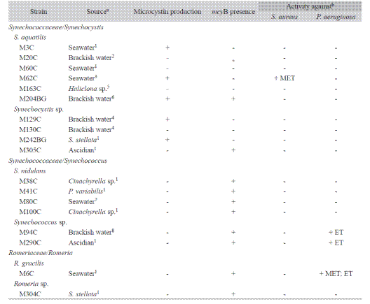
aParaíba state, Brazil: 1- Cabo Branco, João Pessoa; 2- Intermares Lagoon, João Pessoa; 3 - Cabedelo; 4 - Mamanguape River estuary; 5 - Carapibus, João Pessoa.6- Bucatú River estuary; 7 - Acaú, João Pessoa; Rio Grande do Norte state, Brazil: 8 - Pirangí River estuary; b MET - methanolic extract, ET - ethanolic extract.
The production of microcystin analyzed by ELISA was observed in five strains, all belonging to the genus Synechocystis (Table 2): S. aquatilis (M3C - 4.2 Mg·g-1, M62C - 2.5 mg·g-1, M204BG - 8.4 mg·g-1) and Synechocystis spp. (M129C - 5.0 mg·g-1, M242BG - 2.6 mg·g-1).
The presence of mcyB gene was detected in 11 strains belonging to three genera studied: Synechocystis (three strains), Synechococcus (six strains) and Romeria (two strains) (Table 2, Fig. 1).
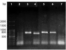
Fig. 1 Products of mcyB gene amplification of cyanobacterial strains isolated from water and tissue of reef organisms of the Brazilian Northeast coast. Lines:1 - 100bp ladder, 2 - Synechocystis aquatilis M3C (mcyB not detected), 3 - Romeria gracilis M6C, 4 - Synechocystis aquatilis M20C, 5 - Synechococcus nidulans M38C, 6 - Synechococcus nidulans M41C, 7 - Negative control. Positive samples: 3, 4, 5 and 6 (PCR product of mcyB gene ~ 800pb).
Antibacterial activity of cyanobacterial strains extracts
The results of antibacterial activity of ethanolic and methanolic extracts are shown in table 2 and table 3. Among the analyzed strains, only four showed inhibition of S. aureus or P. aeruginosa growth, and the inhibition zone of pathogenic bacteria tested ranged between 10.5 and 14.0 mm. Antibacterial activity was observed in strains of Romeria gracilis M6C, Synechocystis aquatilis M62C and Synechococcus sp. (M94C and M290C).
No ethanol extract showed activity against S. aureus; however, ethanolic extracts obtained from three strains inhibited growth of P. aeruginosa (Table 3). Among the methanolic extracts only one strain (M62C) showed inhibitory activity against S. aureus and another strain (M6C) against P. aeruginosa.
Only the Romeria gracilis strain (M6C) showed activity for both, methanolic and ethanolic extracts against P. aeruginosa (Table 3).
Table 3 Activity of cyanobacterial strains extracts against P. aeruginosa and S. aureus
| - | Methanolic extract | Ethanolic extract | |||
|---|---|---|---|---|---|
| Strain | S. aureus | P. aeruginosa | S. aureus | P. aeruginosa | |
| R. gracilis M6C | 0 | 10.5 ± 0.71 | 0 | 11.0 ± 1.41 | |
| S. aquatilis M62C | 11.5 ± 0.71a | 0 | 0 | 0 | |
| Synechococcus sp. M94C | 0 | 0 | 0 | 12.5 ± 0.71 | |
| Synechococcus sp. M290C | 0 | 0 | 0 | 14.0 ± 1.41 | |
aMean ± Standard Deviation.
Growth inhibition zones are expressed in mm.
Discussion
The information on cyanobacterial diversity in most of marine environments and reef organisms, such as studied in the present work, are still limited, in spite of their great importance in benthic and open ocean primary production (Hoffman, 1999; Golubic et al., 2010). Among the marine planktonic cyanobacterial species are two dominant groups: Synechococcus and Prochlorococcus (Hoffman, 1999; Flombaum et al., 2013; Mackey et al., 2015). Cyanobacteria have been found also associated with marine organisms in a range of symbiotic relationships, more explored in sponges (Steindler, Huchon, Avni, & Ilan, 2005; Hirose, Hirose, & Neilan, 2006; Lins-deBarros et al., 2009).
There is also a little information on microcystin production or detection of mcy genes in marine culturable cyanobacteria. Carmichael and Li (2006) reported the production of microcystins by a marine Synechococcus from Salton Sea, and observed that microcystins may show a more common occurrence in marine environments. Some toxins have promising anticancer, antimycobacterial or other anti-disease activities (Gerwick et al., 2008; Ramos et al., 2015).
In our work we detected the microcystins by ELISA technique in the strains of S. aquatilis and Synechocystis spp., and the mcyB gene was detected in only one of these strains.
There are a few reports on the microcystin production by Synechocystis. Nascimento and De Oliveira e Azevedo (1999) reported the production of microcystin by S. aquatilis f. salina isolated from the saline water of coastal Barra lagoon at Maricá, Rio de Janeiro state, Brazil. Magalhães et al. (2003) reported that S. aquatilis produced microcystin in Sepetiba Bay, Brazil, where were observed regular blooms of these cyanobacteria, and the microcystin was found in fish caught in this bay. Several works of Martins, Pereira, Welker, Fastner, and Vasconcelos (2005), Martins, Fernandez, Beiras, and Vasconcelos (2007) and Martins et al. (2008) reported the production of microcystin by the marine cyanobacteria, including genus Synechocystis. Vareli et al. (2012) reported the presence of the hepatotoxic microcystins in the Mediterranean Sea, and he suggested a potential association of microcystins with Synechococcus and/or Synechocystis cyanobacteria.
In the relation to the diference in the detection of microcystin production by ELISA and mcyB presence by PCR approach, it is worth to point out that cyanobacteria usually produce diverse compounds, including pigments that were observed in most of the strains studied in this work (data not shown), which may act as inhibitors in PCR amplification of target genes. Brežna and Piknová (2013) reported that many plant components may act as PCR inhibitors leading to false negative result of PCR-based assay.
The mcyB gene was observed in all tested strains of Synechococcus and Romeria, however, the production of this toxin was not detected by ELISA. Similarly, Frazão, Martins, and Vasconcelos (2010) did not detect the microcystins and other known toxic peptides, using mass spectrometry, by one Leptolyngbya strain and one Oscillatoria strain that showed mcyE gene.
It is known that large fraction of marine bacterial isolates, including cyanobacteria, exhibit antagonistic properties against other pelagic bacteria and antagonistic interactions seem to be very common in the pelagic ocean (Caicedo, Heyduck-Söller, Fischer, & Thöming, 2011; Senhorinho et al., 2015). Several studies have highlighted the importance of marine cyanobacteria as sources of pharmacological agents (Martins et al., 2008; Leão et al., 2013).
The extractions of bioactive compounds produced by cyanobacterial strains are ususally made using solvents such as methanol, ethanol, acetone, among others (Biondi et al., 2008; Madhumathi, Deepa, Jeyachandran, Manoharan, & Vijayakumar, 2011).
In our study we observed differences in antibacterial activity against S. aureus and P. aeruginosa between methanolic and ethanolic extracts of the strains of S. aquatilis, Synechococcus spp. and R. gracilis. Martins et al. (2008) reported inhibition of gram positive bacteria by extracts of marine Synechocystis and Synechococcus, and also observed the variations in the activity of different extracts.
Several strains of cyanobacteria studied in this work showed the microcystin production and antibacterial activity against pathogenic bacteria S. aureus and P. aeuruginosa, showing the potential for future studies of bioactive compounds.

