




 Inglés (pdf)
 Articulo en XML
 Referencias del artículo

 Traducción automática
 Enviar articulo por email
 Citado por SciELO
 Similares en
SciELO
 uBio

 Permalink

Teleosts have developed a great variety of mechanisms to inhabit all existing aquatic environments and feed both on animals and plants (Wilson & Castro, 2011). Despite the differences among species resulting from these ecological variations, the general morphological pattern of teleost's intestine corresponds with that of other vertebrates, which is divided into regions that cannot always be distinguished macroscopically (Cao & Wang, 2009; Leknes, 2009; Cao, Wang, & Song, 2011). Microscopically, in the great majority of fish species, intestinal wall is described by three (Çinar & Şenol, 2006) or four tunics (Olaya et al., 2007; Wilson & Castro, 2011; Xiong et al., 2011), depending on whether the tunica submucosa is considered together with the tunica mucosa or not.
Mucus, the product of the hydratation of mucins, is a common feature in the tunica mucosa of vertebrates; it is secreted by the goblet cells or mucous cells (MC) (Yashpal, Kumari, Mittal, & Mittal, 2007; Díaz, García, Figueroa, & Goldemberg, 2008a; Díaz, García & Goldemberg, 2008b; Tano de la Hoz, Flamini, & Díaz, 2012). These cells secrete a variety of mucins that contribute to the generation of a gelatinous layer, to protect and lubricate the intestinal surface to facilitate the transport of food and reduce the epithelial mechanical stress. Recent studies of molecular biology indicate that MC also participates in signaling pathways leading to a coordinated response for proliferation, differentiation, apoptosis and cell adhesion (Pérez-Sánchez et al., 2013). Several authors have studied the mucins distribution and their role in the process of digestion, in the local immune responses and antimicrobial action for defense against a wide range of injuries (Carraway, Ramsauer, Haq, & Carraway, 2003; Sasaki, Ikeda, & Nakanuma, 2007). The identification of the different types of GCs present in tissue sections can be made using conventional histochemical techniques (Díaz et al., 2008b).
Air-breathing fishes are commonly found in tropical or sub-tropical fresh waters that are ambient ion-poor and prone to hypoxia and hypercarbia (Shartau & Brauner, 2014). The Neotropical catfish Corydoras paleatus Jenyns 1842 is a facultative air-breather and the caudal half of the intestine is involved in gas exchange (Podkowa & Goiakowska-Witalinska, 2002). To date, despite the commercial importance of this species like ornamental fish, there have been few studies on the digestive tract of C. paleatus.
The aim of this study was to characterize the carbohydrate pattern composition of mucins secreted by cells of the tunica mucosa in different regions of the intestine of C. paleatus. Furthermore, lectinhistochemical (LHC) technique is used to locate, identify and differentiate variations in the saccharide residues present in the glycoconjugates (GCs) with the aim to discriminate different cell populations (Barbeito, Ortega, Matiller, Gimeno, & Salvetti, 2013).
Materials and methods
Animals: Twenty-five healthy adult specimens (15 females and 10 males) of C. paleatus (LS: 62.22 ± 1.84 mm for females and 51.70 ± 1.54 mm for males) were used. The field work was carried out in streams situated in La Plata, Buenos Aires, Argentina. The handling, collection and slaughter process of all individuals followed the guidelines of the American Fisheries Society (A.F.S., 2004). Fish were transported alive in plastic bags, and were kept in an aquarium at 22° C and pH 7.0 (Gómez, 1996) for a minimum of three weeks, they were fed at sunset with food for bottom fish (Tetra min tropical tablet, Germany). The animals were slaughtered by an anesthesia overdose using a Eugenol (30 mg/L) (García-Gómez, de la Gándara, & Raja, 2002).
Sampling: The necropsies were carried out immediately after sacrifice, the digestive tract was rapidly removed and samples were fixed by immersing in 10 % buffered formalin before the dehydration in an ethanol series and xylene, and posteriorly embedded in paraffin. Samples of intestine were longitudinally divided into three parts (cranial, middle and caudal portions) considering the shape, transparency, thickness of the wall and the width of the lumen in transverse sections. The cranial portion or intestine 1 (I1) has a typical tubular form and its wall is opaque; the middle area, which was subdivided into two portions, intestine 2 (I2) and intestine 3 (I3), has a broader lumen and its wall became thinner and more translucent than I1; in the caudal portion or intestine 4 (I4), the lumen diameter kept increasing and the wall was even thinner. Histological sections were cut using a sledge microtome, prepared according to standard protocol, and then stained using routine techniques haematoxylin and eosin (H-E) and Masson trichrome staining for morphology (Martoja & Martoja, 1970).
Traditional histochemistry: Sections of tissue were also subjected to histochemical procedures for GCs identification, as detailed in Table 1. The combined technique AB pH 2.5/ PAS allowed the identification of three groups of goblet cells. With regard to this, was calculated the percentage of goblet cells for each group in the different intestinal portions using the following formula:
% of globet cells=
cells number of each group/total number of cells counted.
Table 1: Histochemical procedures for visualizing and identifying GCs in the intestine of C. paleatus
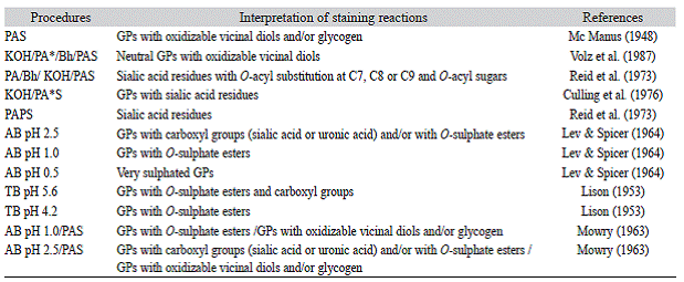
AB: alcian blue; Bh: borohydride; GCs: glycoconjugates; KOH: saponification; PA*: selective periodic acid oxidation; PAS: periodic acid Schiff reagent; PA*S: periodic acid-Schiff at low temperature and low pH; TB: toluidine blue.
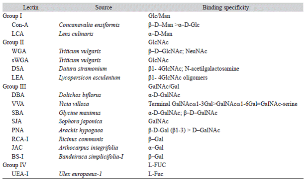
Lectinhistochemistry: Labeling with biotinylated lectins was used to identify specific sugar residues of GCs (Díaz et al., 2008b). Fifty different specific lectins, purchased at Vector Laboratories, Inc. (Burlingame, CA, USA) were employed (Table 2). Paraffin sections were deparaffinised with xylene and decreasing ethanol series, and incubated in 0.03 % H2O2 in methanol for 30 min at room temperature, to inhibit the endogenous peroxidase. Sections then were treated with 0.1 % bovine serum albumin in PBS for 30 min and incubated with fifteen Biotinylated lectins for 2 h at room temperature. After that, sections were washed separately and subsequently treated with an avidin-biotin-peroxidase complex (ABC kit, Vectastain Elite PK 6 200). Diaminobenzidine (DAB) 0.02 % (Biogenex, San Ramón, CA) was used with chromogen. Slides were counterstained with Mayer's haematoxylin. Staining intensity was graded according to a semiquantitative range (-) negative reaction, (1) weak reaction, (2) moderate reaction, (3) strong reaction. Controls for lectin staining included exposure of the specimens to substrates without lectin and incubation of samples using lectins pre-incubated with the corresponding sugar inhibitors.
Micrographs were taken with an Olympus microscope, CX 31 equipped with an Olympus camera U-CMAD3 (Tokyo, Japan).
Results
Digestive tract of C. paleatus had a short oesophagus and a J-shaped stomach localized behind the liver. Between the stomach and the intestine, there was a layer of thick muscle (the gastrointestinal sphincter). The total longitude of the intestine was of 54.9 ± 2.01 mm for females and 45.9 ± 1.76 mm for males.
Histological characterization: Microscopically the general four-layered structure of a vertebrate intestine was observed and showed all the cell types characteristic of the intestinal epithelium (Figure 1). The tunica mucosa of intestine showed a single-layered columnar epithelium (enterocytes) with a well-developed brush border (Figure 1C). This cellular type became gradually less numerous and smaller towards the caudal portions; the brush border of these cells also decreased in height caudally (Figure 1D). The goblet cells or mucous cells (MC), which were arranged among the enterocytes, increased in number towards the caudal portion of the intestine. The most important change that was observed in the intestinal epithelium was related with the extraction of atmospheric oxygen in sectors I3 and I4, transforming the intestine into a complementary air-breathing organ. In these sectors, the epithelium was low cubic (Figure 1D), and interspersed between enterocytes, MC and capillaries that reach the lumen of the mucosa were observed. Underlying the epithelium, a well-developed lamina propria-submucosa consisting of connective tissue; this layer was observed highly vascularized and did not exhibit glands (Figure 1B). No muscularis mucosa was detected, but a thin layer of reticular fibers separating the lamina propria of the tunica submucosa was present. The tunica muscularis was composed of two relatively thick layers of smooth muscle (Figure 1A), among which the myenteric plexus was located. Towards the caudal sector, the outer longitudinal layer became thinner; in the I4 sector, the tunica muscularis was restricted to the circular layer. Tunica serosa was composed of a connective tissue and a simple squamous epithelium (mesothelium).
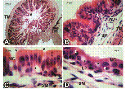
Figure 1: Histology of the cranial, middle and caudal portion of C. paleatus intestine, H-E. A. Cross-section of I1 portion showing of development of villi and the absence of crypts. B. Portion I2 with shorter and thicker villi than those of the I1 portion, and a great development of lamina propria-submucosa. C. Intestinal mucosa of I3 portion, high-cubic enterocytes with microvillous (arrows) was observed, and blood capillaries (stars) located basally to enterocytes. D. Intestinal wall of I4 portion, blood capillaries (stars) interspersed between the enterocytes were observed. BV: blood vessel, E: enterocytes, e: erythrocyte, L: lymphocyte, LP: lamina propria, MC: goblet cells, SM: tunica submucosa, TM: tunica muscularis, V: villus.
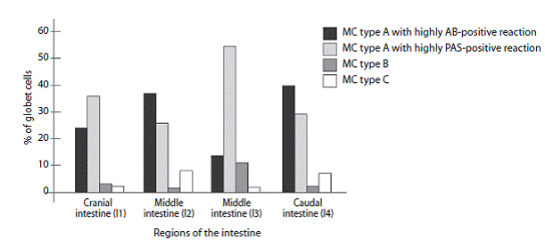
Figure 2: Percentage of goblet cells (MC) with AB pH 2.5/PAS staining shows different regions of the C. paleatus intestine
Histochemical characterization: The implementation of different histochemical techniques to demonstrate the presence of GCs in mucous intestinal showed a similar pattern of distribution in the different regions studied. The structures analyzed were glycocalyx and MC. The results are summarized in Table 3.
Table 3: Histochemical reactions of GCs in different portions of the C. paleatus intestine
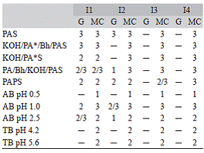
I1: intestine 1; I2: intestine 2; I3: intestine 3; I4: intestine 4. G: glycocalyx; MC: goblet cells. Staining intensity: ─: negative; 1: weakly positive; 2: moderately positive; 3: strongly positive.
The combined technique AB pH 2.5/PAS allowed the identification of three MC types in all intestinal portions (Figure 2). Due to this reason, MC populations were called type A (AB/PAS-positive), type B (only PAS-positive) and type C (only AB-positive). In all sectors there was a predominance of type A, but the intensity of staining varied among the different sectors. Type A cells that were found in I1 and I3 (Figure 3A, Figure 3C) showed a highly PASpositive reaction, this confirmed the presence of GCs with oxidizable vicinal diols in a major proportion. However, in I2 and I4 (Figure 3B, Figure 3D) the type A cells showed a highly AB-positive reaction that evidenced an increase of GCs with weak acid sulfated mucins.
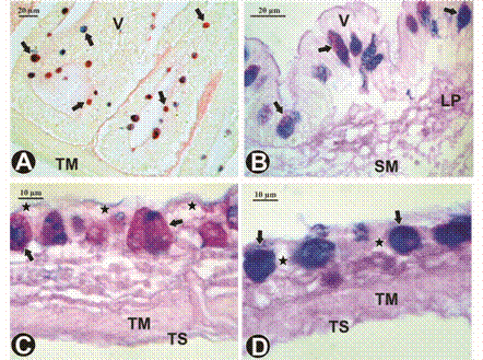
Figure 3: GC histochemistry of the intestine of C. paleatus with a mixed AB pH 2.5/PAS staining. A. Basal surface of the villi of the I1 portion, scarce goblet cells (arrows) AB/PAS-positive reaction. B. Intestinal wall of portion I2, goblet cells of type A (arrows), with a predominance of AB-positive reaction. C. Intestinal wall of portion I3, goblet cells of type A (arrows), with a predominance of PAS-positive reaction. D. Intestinal wall of portion I4, in this sector, the goblet cells are highly AB-positive. : blood capillaries, LP: lamina propria, SM: tunica submucosa, TM: tunica muscularis, TS: tunica serosa, V: villus.
Cranial intestine (I1): The glycocalyx showed a moderately to strong positive reaction with the PAS and all its variants (Figure 4A, Figure 4B). These results revealed the presence of GCs with oxidizable vicinal diols, GCs with sialic acid residues non-substituted or O-acyl substituted in C7, C8 or C9 and O-acyl sugars. Besides, the glycocalyx stained weakly with AB pH 1.0 and moderately with AB pH 2.5 demonstrating the presence of abundant GCs carboxylated and scarce sulfated. MC reacted strongly with all techniques used, showing the presence of a variety of GCs (Figure 4A, Figure 4B). A positive metachromatic reaction with the TB technique at both pHs allowed the identification of GCs with O-sulphate esters and carboxyl groups. This sector reacted positively to the AB technique, which made possible the identification of weak acid sulfated mucins.
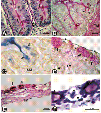
Figure 4: GC histochemistry of the intestine of C. paleatus. A. Basal portion of the intestinal villi, goblet cells (arrows) intensely PAS-positive. B. Glycocalyx and goblet cells (arrows) PA*/Bh/KOH/PAS-positive. C. Goblet cells (arrows) of I2 portion, was moderately labeled with AB pH 1.0 technique. D. Intestinal wall of the I3 portion, abundant goblet cells (arrows) with a rounded shape, note the proximity of the capillaries to the luminal surface (stars), PAPS technique. E. F. Intestinal wall, note the large size of the tunica mucosa in relation to other tunics, and the presence of goblet cells (arrows) and blood capillaries (stars), KOH/PA*/Bh/PAS and TB pH 5.6 technique respectively. E: enterocytes, e: erythrocyte, L: lymphocyte, LP: lamina propria, SM: tunica submucosa, TM: tunica muscularis.
Middle intestine (I2 e I3): All histochemical techniques were positive in the I2 and reactivity gradually diminished towards the I3. Unlike the cranial portion, the glycocalyx showed strong reaction to PAS technique but weak to moderate to PA*/Bh/KOH/PAS and PAPS. The AB technique showed a scarce reaction for GCs with weak sialic acid and a moderate to strong presence of sulfated GCs. As in the I1 the MC reacted strongly with all techniques (Figure 4C, Figure 4D), except to PAPS techniques, which reactivity was moderate in the I2 to strong towards to the I3.
Caudal intestine (I4): The glycocalyx showed negative reaction with all histochemical techniques. These sector also displayed variation in the presence of GCs, the mucosubstances present in MC were stained very strongly with PAS and its variants (Figure 4E), AB at different pHs and TB at both pHs (Figure 4F).
Lectinhistochemical characterization: The application of LHC to detect the presence of GCs in the intestinal mucous, showed a different pattern in each region studied. Glycocalyx and cytoplasm of enterocytes, MC and endothelium were observed in order to establish differences among the physiological regions. No labeling was observed in any of the sectors with DSA and LEA lectins, the enterocytes' cytoplasm showed negative reaction to LCA lectin. The results are summarized in Table 4.
Table 4: Lecting binding in the different portions of the intestine of C. paleatus
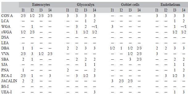
I1: intestine 1; I2: intestine 2; I3: intestine 3; I4: intestine 4. Staining intensity: ─: negative; 1: weakly positive; 2: moderately positive; 3: strongly positive.
Cranial intestine (I1): Intensively positive with JACALIN, RCA-I, sWGA and SBA was found in the apical cytoplasm of enterocytes (Figure 5A); this demonstrated the presence of terminal galactose residues. Also the glycocalyx' GCs strongly reacted with Con-A. MC showed positively a great variety of lectins, although only DBA positivity to these cells was identified in all regions, with a labeled which became more intense from cranial to caudal.
Middle intestine (I2 e I3): In the enterocytes the only lectin which increased binding intensity from I2 to I3 was Con-A. The opposite happened with sWGA, JACALIN, WGA, DBA, VVA, SBA, and RCA-I indicating a gradual decrease in N-acetylglucosamine and N-acetylgalactosamine/galactose residues. Glycocalyx behaved differently from I1, in the middle intestine WGA and RCA-I labeled intensely, but its affinity gradually decreased to the caudal sector, showing that the glycocalyx of I2 (Figure 5B) is rich in N-acetylglucosamine and galactose, but these residues were gradually lost when reaching the caudal region. MC were positive with four lectins, DBA, VVA, SBA and JACALIN (Figure 5C); from these, only the latter remained from moderate to strong labeled in both sectors. The others were increasing their affinity towards the caudal region. All these lectins belong to the group that recognizes N-acetylgalactosamine and galactose. Endothelial cells of the regions I1 and I2 showed a great similarity.
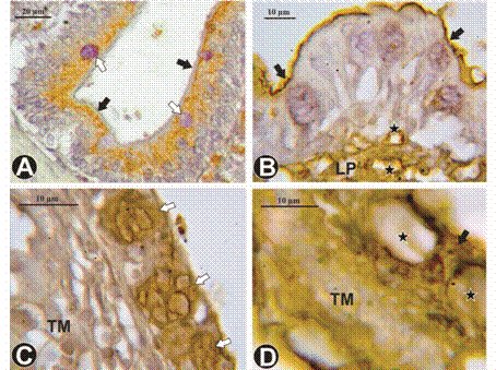
Figure 5: LHC staining of the intestine of C. paleatus. A. SBA lectin binding is present in the enterocytes apical cytoplasm of portion I1, and absent in glycocalyx (black arrows) and goblet cells (white arrows). B. In portion I2, RCA-I lectin binding shows positivity in glycocalyx (black arrows) and capillaries endothelium (stars) of lamina propria. C. Intestinal wall of portion I3, note the heterogeneous positivity of goblet cells (white arrows) with SBA lectin binding. D. Con-A lectin binding is evident in the enterocytes (arrow) located between two blood capillaries (stars). LP: lamina propria, TM: tunica muscularis.
Caudal intestine (I4): Con-A, RCA-I and VVA bind to GCs in the cytoplasm of enterocytes (Figure 5D). Lectins that had a positive reaction for the MC were the same as for I3 but remained constant in labeling intensity with JACALIN. However DBA and VVA labeled more intensively, while SBA decreased its affinity abruptly. All of them belong to the group of lectins that recognizes N-acetylgalactosamine and galactose. LCA, WGA, sWGA, DBA, SBA and SJA showed a similar binding pattern in endothelial cells of the regions I3 and I4.
Discussion
The morphology and histology of the gastrointestinal tract has been described for several teleost species. Despite interspecific differences, this system has showed to share basic structural features (Díaz, García, Devincenti, & Goldemberg, 2003; Díaz et al., 2008a,b; Domeneghini, Arrighi, Radaelli, Bosi, & Veggetti,2005; Fishelson, Golani, Russell, Galil, & Goren, 2011). However, the morphological and functional variations found in C. paleatus demonstrated specializations in this group of vertebrates, not only in relation to its position in the food chain, but also for their habitat. In C. paleatus, histological characteristics of the intestinal mucosa, submucosa, muscular and serosa tunics coincided with other studied teleost (Olaya et al., 2007), with the exception of the I3 and I4 portions. These sectors present modifications in relation to gas exchange such as a very thin wall and the presence of many capillaries located basally, in between, and apically to the epithelial cells. A similar intestinal wall has been observed in teleosts in which the intestine has an accessory respiratory function, like other members of the order Siluriformes (Huebner & Chee, 1978; Podkowa & Goiakowska-Witalinska, 2002; Jucá-Chagas & Boccardo, 2006), and also some members of the family Cobitidae (Jasinski, 1973; Yadav & Singh, 1980; Park & Kim, 2001; Gonçalves, Castro, Pereira-Wilson, Coimbra, & Wilson, 2007).
HC and LHC patterns of the mucosal layer differ among species and in different regions of the digestive tract (Reid, Volz, Cho, & Owen, 1988; Grau, Crespo, Sarasquete, & González de Canales, 1992; Leknes, 2011). In this research, a variety of GCs in the glycocalyx of I1, and a moderated to strong presence of sulfated GCs were observed. This positivity decreased gradually towards the caudal portion of the intestine where it became negative for all techniques in portion I3 and I4. These results confirm the functional differences that these cells possess along the intestine. The glycocalyx, extending in the apical surface of I1 and I2, has a buffer effect on the high acidity from the stomach. Additionally, to protecting the epithelial cells from the action of proteolytic enzymes, the glycocalyx plays a key role in cell-pathogen interactions (Vásquez-Piñeros, Rondón-Barragan, & Eslava-Mocha, 2012). Also, the glycocalyx and the endothelial cells in C. paleatus showed a strong affinity for lectins Con-A and DBA in the whole intestine and UEA-I in the I4 portion. These results demonstrated that glucose/mannose; N-acetilgalactosamine and L-fucose residues would probably have general functions in the intestinal epithelium that do not depend on the sector. Previous studies had also described the role of L-fucose, with an important function in morphogenetic processes of the plasmatic membranes, and increasing the resistance against bacterial populations in the apical border of enterocytes (Bennett, Leblond, & Haddad, 1974; Vásquez-Piñeros et al., 2012). Enterocytes cytoplasm showed affinities to the four groups of lectins, but only Con-A and VVA lectins bound from moderately to intensively in all sectors of the intestine. The enterocytes of Paralichthys olivaceus (Jung, Ahn, Lee, Go, & Shin, 2002) and Oncorhynchus mykiss (Marchetti et al., 2006) showed a similar LHC pattern. The LHC method mainly revealed the presence of glucose/mannose and N-acetylgalactosamine residues in the GCs that are produced by the enterocytes and transported to the cell surface along the intestine, and also a weak presence of N-acetylglucosamine and 1.3 ß N-acetylgalactosamine terminal residues. The HC and LHC patterns of C. paleatus in I1 and I2 were similar between them, as well as those of I3 and I4; these could indicate that enterocytes belong to two different cell populations.
The presence of MC is a common feature in the intestinal epithelium of teleosts (Grau et al., 1992; Çinar & Şenol, 2006; Hernández, Pérez Gianeselli, & Domitrovic, 2009). In the I1 of C. paleatus, the MC were scarce and had a low content of highly sulfated mucins and a high content of weak acid mucins. This result agrees with those obtained previously for teleosts of the orders Perciformes, Cypriniformes, Esociformes, Siluriformes, Beloniformes and Salmoniformes (Reifel & Travill, 1979; Tibbets, 1997; Podkowa & Goiakowska-Witalinska, 2002; Marchetti et al., 2006; Hernández et al., 2009). In this catfish, the AB combined techniques allowed the identification of three types of MC. In I1 and I3 the carboxylated groups were predominant, in contrast with the abundance the sulphated groups found in I2 and I4. According to the observations made by Yadav and Singh (1980), Podkowa and Goiakowska-Witalinska (2002) and Çinar and Şenol (2006), the caudal portion of the intestine contains acidic glycosaminoglycans with high affinity to water, which would be necessary for the maintenance of body moisture. The presence of different types of MC has already been determined in other groups of fishes (Hung, Groff, Lutes, & Alkins, 1990). Narasimham and Parvatheswarao (1974) and Domeneghini, Stranini, and Veggetti (1998) have suggested that intestinal mucins possibly play a role in osmoregulation. Recently, Pérez-Sánchez et al. (2013) have shown that MUC18, an intestinal mucin that binds to membranes, plays a crucial role in protection against pathogen agents together with other biological processes such as osmoregulation and ion exchange. In addition, Mittal, Whitear, and Agarwal (1980) and Mittal, Pinky and Mittal (2002) observed that the mucus has a remarkable power to precipitate the mud in suspension, keeping the epithelium clean for respiration. Even though these studies were done in skin and opercule, this function could be applied to the intestine of C. paleatus since it is a benthic fish that incorporates suspended matter together with water, making it difficult to breathe at the intestinal level.
In conclusion, this study has shown that GCs produced along the intestine of C. paleatus exhibit a high complexity degree associated with the multiple functions performed by the mucus in the digestive tract. Furthermore we found distinctive features of the mucosa in relation to gas exchange such as a very thin wall, presence of many capillaries interspersed between enterocytes in direct contact with the lumen and a decreased of the GCs in the glycocalyx that relate more with their respiratory function than with digestive function. The information generated here may be a relevant biological tool for comparing and analyzing the possible glycosidic changes in the intestinal mucus under different conditions, such as changes in diet or different pathological stages.

