<?xml version="1.0" encoding="ISO-8859-1"?><article xmlns:mml="http://www.w3.org/1998/Math/MathML" xmlns:xlink="http://www.w3.org/1999/xlink" xmlns:xsi="http://www.w3.org/2001/XMLSchema-instance">
<front>
<journal-meta>
<journal-id>0034-7744</journal-id>
<journal-title><![CDATA[Revista de Biología Tropical]]></journal-title>
<abbrev-journal-title><![CDATA[Rev. biol. trop]]></abbrev-journal-title>
<issn>0034-7744</issn>
<publisher>
<publisher-name><![CDATA[Universidad de Costa Rica]]></publisher-name>
</publisher>
</journal-meta>
<article-meta>
<article-id>S0034-77442002000200034</article-id>
<title-group>
<article-title xml:lang="en"><![CDATA[Angiostrongylus costaricensis n. sp. (Nematoda: Metastrongyloidea), a new lungworm occurring in man in Costa Rica]]></article-title>
</title-group>
<contrib-group>
<contrib contrib-type="author">
<name>
<surname><![CDATA[Morera]]></surname>
<given-names><![CDATA[Pedro]]></given-names>
</name>
</contrib>
<contrib contrib-type="author">
<name>
<surname><![CDATA[Céspedesu]]></surname>
<given-names><![CDATA[Rodolfo]]></given-names>
</name>
</contrib>
</contrib-group>
<aff id="A">
<institution><![CDATA[,  ]]></institution>
<addr-line><![CDATA[ ]]></addr-line>
</aff>
<pub-date pub-type="pub">
<day>00</day>
<month>06</month>
<year>2002</year>
</pub-date>
<pub-date pub-type="epub">
<day>00</day>
<month>06</month>
<year>2002</year>
</pub-date>
<volume>50</volume>
<numero>2</numero>
<fpage>783</fpage>
<lpage>796</lpage>
<copyright-statement/>
<copyright-year/>
<self-uri xlink:href="http://www.scielo.sa.cr/scielo.php?script=sci_arttext&amp;pid=S0034-77442002000200034&amp;lng=en&amp;nrm=iso"></self-uri><self-uri xlink:href="http://www.scielo.sa.cr/scielo.php?script=sci_abstract&amp;pid=S0034-77442002000200034&amp;lng=en&amp;nrm=iso"></self-uri><self-uri xlink:href="http://www.scielo.sa.cr/scielo.php?script=sci_pdf&amp;pid=S0034-77442002000200034&amp;lng=en&amp;nrm=iso"></self-uri><abstract abstract-type="short" xml:lang="en"><p><![CDATA[Angiostrongylus costaricensis n. sp. is described from Costa Rica where it produces lesions in the abdominal cavity of man. It can be distinguished from other species of the genus on the basis of its size, the length of the spicules, the position of the vulva and the morphology and position of the bursal rays. The parasite localizes in the small mesenteric arteries, especially in the ileocecal region, where it produces arteritis and thrombosis. Eggs in various stages of embryonation were found scattered in the tissues of the intestinal wall and regional lymph nodes, eliciting a granulomatous inflammatory reaction with in. tense eosinophilic infiltration]]></p></abstract>
<abstract abstract-type="short" xml:lang="es"><p><![CDATA[Los autores hacen la descripción de Angiostrongylus costaricensis n. sp., un nuevo metastrongilideo encontrado en Costa Rica, que produce lesiones en el hombre. Esta nueva especie se puede distinguir de las otras Catorce descritas hasta ahora, en base a su tamaño, la longitud de las espículas, la posición de la vulva y la morfología y disposición de los rayos bursales. A. costaricensis es la segunda especie del género de importancia en parasitología humana. La otra especie patógena para el hombre, A. cantonensis, es responsable de una forma de meningoencefalitis eosinofílica. Los parásitos adultos, machos y hembras, se localizan en las arterias del mesenterio y de la pared intestinal, especialmente de la región ileocecal en donde provocan fenómenos inflamatorios y trombóticos que llevan a grados diversos de necrosis. La presencia de huevos en varios estados de embrionación, en el tejido de la pared intestinal y de los ganglios linfáticos regionales, produce una reacción inflamatoria granulomatosa con intensa infiltración eosinofilica. La pared intestinal se presenta engrosada, llegándose a producir cuadros de suboclusión u oclusión que obligan a intervenciones quirúrgicas de emergencia. La presencia 4e huevos en los tejidos humanos sugiere una mejor adaptación de A. costaricensis en el hombre, en comparación con A. cantonensis, que no llega a alcanzar el estado adulto en los tejidos encefálicos humanos. Sin embargo en los casos en que hemos encontrado larvas completamente formadas, ellas siempre están encerradas en la cáscara del huevo, lo que sugiere la imposibilidad de que las mismas lleguen a la luz intestinal y regresen al suelo para completar su ciclo de vida]]></p></abstract>
</article-meta>
</front><body><![CDATA[ <div style="text-align: center;"><span  style="font-family: arial; font-weight: bold;">Angiostrongylus costaricensis n. sp. (Nematoda: </span><br  style="font-family: arial; font-weight: bold;"> <span style="font-family: arial; font-weight: bold;">Metastrongyloidea), a new lungworm occurring in </span><br  style="font-family: arial; font-weight: bold;"> <span style="font-family: arial; font-weight: bold;">man in Costa Rica</span>    <br> </div> <font face="Arial" size="2"></font>     <p align="center"><font face="Arial" size="2">by</font></p>     <p align="center"></p>     <p align="center"><font face="Arial" size="2">Pedro Morera <a  name="a01"></a><a href="#a02">*</a>, <a href="#a02">***</a> and Rodolfo Céspedesu <a href="#a02">**</a>, <a href="#a02">***</a>    <br> (Received for publication November 15, 1970)</font></p>     <p><font face="Arial" size="2">Fourteen species of the genus <i>Angiostrongyilus, </i>created by KAMENSKY <a href="#18">(18)</a>, have been described, but among these only <i>A. cantonemis </i>(Chen, 1935) Dougherty, 1946 has been incriminated as a causal agent of disease in man. The first investigators who found this parasite in humans were Nomura and Lin in 1945 (BEAVER and ROSEN, <a href="#10">10</a>). However, because the paper was published in Japanese, it failed to appear in the standard indexing and abstracting journals and this finding remained unknown until ROSEN <i>et al. </i>(<a href="#24">24</a>, <a  href="#25">25</a>), HORIO and ALlCATA <a href="#17">(17)</a> and AUCATA (<a href="#1">1</a>, <a href="#2">2</a>) established the real importance of this work by attributing eosinophilic meningitis to this parasite. Recent studies have shown that parasitized rats are found in most tropical zones, except those of Africa and the Americas (AUCATA, <a  href="#6">6</a>). AUCATA <a href="#3">(3)</a> searched unsuccessfully for <i>A. cantonensis </i>in Costa Rica. However, for several years we have had the opportunity to study a clinico-pathological picture, observed mainly in children, characterized by the formation of granulomas with heavy eosinophilic infiltration in the abdominal cavity. We propose the name <i>Angiostr01zgylfls costaricensis, </i>n. sp. for the etiological agent of this disease, because this parasite has many morphological characteristics that distinguish it from the other species of the genus.</font></p> <font face="Arial" size="2"><b>     <p>Material and Methods</p> </b> </font>     <p style="text-align: left;"><font face="Arial" size="2">Surgical specimens obtained from several hospitals of the country were used in this study. From fixed material the worms were obtained by dissecting the small arteries of the intestinal wall and in unfixed specimens they were liberated by digestion with pepsin. Drawings were made with the aid of a camera lucida. Measurements are given in millimeters unless otherwise specified.</font></p> <font face="Arial" size="2"><i></i></font>     <p align="center"><font face="Arial" size="2"><i>Angiostrongylus costaricensis </i>n. sp.    ]]></body>
<body><![CDATA[<br> (Figs. 1- 7)</font></p>     <p><font face="Arial" size="2">DESCRIPTION: Angiostrongylinae, Böhm and Gebauer, 1934; <i>Angiostrongylus </i>Kamensky, 1905.</font></p>     <p><font face="Arial" size="2">
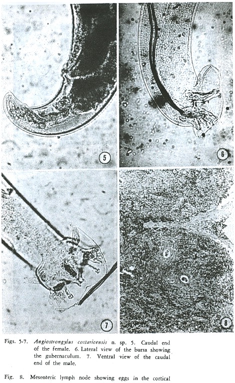
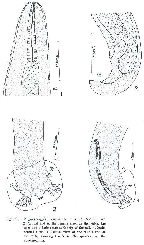
Body filiform in both sexes, tapering toward both ends. The caudal end of the male curves ventrally, with a well developed copulatory bursa; in the female thc caudal end is roughly conical and slightly curved. Cuticle transversely striated and thicker toward both ends. Excretory pore immediately posterior to the base of the club-shaped esophagus.    <br> </font></p>     <p style="text-align: center;"><font face="Arial" size="2"><a  name="f1-4"></a><img src="/img/fbpe/rbt/v50n2/2831i1.JPG" title="" alt=""  style="width: 480px; height: 815px;">    
<br> </font></p>     <p><font face="Arial" size="2">MALE: (3 specimens): Body length 15.0 to 17.9; width at the esophageal-intestinal junction 0.079 to 0.093: esophagus 0.160 to 0.180. Spicules slender, equal, striated, ending in two hook-shaped formations, 0.267 to 0.297 in length. Gubernaculum present, 0.045 long. This structure was not identified in a preliminary study (MORERA, <a href="#21">21</a>) . In the copulatory bursa ventral rays fused except for the distal fifth, with bud-shaped endings; the ventrolateral is slightly longer than the ventroventral. The lateral rays arise from a common trunk and are of similar size, the posterolateral and mediolateral are fused in the proximal half and the externolateral separates from these after its emergence from the common trunk. The externodorsal ray arises near the base of the laterals and is separate from the dorsal; it is long, slender, and larger at thc distal end. The dorsal ray is thick, short, terminating in three tips; the central one is short, knob-like, and the laterals beak-shaped.</font></p>     <p><font face="Arial" size="2">FEMALE (1 specimen): Body length 26.9; width at base of esophagus 0.108; esophagus 0.165. "Barber-pole" appearance of the reproductive apparatus coiled around the intestine, typical of the genus, not prominent in the fixed material. Vulva situated 0.175 and anus 0.053 from tip of tail; caudal end almost cone-shaped; tip of tail bears a small refringent spine.</font></p>     <p><font face="Arial" size="2">HOST: Man is not considered to be the normal host; animal hosts are as yet unknown. Normal habitat: unknown. In man the worms are localized in small branches of the mesenteric artery.</font></p>     <p><font face="Arial" size="2">Type specimens: Holotype male, allotype female, Department of Parasitology, University of Costa Rica.</font></p> <font face="Arial" size="2"><b>     ]]></body>
<body><![CDATA[<p>Discussion</p> </b> </font>     <p><font face="Arial" size="2">In 1967, ASH <a href="#7">(7)</a> mentioned ten species of the genus <i>Angiostrongylus, </i>in addition to <i>A. michiganensis </i>described by him; he did not mention <i>A. sciuri </i>described by MERDIVENCI <a href="#20">(20)</a>. More recently two other species have been described: <i>A. sandarsae, </i>Alicata, 1968 and <i>A. mackerrasae, </i>Bhaibulaya, 1968.</font></p>     <p><font face="Arial" size="2">A comparative study of these fourteen species with the parasite found in Costa Rica revealed many characteristic differences whose point by point description would be too long to include here. Therefore, we have limited ourselves only to the description of the most important differences, which in out opinion justify the establishment of a new species. In the first place, on the basis of the parasite size, we are able to ignore all those whose length is less than one half that of <i>A. costaricensis: A. blarini, </i>Ogren, 1954; <i>A. ondatrae, </i>Schultz, Orlow and Kutass, 1933; <i>A. soricis, </i>Soltys, 1954; and <i>A. michiganensis, </i>Ash, 1967.</font></p>     <p><font face="Arial" size="2">The length of the spicules, an important taxonomic character, permits us to disregard all those species in which these structures measure more than 0.400; this dimension being much greater than the maximum of 0.297 found in <i>A. costaricensis. </i>These species are <i>A. cantonensis, </i>Chen, 1935; <i>A. gubernaculatus, </i>Dougherty, 1946; <i>A. tateronae, </i>Baylis, 1928; <i>A. chaballdi, </i>Biocca, 1957; <i>A. sciuri </i>and <i>A. mackerrasae.</i></font></p>     <p><font face="Arial" size="2">The spicules of <i>A. vasorum </i>(Baillet, 1866) Kamensky, 1905 (considered by most authors to be synonymous with <i>A. raillieti, </i>Travassos, 1927), although a little shorter than 0.400, are still longer than those of <i>A. costaricensis </i>the original description by Baillet (1866) and the redescription by CABRAL. GONÇALVES <a href="#13">(13)</a> allow us to establish other important differences; these are that the vulva is further from the tip of the tail in <i>A. vasorum </i>and that the tail is roughly conical in <i>A. costaricensis </i>and rounded in the other two. <i>A. sandarsae </i>has relatively longer spicules and a notably longer esophagus. There were also .appreciable differences in the morphology and distribution of the bursa! rays, especially the size relationship between the lateroventral and the ventroventral, and in the form and origin of the externodorsal.</font></p>     <p><font face="Arial" size="2">We think that <i>A. tm, </i>Yamaguti, 1941, is not an acceptable species, because until now a mate has not been described. </font></p>     <p><font face="Arial" size="2">An important characteristic is the presence of a conspicuous spine at the tip of the tail of the female of <i>A. costaricensis. </i>A similar structure occurs only in <i>A. mackerrasae.    <br> </i></font></p>     <p style="text-align: center;"><font face="Arial" size="2"><i><a  name="f5-8"></a><img src="/img/fbpe/rbt/v50n2/2831i2.JPG" title="" alt=""  style="width: 471px; height: 767px;">    
<br> </i></font></p>     ]]></body>
<body><![CDATA[<p><font face="Arial" size="2">Another important characteristic is the presence of a gubernaculum in <i>A. costaricensis. </i>The first species described with this structure was <i>A. ondatrae,. </i>however, BAYLIS <a href="#9">(9)</a> in the description of <i>A. tateronae </i>mentioned that the dorsal wall of the cloaca appeared thickened and chitinised. Although he interpreted this area as a telamon, it is more than likely a gubernaculum.</font></p>     <p><font face="Arial" size="2">In 1946, DOUGHERTY <a href="#16">(16)</a> in describing <i>A. gubernaculatus, </i>stated erroneously that it was the first species that had a gubernaculum. Recently, ALICATA <a  href="#4">(4)</a> described another species with a gubernaculum, <i>A. sandarsae, </i>and later the same author described this structure in <i>A. cantonensis </i>(AUCATA, <a href="#5">5</a>). The presence of a gubernaculum in <i>A. gubemaculatus </i>and <i>A. ondatrae </i>served as the basis on which SKRJABIN <i>et al. </i><a href="#27">(27)</a> classified these parasites into the genera <i>Angiocaulus </i>and <i>Rodentocautus, </i>respectively. The latter had been originally classified in this way by SCHULTZ <i>et al. </i><a href="#26">(26)</a>. Working with Ash (unpubIished data), it was found that careful examination of <i>A. vasorum </i>revealed the presence of a gubernaculum and recently, Bhaibulaya has found a gubernaculum in <i>A. mackerrasae </i>(ASH, personal communication).</font></p>     <p><font face="Arial" size="2">
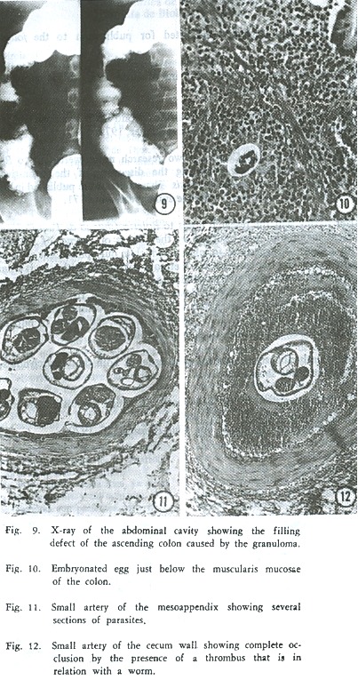
In 1952, the first Costa Rican case of the disease caused by this parasite was observed (CÉSPEDES <i>et al., </i>14; MORERA, <a href="#21">21</a>) and since then, more than 70 cases have been studied. The main symptoms (ROBLES <i>et al., </i><a href="#23">23</a>) were abdominal pain, usually localized in the right iliac fossa. Fever was usually high and sometimes lasting up to two months. Anorexia was often observed and one third of the patients experienced vomiting. The physical examination showed an intra-abdominal mass which could cause confusion with malignant tumors or acute appendicitis of other etiology. Rectal examination was usually painful and sometimes the tumoral mass could be palpated. The most important laboratory findings were leucocytosis ranging from 10,000 to 52,000 per mm<sup>3</sup> with eosinophilia from 11 to 81 per cent. In a few cases, eosinophilia was lower than 10 per cent. X-ray examination showed alteration of the intestinal wall as demonstrated by a filling defect (<a href="#f9-12">Fig. 9</a>), spasticity and irritability (Stierling's sign) as well as a festooned aspect of the mucosa.    <br> </font></p>     <p style="text-align: center;"><font face="Arial" size="2"><a  name="f9-12"></a><img src="/img/fbpe/rbt/v50n2/2831i3.JPG" title="" alt=""  style="width: 403px; height: 765px;">    
<br> </font></p>     <p><font face="Arial" size="2">Anatomo-pathoIogical studies revealed that lesions were, in most cases, localized in the appendix, but some were found to extend to the terminal portion of the ileum, cecum, fue first part of the ascending colon, and to the regional lymph nodes (14). The main characteristic was thickening and hardening of the edematous intestinal wall, with milliary yellowish granulomatous infiltration. These lesions could produce incomplete or complete obstruction. In some cases, necrotic processes could produce perforation. Less frequently, lesions were observed in the transverse and descending colon and sigmoid. Histologically the lesions appeared as a granulomatous inflammation with massive eosinophilic infiltration, involving all the layers of the intestinal wall. This' same inflammatory process was observed to aHect the regional lymph nodes. Thin-shelled, oval, embryonated eggs were scattered in the lesion <a  href="#f5-8">(Fig. 8)</a>. Occasionally, the embryos reached the larval stage, but the larvae remained enclosed within the egg shell <a  href="#f9-12">(Fig. 10)</a>. In cases of longer evolution, the eggs appeared inside the giant cells. Sections of the adult parasite could be found in both mesenteric arteries and their intramural branches <a  href="#f9-12">(Fig. 11)</a>. These worms sometimes produced arteritis and thrombosis <a href="#f9-12">(Fig. 12)</a>.</font></p> <font face="Arial" size="2"><i></i></font>     <p><font face="Arial" size="2"><i>A. costaricmsis </i>is the second species of the genus to be found in man and there is a cIear difference between the disease produced by it and the eosinophilic meningoencephalitis produced by <i>A. canlonensis. </i>We believe this difference is caused by different life cycles, known only for <i>A. cantonensis </i>(MACKERRAS and SANDARS, <a href="#19">19</a>). In any case, <i>A. costaricensis </i>appears to adapt better to man, as demonstrated by the presence of adult worms in the arteries, and embryonated eggs in the tissues. However, the larvae apparently do not hatch, which suggests the impossibility of these larvae ever entering the intestinal lumen and returning to the soil to complete their life cycle. In the human cases in which we have had the opportunity to screen the feces for larvae, the results were negative.</font></p> <font face="Arial" size="2"><b>     <p>Acknowledgements</p> </b> </font>     <p><font face="Arial" size="2">The authors are indebted to Dr. Lawrence R. Ash, School of Public Health, UCLA, for his kindness and helpful criticism in preparation of the texto We also express our appreciation to Dr. Alfonso Trejos, Dr. Alan Altman and Dr. Thomas Lau for their help in preparation of the English manuscript.</font></p> <font face="Arial" size="2"><b>     ]]></body>
<body><![CDATA[<p>Summary</p> </b><i> </i></font>     <p><font face="Arial" size="2"><i>Angiostrongylus costaricensis </i>n. sp. is described from Costa Rica where it produces lesions in the abdominal cavity of man. It can be distinguished from other species of the genus on the basis of its size, the length of the spicules, the position of the vulva and the morphology and position of the bursal rays. The parasite localizes in the small mesenteric arteries, especially in the ileocecal region, where it produces arteritis and thrombosis. Eggs in various stages of embryonation were found scattered in the tissues of the intestinal wall and regional lymph nodes, eliciting a granulomatous inflammatory reaction with in. tense eosinophilic infiltration.</font></p> <font face="Arial" size="2"><b>     <p>Resumen</p> </b> </font>     <p><font face="Arial" size="2">Los autores hacen la descripción de <i>Angiostrongylus costaricensis </i>n. sp., un nuevo metastrongilideo encontrado en Costa Rica, que produce lesiones en el hombre. Esta nueva especie se puede distinguir de las otras Catorce descritas hasta ahora, en base a su tamaño, la longitud de las espículas, la posición de la vulva y la morfología y disposición de los rayos bursales. <i>A. costaricensis </i>es la segunda especie del género de importancia en parasitología humana. La otra especie patógena para el hombre, <i>A. cantonensis, </i>es responsable de una forma de meningoencefalitis eosinofílica.</font></p>     <p><font face="Arial" size="2">Los parásitos adultos, machos y hembras, se localizan en las arterias del mesenterio y de la pared intestinal, especialmente de la región ileocecal en donde provocan fenómenos inflamatorios y trombóticos que llevan a grados diversos de necrosis. La presencia de huevos en varios estados de embrionación, en el tejido de la pared intestinal y de los ganglios linfáticos regionales, produce una reacción inflamatoria granulomatosa con intensa infiltración eosinofilica. La pared intestinal se presenta engrosada, llegándose a producir cuadros de suboclusión u oclusión que obligan a intervenciones quirúrgicas de emergencia.</font></p>     <p><font face="Arial" size="2">La presencia 4e huevos en los tejidos humanos sugiere una mejor adaptación de <i>A. costaricensis </i>en el hombre, en comparación con <i>A. cantonensis, </i>que no llega a alcanzar el estado adulto en los tejidos encefálicos humanos. Sin embargo en los casos en que hemos encontrado larvas completamente formadas, ellas siempre están encerradas en la cáscara del huevo, lo que sugiere la imposibilidad de que las mismas lleguen a la luz intestinal y regresen al suelo para completar su ciclo de vida.</font></p> <font face="Arial" size="2"><b>     <p>    <br> Literature Cited</p> </b> </font>     <!-- ref --><p><font face="Arial" size="2"><a name="1"></a>1. ALICATA, J. E. 1961. A cause of parasitic meningitis in the Pacific-rat lungworms. <i>Hawaii; Farm. Bur</i>. J., 1-2 (Special Number).</font>&nbsp;&nbsp;&nbsp;&nbsp;&nbsp;&nbsp;&nbsp;&nbsp;[&#160;<a href="javascript:void(0);" onclick="javascript: window.open('/scielo.php?script=sci_nlinks&ref=1217571&pid=S0034-7744200200020003400001&lng=','','width=640,height=500,resizable=yes,scrollbars=1,menubar=yes,');">Links</a>&#160;]<!-- end-ref --><!-- ref --><p><font face="Arial" size="2"><a name="2"></a>2. ALICATA. J. E. 1962. <i>Angiostrongylus cantonensis </i>(Nematoda: Metastrongylidae) as a causative agent of eosinophilie meningitis of man in Hawaii and Tahiti. <i>Canad. J.</i> <i>Zool., </i>40: 5-8.</font>&nbsp;&nbsp;&nbsp;&nbsp;&nbsp;&nbsp;&nbsp;&nbsp;[&#160;<a href="javascript:void(0);" onclick="javascript: window.open('/scielo.php?script=sci_nlinks&ref=1217572&pid=S0034-7744200200020003400002&lng=','','width=640,height=500,resizable=yes,scrollbars=1,menubar=yes,');">Links</a>&#160;]<!-- end-ref --><!-- ref --><p><font face="Arial" size="2"><a name="3"></a>3. ALICATA. J. E. 1967. Absence of <i>Angiostrongylus cantonensis </i>among rodents in parts of Central America and South America. <i>J. Parasitol.,</i> 54: 1118.</font>&nbsp;&nbsp;&nbsp;&nbsp;&nbsp;&nbsp;&nbsp;&nbsp;[&#160;<a href="javascript:void(0);" onclick="javascript: window.open('/scielo.php?script=sci_nlinks&ref=1217573&pid=S0034-7744200200020003400003&lng=','','width=640,height=500,resizable=yes,scrollbars=1,menubar=yes,');">Links</a>&#160;]<!-- end-ref --><!-- ref --><p><font face="Arial" size="2"><a name="4"></a>4. ALlCATA. J. E. 1968a. <i>Angiostrongylus sandarse</i> sp. n. (Nematoda: Metastrongyloidea) a lungworm of rodents in Mozambique, East Africa. <i>J. Parasitol.,</i> 54: 896-899.</font>&nbsp;&nbsp;&nbsp;&nbsp;&nbsp;&nbsp;&nbsp;&nbsp;[&#160;<a href="javascript:void(0);" onclick="javascript: window.open('/scielo.php?script=sci_nlinks&ref=1217574&pid=S0034-7744200200020003400004&lng=','','width=640,height=500,resizable=yes,scrollbars=1,menubar=yes,');">Links</a>&#160;]<!-- end-ref --><!-- ref --><p><font face="Arial" size="2"><a name="5"></a>5. ALICATA, J. E. 1968b. The gubernaculum of <i>Angiostrongylus cantonesis </i>(Chen). J. <i>Parasitol., </i>54: 1193.</font>&nbsp;&nbsp;&nbsp;&nbsp;&nbsp;&nbsp;&nbsp;&nbsp;[&#160;<a href="javascript:void(0);" onclick="javascript: window.open('/scielo.php?script=sci_nlinks&ref=1217575&pid=S0034-7744200200020003400005&lng=','','width=640,height=500,resizable=yes,scrollbars=1,menubar=yes,');">Links</a>&#160;]<!-- end-ref --><!-- ref --><p><font face="Arial" size="2"><a name="6"></a>6. ALICATA. J. E. 1969. Present status of <i>Angiostrongylus cantonensis </i>infection in man and animals in the tropics. <i>J. Trop. Med. Hyg., </i>72: 53-63.</font>&nbsp;&nbsp;&nbsp;&nbsp;&nbsp;&nbsp;&nbsp;&nbsp;[&#160;<a href="javascript:void(0);" onclick="javascript: window.open('/scielo.php?script=sci_nlinks&ref=1217576&pid=S0034-7744200200020003400006&lng=','','width=640,height=500,resizable=yes,scrollbars=1,menubar=yes,');">Links</a>&#160;]<!-- end-ref --><!-- ref --><p><font face="Arial" size="2"><a name="7"></a>7. ASH, L. R. 1967. <i>Angiostrongylus michiganensis </i>sp. n. (Nematoda: Metastrongyloidea). a lungworm occurring in the shrew, <i>Sorex cinereus, </i>in Miehigan. <i>J. Parasitol</i>. 53: 625-629.</font>&nbsp;&nbsp;&nbsp;&nbsp;&nbsp;&nbsp;&nbsp;&nbsp;[&#160;<a href="javascript:void(0);" onclick="javascript: window.open('/scielo.php?script=sci_nlinks&ref=1217577&pid=S0034-7744200200020003400007&lng=','','width=640,height=500,resizable=yes,scrollbars=1,menubar=yes,');">Links</a>&#160;]<!-- end-ref --><!-- ref --><p><font face="Arial" size="2"><a name="8"></a>8. BAILLET, M. C. 1866. <i>Nouveau Dictionarie Pratique de Médicine, Chirurgie et d'Higiène Vétérinaries</i>, T. VIII: 587-588.</font>&nbsp;&nbsp;&nbsp;&nbsp;&nbsp;&nbsp;&nbsp;&nbsp;[&#160;<a href="javascript:void(0);" onclick="javascript: window.open('/scielo.php?script=sci_nlinks&ref=1217578&pid=S0034-7744200200020003400008&lng=','','width=640,height=500,resizable=yes,scrollbars=1,menubar=yes,');">Links</a>&#160;]<!-- end-ref --><!-- ref --><p><font face="Arial" size="2"><a name="9"></a>9. BAYLIS, H. A. 1928. On a collection of nematodes from Nigerian mammals (chiefly rodents). <i>Parasitology, </i>20: 280-304.</font>&nbsp;&nbsp;&nbsp;&nbsp;&nbsp;&nbsp;&nbsp;&nbsp;[&#160;<a href="javascript:void(0);" onclick="javascript: window.open('/scielo.php?script=sci_nlinks&ref=1217579&pid=S0034-7744200200020003400009&lng=','','width=640,height=500,resizable=yes,scrollbars=1,menubar=yes,');">Links</a>&#160;]<!-- end-ref --><!-- ref --><p><font face="Arial" size="2"><a name="10"></a>10. BEAVER, P. C., &amp; L. ROSEN 1964. Memorandum on the first report of <i>Angiostrongylus </i>in man by Nomura and Lin, 1945. <i>Amer. J. Trop. Med. Hyg., </i>13: 589-590.</font>&nbsp;&nbsp;&nbsp;&nbsp;&nbsp;&nbsp;&nbsp;&nbsp;[&#160;<a href="javascript:void(0);" onclick="javascript: window.open('/scielo.php?script=sci_nlinks&ref=1217580&pid=S0034-7744200200020003400010&lng=','','width=640,height=500,resizable=yes,scrollbars=1,menubar=yes,');">Links</a>&#160;]<!-- end-ref --><!-- ref --><p><font face="Arial" size="2"><a name="11"></a>11. BHAlBULAYA, M. 1968. A new species of <i>Angiostrongylus </i>in an Australian rat, <i>Rattus fuscipes</i> <i>Parasitology, </i>58: 789-799</font>&nbsp;&nbsp;&nbsp;&nbsp;&nbsp;&nbsp;&nbsp;&nbsp;[&#160;<a href="javascript:void(0);" onclick="javascript: window.open('/scielo.php?script=sci_nlinks&ref=1217581&pid=S0034-7744200200020003400011&lng=','','width=640,height=500,resizable=yes,scrollbars=1,menubar=yes,');">Links</a>&#160;]<!-- end-ref --><!-- ref --><p><font face="Arial" size="2"><a name="12"></a>12. BIOCCA, E. 1957. <i>Angiostrongylus chabaudi </i>n. sp. parassita del <i>Cuore </i>e dei vasi polmonari del gatto selvatico <i>(Felis silvestris). </i>R. <i>Accad. </i>Naz. <i>Lincet, </i>22: 526-532.</font>&nbsp;&nbsp;&nbsp;&nbsp;&nbsp;&nbsp;&nbsp;&nbsp;[&#160;<a href="javascript:void(0);" onclick="javascript: window.open('/scielo.php?script=sci_nlinks&ref=1217582&pid=S0034-7744200200020003400012&lng=','','width=640,height=500,resizable=yes,scrollbars=1,menubar=yes,');">Links</a>&#160;]<!-- end-ref --><!-- ref --><p><font face="Arial" size="2"><a name="13"></a>13. CABRAL.GONCALVES, P. 1961. <i>Angiostrongylus vasorum </i>(Baillet, 1866), navo parasito de câo no <i>Río </i>Grande do Sul (Brasil) (Nematoda: Metasuongylidae) Rev. Par. <i>Agron.Veler., </i>P. <i>Alegre,</i> 4: 35-40.</font>&nbsp;&nbsp;&nbsp;&nbsp;&nbsp;&nbsp;&nbsp;&nbsp;[&#160;<a href="javascript:void(0);" onclick="javascript: window.open('/scielo.php?script=sci_nlinks&ref=1217583&pid=S0034-7744200200020003400013&lng=','','width=640,height=500,resizable=yes,scrollbars=1,menubar=yes,');">Links</a>&#160;]<!-- end-ref --><!-- ref --><p><font face="Arial" size="2"><a name="14"></a>14. CÉSPEDES, R., J. SALAS, S. MEKBEL, L. TROPER, F. MÜLLNER &amp; P. MORERA 1967. Grariulomas entéricos <i>y </i>linfáticos con intensa eosinofilia tisular producidos por un estrongilideo (Strongylata) <i>Acta </i>Méti. <i>Costarric., </i>10: 235.255.</font>&nbsp;&nbsp;&nbsp;&nbsp;&nbsp;&nbsp;&nbsp;&nbsp;[&#160;<a href="javascript:void(0);" onclick="javascript: window.open('/scielo.php?script=sci_nlinks&ref=1217584&pid=S0034-7744200200020003400014&lng=','','width=640,height=500,resizable=yes,scrollbars=1,menubar=yes,');">Links</a>&#160;]<!-- end-ref --><!-- ref --><p><font face="Arial" size="2"><a name="15"></a>15. CHEN, H. T. 1935. Un nouveau nematode pulmonaire, <i>Pulmonema cantonensis, </i>n. g., n. sp. des rats de Canton. <i>Ann. Parasit. Hum. Comp., </i>13: 312.317.</font>&nbsp;&nbsp;&nbsp;&nbsp;&nbsp;&nbsp;&nbsp;&nbsp;[&#160;<a href="javascript:void(0);" onclick="javascript: window.open('/scielo.php?script=sci_nlinks&ref=1217585&pid=S0034-7744200200020003400015&lng=','','width=640,height=500,resizable=yes,scrollbars=1,menubar=yes,');">Links</a>&#160;]<!-- end-ref --><!-- ref --><p><font face="Arial" size="2"><a name="16"></a>16. DOUGHERTY, E. C. 1946. The genus <i>Aelurostrongylus </i>Cameron, 1927 (Nematoda: Metasuongylidae), and its relation with description of <i>Parafilaroides, </i>gen. nov. and <i>Angiostrongylus gubernaculatus </i>sp. nov. Proc. <i>Helminth. </i>Soc. <i>Wash., </i>13: 16.26.</font>&nbsp;&nbsp;&nbsp;&nbsp;&nbsp;&nbsp;&nbsp;&nbsp;[&#160;<a href="javascript:void(0);" onclick="javascript: window.open('/scielo.php?script=sci_nlinks&ref=1217586&pid=S0034-7744200200020003400016&lng=','','width=640,height=500,resizable=yes,scrollbars=1,menubar=yes,');">Links</a>&#160;]<!-- end-ref --><!-- ref --><p><font face="Arial" size="2"><a name="17"></a>17. HORIO, S. R., &amp; <i>L. </i>E. ALICATA 1961. Parasitic meningoencephalitis in Hawaii, a new parasitic disease of man. Hawaii <i>Med. </i>J. 21: 139-140.</font>&nbsp;&nbsp;&nbsp;&nbsp;&nbsp;&nbsp;&nbsp;&nbsp;[&#160;<a href="javascript:void(0);" onclick="javascript: window.open('/scielo.php?script=sci_nlinks&ref=1217587&pid=S0034-7744200200020003400017&lng=','','width=640,height=500,resizable=yes,scrollbars=1,menubar=yes,');">Links</a>&#160;]<!-- end-ref --><!-- ref --><p><font face="Arial" size="2"><a name="18"></a>18. KAMENSKY, S. N. 1905. The systematic position of the genera <i>Haemostrongylus </i>Wost. and <i>Proloslrongylus </i>g. n. among the other Strongylidae. Sborn. <i>Trud. Charkov Vet. Inst. </i>7: 17.50 (in Russian).</font>&nbsp;&nbsp;&nbsp;&nbsp;&nbsp;&nbsp;&nbsp;&nbsp;[&#160;<a href="javascript:void(0);" onclick="javascript: window.open('/scielo.php?script=sci_nlinks&ref=1217588&pid=S0034-7744200200020003400018&lng=','','width=640,height=500,resizable=yes,scrollbars=1,menubar=yes,');">Links</a>&#160;]<!-- end-ref --><!-- ref --><p><font face="Arial" size="2"><a name="19"></a>19. MACKERRAS, MARY J., &amp; DOROTHY F. SANDARS. 1955. The life history of the rat lungworm <i>Angiostrongylus cantonensis </i>(Chen) (Nematoda: Metasuongylidae). <i>Austr. J</i>. <i>Zool.</i> 3: 1-21.</font>&nbsp;&nbsp;&nbsp;&nbsp;&nbsp;&nbsp;&nbsp;&nbsp;[&#160;<a href="javascript:void(0);" onclick="javascript: window.open('/scielo.php?script=sci_nlinks&ref=1217589&pid=S0034-7744200200020003400019&lng=','','width=640,height=500,resizable=yes,scrollbars=1,menubar=yes,');">Links</a>&#160;]<!-- end-ref --><!-- ref --><p><font face="Arial" size="2"><a name="20"></a>20. MERDIVENO, A. 1964. A new lungworm <i>Angiostrongylus sciuri </i>n. sp. parasiting in the Venae Pulmonales of the squirred, <i>Sciurus </i>vulgaris. Rev. Fac. Sci. Univ. Instanbul<i>. </i>Ser. <i>B-Sciences Naturelles </i>29: 155-158.</font>&nbsp;&nbsp;&nbsp;&nbsp;&nbsp;&nbsp;&nbsp;&nbsp;[&#160;<a href="javascript:void(0);" onclick="javascript: window.open('/scielo.php?script=sci_nlinks&ref=1217590&pid=S0034-7744200200020003400020&lng=','','width=640,height=500,resizable=yes,scrollbars=1,menubar=yes,');">Links</a>&#160;]<!-- end-ref --><!-- ref --><p><font face="Arial" size="2"><a name="21"></a>21. MORERA, P. 1967. Granulomas entéricos <i>y </i>linfáticos con intensa eosinofilia tisutar producidos por un esuongilideo (Strongylata; Railliet <i>y </i>Henry, 1913).II. Aspecto Parasitológico (Nota Previa) <i>Acta </i>Méd. <i>Costarric, </i>10: 257.265.</font>&nbsp;&nbsp;&nbsp;&nbsp;&nbsp;&nbsp;&nbsp;&nbsp;[&#160;<a href="javascript:void(0);" onclick="javascript: window.open('/scielo.php?script=sci_nlinks&ref=1217591&pid=S0034-7744200200020003400021&lng=','','width=640,height=500,resizable=yes,scrollbars=1,menubar=yes,');">Links</a>&#160;]<!-- end-ref --><!-- ref --><p><font face="Arial" size="2"><a name="22"></a>22. OGREN, R. E. 1954. A lungworm, <i>Angiostrongylus blarini, </i>n. sp. from the short-tailed shrew, with observations on the histopathology and life cycle J Parasitol 40: 681-684.</font>&nbsp;&nbsp;&nbsp;&nbsp;&nbsp;&nbsp;&nbsp;&nbsp;[&#160;<a href="javascript:void(0);" onclick="javascript: window.open('/scielo.php?script=sci_nlinks&ref=1217592&pid=S0034-7744200200020003400022&lng=','','width=640,height=500,resizable=yes,scrollbars=1,menubar=yes,');">Links</a>&#160;]<!-- end-ref --><!-- ref --><p><font face="Arial" size="2"><a name="23"></a>23. ROBLES, G., R. LORÍA, F. LOBO, A. ROBLES, S. VALLE &amp; C. CORDERO 1968. Granuloma eosinofílico parasitario intestinal. <i>Rev. Méd. Hosp. Nal. Niños,</i> 3: 67-80.</font>&nbsp;&nbsp;&nbsp;&nbsp;&nbsp;&nbsp;&nbsp;&nbsp;[&#160;<a href="javascript:void(0);" onclick="javascript: window.open('/scielo.php?script=sci_nlinks&ref=1217593&pid=S0034-7744200200020003400023&lng=','','width=640,height=500,resizable=yes,scrollbars=1,menubar=yes,');">Links</a>&#160;]<!-- end-ref --><!-- ref --><p><font face="Arial" size="2"><a name="24"></a>24. ROSEN, L, J. LAIGRET &amp; S. BORlES 1961. Observations on an outbreak of eosinophilic meningitis on Tahiti, French Polynesia. <i>Amer. </i>J. <i>Hyg., </i>74: 26-41.</font>&nbsp;&nbsp;&nbsp;&nbsp;&nbsp;&nbsp;&nbsp;&nbsp;[&#160;<a href="javascript:void(0);" onclick="javascript: window.open('/scielo.php?script=sci_nlinks&ref=1217594&pid=S0034-7744200200020003400024&lng=','','width=640,height=500,resizable=yes,scrollbars=1,menubar=yes,');">Links</a>&#160;]<!-- end-ref --><!-- ref --><p><font face="Arial" size="2"><a name="25"></a>25. ROSEN, L., R. CHAPELL, G. L. LAQUEUR, G. D. WALLACE &amp; P. P. WEINSTEIN 1962. Eosinophilic meningoencephalitis caused by a metastrongylid lungworm of rats. J. <i>Amer. Med. Assoc., </i>179: 620-624.</font>&nbsp;&nbsp;&nbsp;&nbsp;&nbsp;&nbsp;&nbsp;&nbsp;[&#160;<a href="javascript:void(0);" onclick="javascript: window.open('/scielo.php?script=sci_nlinks&ref=1217595&pid=S0034-7744200200020003400025&lng=','','width=640,height=500,resizable=yes,scrollbars=1,menubar=yes,');">Links</a>&#160;]<!-- end-ref --><!-- ref --><p><font face="Arial" size="2"><a name="26"></a>26. SCHULTZ, R. ED., VON, W. ORLOW &amp; A. J. KUTASS 1933. Zur Systematik der Subfamilie Synthetocaulinae Skrj. 1932 nebst Beschreibuns einiger neuer Gattungen und Mea. <i>Zool. Anz., </i>102: 303-310.</font>&nbsp;&nbsp;&nbsp;&nbsp;&nbsp;&nbsp;&nbsp;&nbsp;[&#160;<a href="javascript:void(0);" onclick="javascript: window.open('/scielo.php?script=sci_nlinks&ref=1217596&pid=S0034-7744200200020003400026&lng=','','width=640,height=500,resizable=yes,scrollbars=1,menubar=yes,');">Links</a>&#160;]<!-- end-ref --><!-- ref --><p><font face="Arial" size="2"><a name="27"></a>27. SKRJABIN, K. l., N. P. SHJKHOBALOVA, R. S. SCHULTZ, T. l. POPOVA, S. N. Boev &amp; S. L. DELYAMURE 1961. <i>Key to parasitic nematodes, </i>Vol. III. Strongylats (Transl. of 1952), 890 pp. Washington D. C.</font>&nbsp;&nbsp;&nbsp;&nbsp;&nbsp;&nbsp;&nbsp;&nbsp;[&#160;<a href="javascript:void(0);" onclick="javascript: window.open('/scielo.php?script=sci_nlinks&ref=1217597&pid=S0034-7744200200020003400027&lng=','','width=640,height=500,resizable=yes,scrollbars=1,menubar=yes,');">Links</a>&#160;]<!-- end-ref --><!-- ref --><p><font face="Arial" size="2"><a name="28"></a>28. SOLTYS, A. 1954. Helmintofauna ryjówkowatych (Soricidae) Bialowieskiego Parkv Narodowego. <i>Acta Parasit. PoI., </i>1: 353-402.</font>&nbsp;&nbsp;&nbsp;&nbsp;&nbsp;&nbsp;&nbsp;&nbsp;[&#160;<a href="javascript:void(0);" onclick="javascript: window.open('/scielo.php?script=sci_nlinks&ref=1217598&pid=S0034-7744200200020003400028&lng=','','width=640,height=500,resizable=yes,scrollbars=1,menubar=yes,');">Links</a>&#160;]<!-- end-ref --><!-- ref --><p><font face="Arial" size="2"><a name="29"></a>29. TRAVASSOS, L. 1927. Nematodeos novos. <i>Bol. Biol Sâo Paulo, </i>6: 52.61.</font>&nbsp;&nbsp;&nbsp;&nbsp;&nbsp;&nbsp;&nbsp;&nbsp;[&#160;<a href="javascript:void(0);" onclick="javascript: window.open('/scielo.php?script=sci_nlinks&ref=1217599&pid=S0034-7744200200020003400029&lng=','','width=640,height=500,resizable=yes,scrollbars=1,menubar=yes,');">Links</a>&#160;]<!-- end-ref --><!-- ref --><p><font face="Arial" size="2"><a name="30"></a>30. YAMAGUTI, S. 1941. Studies on the helminth fauna of Japan. Part 35. Mammalian nematodes, II. <i>Jap. </i>J. <i>Zool., </i>9: 409-439.</font>&nbsp;&nbsp;&nbsp;&nbsp;&nbsp;&nbsp;&nbsp;&nbsp;[&#160;<a href="javascript:void(0);" onclick="javascript: window.open('/scielo.php?script=sci_nlinks&ref=1217600&pid=S0034-7744200200020003400030&lng=','','width=640,height=500,resizable=yes,scrollbars=1,menubar=yes,');">Links</a>&#160;]<!-- end-ref --><p align="center">Addendum</p> </b> </font>     <p><font face="Arial" size="2">1. This work was originally submitted for publication to the <i>journal of Parasitology.</i></font></p>     ]]></body>
<body><![CDATA[<p><font face="Arial" size="2">2. Subsequently the definitive and intermediate hosts of <i>Angiostrongylus costaricensis </i>were found.</font></p>     <p><font face="Arial" size="2">3. The complete study was presented at the Second Latin American Congress of Parasitology held in Mexico City in September, 1970.</font></p>     <p><font face="Arial" size="2">4. Immediately after the Congress, two research notes were sent to <i>Boletín Chileno de Parasitología </i>reporting the discovery of the definitive and intermediate hosts of <i>A. costaricensis. </i>These notes were published in volume 25, 1970, which was behind schedule and appeared in 1971.</font></p>     <p><font face="Arial" size="2">5. After having sent the research notes to <i>Boletín Chileno de Parasitología </i>the present manuscript was returned by the editors of <i>Journal of Parasitology </i>with a series of suggestions for changes which in our opinion not only altered its form, but also its content. We therefore decided to withdraw it from this journal and submitted it for publication in <i>Revista de Biología Tropical, </i>published by the University of Costa Rica, which at the time was in press (volume 18, 1970). However, this volume also fell behind schedule and was subsequently published in 1971, bearing this date, actual date of publication and not that of 1970 which correponds to volume 18.</font></p>     <p><font face="Arial" size="2">6. Consequently the actual chronological order of both the research notes, and the present study should be: </font></p>     <p><font face="Arial" size="2">a. The description of the new species in which the hosts were unknown; <i>Rev. Biol. Trop.; </i>18, 1971; and</font></p>     <p><font face="Arial" size="2">b. discovery of these hosts, <i>Bol. Chil Parasitol. </i>25, 1970.</font></p>     <p><font face="Arial" size="2"><a name="a02"></a><a href="#a01">*</a> Department of Parasitology, University of Costa Rica.</font></p>     <p><font face="Arial" size="2"><a href="#a01">**</a> Department of Pathology, University of Costa Rica. </font></p>     <p><font face="Arial" size="2"><a href="#a01">**</a> Division of Pathology, San Juan de Dios Hospital, San José.</font></p>     ]]></body>
<body><![CDATA[ ]]></body><back>
<ref-list>
<ref id="B1">
<label>1</label><nlm-citation citation-type="journal">
<person-group person-group-type="author">
<name>
<surname><![CDATA[ALICATA]]></surname>
<given-names><![CDATA[J. E]]></given-names>
</name>
</person-group>
<article-title xml:lang="en"><![CDATA[A cause of parasitic meningitis in the Pacific-rat lungworms]]></article-title>
<source><![CDATA[Hawaii; Farm. Bur. J]]></source>
<year>1961</year>
<volume>1-2</volume>
<numero>Special Number</numero>
<issue>Special Number</issue>
</nlm-citation>
</ref>
<ref id="B2">
<label>2</label><nlm-citation citation-type="journal">
<person-group person-group-type="author">
<name>
<surname><![CDATA[ALICATA]]></surname>
<given-names><![CDATA[J. E]]></given-names>
</name>
</person-group>
<article-title xml:lang="en"><![CDATA[Angiostrongylus cantonensis (Nematoda: Metastrongylidae) as a causative agent of eosinophilie meningitis of man in Hawaii and Tahiti]]></article-title>
<source><![CDATA[Canad. J. Zool]]></source>
<year>1962</year>
<volume>40</volume>
<page-range>5-8</page-range></nlm-citation>
</ref>
<ref id="B3">
<label>3</label><nlm-citation citation-type="journal">
<person-group person-group-type="author">
<name>
<surname><![CDATA[ALICATA]]></surname>
<given-names><![CDATA[J. E]]></given-names>
</name>
</person-group>
<article-title xml:lang="en"><![CDATA[Absence of Angiostrongylus cantonensis among rodents in parts of Central America and South America]]></article-title>
<source><![CDATA[J. Parasitol]]></source>
<year>1967</year>
<volume>54</volume>
<page-range>1118</page-range></nlm-citation>
</ref>
<ref id="B4">
<label>4</label><nlm-citation citation-type="journal">
<person-group person-group-type="author">
<name>
<surname><![CDATA[ALlCATA]]></surname>
<given-names><![CDATA[J. E]]></given-names>
</name>
</person-group>
<article-title xml:lang="en"><![CDATA[Angiostrongylus sandarse sp. n. (Nematoda: Metastrongyloidea) a lungworm of rodents in Mozambique, East Africa]]></article-title>
<source><![CDATA[J. Parasitol]]></source>
<year>1968</year>
<volume>54</volume>
<page-range>896-899</page-range></nlm-citation>
</ref>
<ref id="B5">
<label>5</label><nlm-citation citation-type="journal">
<person-group person-group-type="author">
<name>
<surname><![CDATA[ALICATA]]></surname>
<given-names><![CDATA[J. E]]></given-names>
</name>
</person-group>
<article-title xml:lang="en"><![CDATA[The gubernaculum of Angiostrongylus cantonesis (Chen)]]></article-title>
<source><![CDATA[J. Parasitol]]></source>
<year>1968</year>
<volume>54</volume>
<page-range>1193</page-range></nlm-citation>
</ref>
<ref id="B6">
<label>6</label><nlm-citation citation-type="journal">
<person-group person-group-type="author">
<name>
<surname><![CDATA[ALICATA]]></surname>
<given-names><![CDATA[J. E]]></given-names>
</name>
</person-group>
<article-title xml:lang="en"><![CDATA[Present status of Angiostrongylus cantonensis infection in man and animals in the tropics]]></article-title>
<source><![CDATA[J. Trop. Med. Hyg]]></source>
<year>1969</year>
<volume>72</volume>
<page-range>53-63</page-range></nlm-citation>
</ref>
<ref id="B7">
<label>7</label><nlm-citation citation-type="journal">
<person-group person-group-type="author">
<name>
<surname><![CDATA[ASH]]></surname>
<given-names><![CDATA[L. R]]></given-names>
</name>
</person-group>
<article-title xml:lang="en"><![CDATA[Angiostrongylus michiganensis sp. n. (Nematoda: Metastrongyloidea). a lungworm occurring in the shrew, Sorex cinereus, in Miehigan]]></article-title>
<source><![CDATA[J. Parasitol]]></source>
<year>1967</year>
<volume>53</volume>
<page-range>625-629</page-range></nlm-citation>
</ref>
<ref id="B8">
<label>8</label><nlm-citation citation-type="">
<person-group person-group-type="author">
<name>
<surname><![CDATA[BAILLET]]></surname>
<given-names><![CDATA[M. C]]></given-names>
</name>
</person-group>
<source><![CDATA[Nouveau Dictionarie Pratique de Médicine, Chirurgie et d'Higiène Vétérinaries, T]]></source>
<year>1866</year>
<volume>VIII</volume>
<page-range>587-588</page-range></nlm-citation>
</ref>
<ref id="B9">
<label>9</label><nlm-citation citation-type="journal">
<person-group person-group-type="author">
<name>
<surname><![CDATA[BAYLIS]]></surname>
<given-names><![CDATA[H. A]]></given-names>
</name>
</person-group>
<article-title xml:lang="en"><![CDATA[On a collection of nematodes from Nigerian mammals (chiefly rodents)]]></article-title>
<source><![CDATA[Parasitology]]></source>
<year>1928</year>
<volume>20</volume>
<page-range>280-304</page-range></nlm-citation>
</ref>
<ref id="B10">
<label>10</label><nlm-citation citation-type="journal">
<person-group person-group-type="author">
<name>
<surname><![CDATA[BEAVER]]></surname>
<given-names><![CDATA[P. C]]></given-names>
</name>
<name>
<surname><![CDATA[ROSEN]]></surname>
<given-names><![CDATA[L]]></given-names>
</name>
</person-group>
<article-title xml:lang="en"><![CDATA[Memorandum on the first report of Angiostrongylus in man by Nomura and Lin, 1945]]></article-title>
<source><![CDATA[Amer. J. Trop. Med. Hyg]]></source>
<year>1964</year>
<volume>13</volume>
<page-range>589-590</page-range></nlm-citation>
</ref>
<ref id="B11">
<label>11</label><nlm-citation citation-type="journal">
<person-group person-group-type="author">
<name>
<surname><![CDATA[BHAlBULAYA]]></surname>
<given-names><![CDATA[M]]></given-names>
</name>
</person-group>
<article-title xml:lang="en"><![CDATA[A new species of Angiostrongylus in an Australian rat]]></article-title>
<source><![CDATA[Rattus fuscipes Parasitology]]></source>
<year>1968</year>
<volume>58</volume>
<page-range>789-799</page-range></nlm-citation>
</ref>
<ref id="B12">
<label>12</label><nlm-citation citation-type="journal">
<person-group person-group-type="author">
<name>
<surname><![CDATA[BIOCCA]]></surname>
<given-names><![CDATA[E]]></given-names>
</name>
</person-group>
<article-title xml:lang="en"><![CDATA[Angiostrongylus chabaudi n. sp. parassita del Cuore e dei vasi polmonari del gatto selvatico (Felis silvestris)]]></article-title>
<source><![CDATA[R. Accad. Naz. Lincet]]></source>
<year>1957</year>
<volume>22</volume>
<page-range>526-532</page-range></nlm-citation>
</ref>
<ref id="B13">
<label>13</label><nlm-citation citation-type="journal">
<person-group person-group-type="author">
<name>
<surname><![CDATA[CABRAL.GONCALVES]]></surname>
<given-names><![CDATA[P]]></given-names>
</name>
</person-group>
<article-title xml:lang="pt"><![CDATA[Angiostrongylus vasorum (Baillet, 1866), navo parasito de câo no Río Grande do Sul (Brasil) (Nematoda: Metasuongylidae)]]></article-title>
<source><![CDATA[Rev. Par. Agron.Veler., P. Alegre]]></source>
<year>1961</year>
<volume>4</volume>
<page-range>35-40</page-range></nlm-citation>
</ref>
<ref id="B14">
<label>14</label><nlm-citation citation-type="journal">
<person-group person-group-type="author">
<name>
<surname><![CDATA[CÉSPEDES]]></surname>
<given-names><![CDATA[R]]></given-names>
</name>
<name>
<surname><![CDATA[SALAS]]></surname>
<given-names><![CDATA[J]]></given-names>
</name>
<name>
<surname><![CDATA[MEKBEL]]></surname>
<given-names><![CDATA[S]]></given-names>
</name>
<name>
<surname><![CDATA[TROPER]]></surname>
<given-names><![CDATA[L]]></given-names>
</name>
<name>
<surname><![CDATA[MÜLLNER]]></surname>
<given-names><![CDATA[F]]></given-names>
</name>
<name>
<surname><![CDATA[MORERA]]></surname>
<given-names><![CDATA[P]]></given-names>
</name>
</person-group>
<article-title xml:lang="es"><![CDATA[Grariulomas entéricos y linfáticos con intensa eosinofilia tisular producidos por un estrongilideo (Strongylata)]]></article-title>
<source><![CDATA[Acta Méti. Costarric]]></source>
<year>1967</year>
<volume>10</volume>
<page-range>235.255</page-range></nlm-citation>
</ref>
<ref id="B15">
<label>15</label><nlm-citation citation-type="journal">
<person-group person-group-type="author">
<name>
<surname><![CDATA[CHEN]]></surname>
<given-names><![CDATA[H. T]]></given-names>
</name>
</person-group>
<article-title xml:lang="en"><![CDATA[Un nouveau nematode pulmonaire, Pulmonema cantonensis, n. g., n. sp. des rats de Canton]]></article-title>
<source><![CDATA[Ann. Parasit. Hum. Comp]]></source>
<year>1935</year>
<volume>13</volume>
<page-range>312.317</page-range></nlm-citation>
</ref>
<ref id="B16">
<label>16</label><nlm-citation citation-type="journal">
<person-group person-group-type="author">
<name>
<surname><![CDATA[DOUGHERTY]]></surname>
<given-names><![CDATA[E. C]]></given-names>
</name>
</person-group>
<article-title xml:lang="en"><![CDATA[The genus Aelurostrongylus Cameron, 1927 (Nematoda: Metasuongylidae), and its relation with description of Parafilaroides, gen. nov. and Angiostrongylus gubernaculatus sp. nov]]></article-title>
<source><![CDATA[Proc. Helminth. Soc. Wash]]></source>
<year>1946</year>
<volume>13</volume>
<page-range>16.26</page-range></nlm-citation>
</ref>
<ref id="B17">
<label>17</label><nlm-citation citation-type="journal">
<person-group person-group-type="author">
<name>
<surname><![CDATA[HORIO]]></surname>
<given-names><![CDATA[S. R]]></given-names>
</name>
<name>
<surname><![CDATA[ALICATA]]></surname>
<given-names><![CDATA[L. E]]></given-names>
</name>
</person-group>
<article-title xml:lang="en"><![CDATA[Parasitic meningoencephalitis in Hawaii, a new parasitic disease of man]]></article-title>
<source><![CDATA[Hawaii Med. J]]></source>
<year>1961</year>
<volume>21</volume>
<page-range>139-140</page-range></nlm-citation>
</ref>
<ref id="B18">
<label>18</label><nlm-citation citation-type="journal">
<person-group person-group-type="author">
<name>
<surname><![CDATA[KAMENSKY]]></surname>
<given-names><![CDATA[S. N]]></given-names>
</name>
</person-group>
<article-title xml:lang="en"><![CDATA[The systematic position of the genera Haemostrongylus Wost. and Proloslrongylus g. n. among the other Strongylidae]]></article-title>
<source><![CDATA[Sborn. Trud. Charkov Vet. Inst]]></source>
<year>1905</year>
<volume>7</volume>
<page-range>17.50</page-range></nlm-citation>
</ref>
<ref id="B19">
<label>19</label><nlm-citation citation-type="journal">
<person-group person-group-type="author">
<name>
<surname><![CDATA[MACKERRAS]]></surname>
<given-names><![CDATA[MARY J]]></given-names>
</name>
<name>
<surname><![CDATA[SANDARS]]></surname>
<given-names><![CDATA[DOROTHY F]]></given-names>
</name>
</person-group>
<article-title xml:lang="en"><![CDATA[The life history of the rat lungworm Angiostrongylus cantonensis (Chen) (Nematoda: Metasuongylidae)]]></article-title>
<source><![CDATA[Austr. J. Zool]]></source>
<year>1955</year>
<volume>3</volume>
<page-range>1-21</page-range></nlm-citation>
</ref>
<ref id="B20">
<label>20</label><nlm-citation citation-type="journal">
<person-group person-group-type="author">
<name>
<surname><![CDATA[MERDIVENO]]></surname>
<given-names><![CDATA[A]]></given-names>
</name>
</person-group>
<article-title xml:lang="en"><![CDATA[A new lungworm Angiostrongylus sciuri n. sp. parasiting in the Venae Pulmonales of the squirred, Sciurus vulgaris]]></article-title>
<source><![CDATA[Rev. Fac. Sci. Univ. Instanbul. Ser. B-Sciences Naturelles]]></source>
<year>1964</year>
<volume>29</volume>
<page-range>155-158</page-range></nlm-citation>
</ref>
<ref id="B21">
<label>21</label><nlm-citation citation-type="journal">
<person-group person-group-type="author">
<name>
<surname><![CDATA[MORERA]]></surname>
<given-names><![CDATA[P]]></given-names>
</name>
</person-group>
<article-title xml:lang="es"><![CDATA[Granulomas entéricos y linfáticos con intensa eosinofilia tisutar producidos por un esuongilideo (Strongylata; Railliet y Henry, 1913).II: Aspecto Parasitológico (Nota Previa)]]></article-title>
<source><![CDATA[Acta Méd. Costarric]]></source>
<year>1967</year>
<volume>10</volume>
<page-range>257.265</page-range></nlm-citation>
</ref>
<ref id="B22">
<label>22</label><nlm-citation citation-type="journal">
<person-group person-group-type="author">
<name>
<surname><![CDATA[OGREN]]></surname>
<given-names><![CDATA[R. E]]></given-names>
</name>
</person-group>
<article-title xml:lang="en"><![CDATA[A lungworm, Angiostrongylus blarini, n. sp. from the short-tailed shrew, with observations on the histopathology and life cycle]]></article-title>
<source><![CDATA[J Parasitol]]></source>
<year>1954</year>
<volume>40</volume>
<page-range>681-684</page-range></nlm-citation>
</ref>
<ref id="B23">
<label>23</label><nlm-citation citation-type="journal">
<person-group person-group-type="author">
<name>
<surname><![CDATA[ROBLES]]></surname>
<given-names><![CDATA[G]]></given-names>
</name>
<name>
<surname><![CDATA[LORÍA]]></surname>
<given-names><![CDATA[R]]></given-names>
</name>
<name>
<surname><![CDATA[LOBO]]></surname>
<given-names><![CDATA[F]]></given-names>
</name>
<name>
<surname><![CDATA[ROBLES]]></surname>
<given-names><![CDATA[A]]></given-names>
</name>
<name>
<surname><![CDATA[VALLE]]></surname>
<given-names><![CDATA[S]]></given-names>
</name>
<name>
<surname><![CDATA[CORDERO]]></surname>
<given-names><![CDATA[C]]></given-names>
</name>
</person-group>
<article-title xml:lang="en"><![CDATA[Granuloma eosinofílico parasitario intestinal]]></article-title>
<source><![CDATA[Rev. Méd. Hosp. Nal. Niños]]></source>
<year>1968</year>
<volume>3</volume>
<page-range>67-80</page-range></nlm-citation>
</ref>
<ref id="B24">
<label>24</label><nlm-citation citation-type="journal">
<person-group person-group-type="author">
<name>
<surname><![CDATA[ROSEN]]></surname>
<given-names><![CDATA[L]]></given-names>
</name>
<name>
<surname><![CDATA[LAIGRET]]></surname>
<given-names><![CDATA[J]]></given-names>
</name>
<name>
<surname><![CDATA[BORlES]]></surname>
<given-names><![CDATA[S]]></given-names>
</name>
</person-group>
<article-title xml:lang="en"><![CDATA[Observations on an outbreak of eosinophilic meningitis on Tahiti, French Polynesia]]></article-title>
<source><![CDATA[Amer. J. Hyg]]></source>
<year>1961</year>
<volume>74</volume>
<page-range>26-41</page-range></nlm-citation>
</ref>
<ref id="B25">
<label>25</label><nlm-citation citation-type="journal">
<person-group person-group-type="author">
<name>
<surname><![CDATA[ROSEN]]></surname>
<given-names><![CDATA[L]]></given-names>
</name>
<name>
<surname><![CDATA[CHAPELL]]></surname>
<given-names><![CDATA[R]]></given-names>
</name>
<name>
<surname><![CDATA[LAQUEUR]]></surname>
<given-names><![CDATA[G. L]]></given-names>
</name>
<name>
<surname><![CDATA[WALLACE]]></surname>
<given-names><![CDATA[G. D]]></given-names>
</name>
<name>
<surname><![CDATA[WEINSTEIN]]></surname>
<given-names><![CDATA[P. P]]></given-names>
</name>
</person-group>
<article-title xml:lang="en"><![CDATA[Eosinophilic meningoencephalitis caused by a metastrongylid lungworm of rats]]></article-title>
<source><![CDATA[J. Amer. Med. Assoc]]></source>
<year>1962</year>
<volume>179</volume>
<page-range>620-624</page-range></nlm-citation>
</ref>
<ref id="B26">
<label>26</label><nlm-citation citation-type="journal">
<person-group person-group-type="author">
<name>
<surname><![CDATA[SCHULTZ]]></surname>
<given-names><![CDATA[R. ED]]></given-names>
</name>
<name>
<surname><![CDATA[ORLOW]]></surname>
<given-names><![CDATA[VON, W]]></given-names>
</name>
<name>
<surname><![CDATA[KUTASS]]></surname>
<given-names><![CDATA[A. J]]></given-names>
</name>
</person-group>
<article-title xml:lang="fr"><![CDATA[Zur Systematik der Subfamilie Synthetocaulinae Skrj: 1932 nebst Beschreibuns einiger neuer Gattungen und Mea]]></article-title>
<source><![CDATA[Zool. Anz]]></source>
<year>1933</year>
<volume>102</volume>
<page-range>303-310</page-range></nlm-citation>
</ref>
<ref id="B27">
<label>27</label><nlm-citation citation-type="">
<person-group person-group-type="author">
<name>
<surname><![CDATA[SKRJABIN]]></surname>
<given-names><![CDATA[K. l]]></given-names>
</name>
<name>
<surname><![CDATA[SHJKHOBALOVA]]></surname>
<given-names><![CDATA[N. P]]></given-names>
</name>
<name>
<surname><![CDATA[SCHULTZ]]></surname>
<given-names><![CDATA[R. S]]></given-names>
</name>
<name>
<surname><![CDATA[POPOVA]]></surname>
<given-names><![CDATA[T. l]]></given-names>
</name>
<name>
<surname><![CDATA[Boev]]></surname>
<given-names><![CDATA[S. N.]]></given-names>
</name>
<name>
<surname><![CDATA[DELYAMURE]]></surname>
<given-names><![CDATA[S. L]]></given-names>
</name>
</person-group>
<source><![CDATA[Key to parasitic nematodes, Vol. III: Strongylats (Transl. of 1952)]]></source>
<year>1961</year>
<page-range>890</page-range><publisher-loc><![CDATA[^eWashington D. C Washington D. C]]></publisher-loc>
</nlm-citation>
</ref>
<ref id="B28">
<label>28</label><nlm-citation citation-type="journal">
<person-group person-group-type="author">
<name>
<surname><![CDATA[SOLTYS]]></surname>
<given-names><![CDATA[A]]></given-names>
</name>
</person-group>
<article-title xml:lang="de"><![CDATA[Helmintofauna ryjówkowatych (Soricidae) Bialowieskiego Parkv Narodowego]]></article-title>
<source><![CDATA[Acta Parasit. PoI]]></source>
<year>1954</year>
<volume>1</volume>
<page-range>353-402</page-range></nlm-citation>
</ref>
<ref id="B29">
<label>29</label><nlm-citation citation-type="journal">
<person-group person-group-type="author">
<name>
<surname><![CDATA[TRAVASSOS]]></surname>
<given-names><![CDATA[L]]></given-names>
</name>
</person-group>
<article-title xml:lang="pt"><![CDATA[Nematodeos novos]]></article-title>
<source><![CDATA[Bol. Biol Sâo Paulo]]></source>
<year>1927</year>
<volume>6</volume>
<page-range>52.61</page-range></nlm-citation>
</ref>
<ref id="B30">
<label>30</label><nlm-citation citation-type="journal">
<person-group person-group-type="author">
<name>
<surname><![CDATA[YAMAGUTI]]></surname>
<given-names><![CDATA[S]]></given-names>
</name>
</person-group>
<article-title xml:lang="en"><![CDATA[Studies on the helminth fauna of Japan. Part 35: Mammalian nematodes, II]]></article-title>
<source><![CDATA[Jap. J. Zool]]></source>
<year>1941</year>
<volume>9</volume>
<page-range>409-439</page-range></nlm-citation>
</ref>
</ref-list>
</back>
</article>
