<?xml version="1.0" encoding="ISO-8859-1"?><article xmlns:mml="http://www.w3.org/1998/Math/MathML" xmlns:xlink="http://www.w3.org/1999/xlink" xmlns:xsi="http://www.w3.org/2001/XMLSchema-instance">
<front>
<journal-meta>
<journal-id>1409-4142</journal-id>
<journal-title><![CDATA[Revista Costarricense de Cardiología]]></journal-title>
<abbrev-journal-title><![CDATA[Rev. costarric. cardiol]]></abbrev-journal-title>
<issn>1409-4142</issn>
<publisher>
<publisher-name><![CDATA[Asociación Costarricense de Cardiología]]></publisher-name>
</publisher>
</journal-meta>
<article-meta>
<article-id>S1409-41422000000200004</article-id>
<title-group>
<article-title xml:lang="es"><![CDATA[Corrección quirúrgica de la coartación de la aorta: experiencia quirúrgica durante 30 años en el Hospital México]]></article-title>
</title-group>
<contrib-group>
<contrib contrib-type="author">
<name>
<surname><![CDATA[Induni]]></surname>
<given-names><![CDATA[Eduardo]]></given-names>
</name>
<xref ref-type="aff" rid="A01"/>
</contrib>
<contrib contrib-type="author">
<name>
<surname><![CDATA[Pucci]]></surname>
<given-names><![CDATA[Juan]]></given-names>
</name>
<xref ref-type="aff" rid="A01"/>
</contrib>
<contrib contrib-type="author">
<name>
<surname><![CDATA[Soto]]></surname>
<given-names><![CDATA[Longino]]></given-names>
</name>
<xref ref-type="aff" rid="A01"/>
</contrib>
<contrib contrib-type="author">
<name>
<surname><![CDATA[Alvarado]]></surname>
<given-names><![CDATA[Manuel]]></given-names>
</name>
<xref ref-type="aff" rid="A01"/>
</contrib>
<contrib contrib-type="author">
<name>
<surname><![CDATA[Mendez]]></surname>
<given-names><![CDATA[Edgar]]></given-names>
</name>
<xref ref-type="aff" rid="A01"/>
</contrib>
<contrib contrib-type="author">
<name>
<surname><![CDATA[Salazar]]></surname>
<given-names><![CDATA[Carlos]]></given-names>
</name>
<xref ref-type="aff" rid="A01"/>
</contrib>
<contrib contrib-type="author">
<name>
<surname><![CDATA[Robelo]]></surname>
<given-names><![CDATA[Bayardo]]></given-names>
</name>
<xref ref-type="aff" rid="A01"/>
</contrib>
</contrib-group>
<aff id="A01">
<institution><![CDATA[,CCSS Hospital México ]]></institution>
<addr-line><![CDATA[San José ]]></addr-line>
<country>Costa Rica</country>
</aff>
<pub-date pub-type="pub">
<day>00</day>
<month>08</month>
<year>2000</year>
</pub-date>
<pub-date pub-type="epub">
<day>00</day>
<month>08</month>
<year>2000</year>
</pub-date>
<volume>2</volume>
<numero>2</numero>
<fpage>11</fpage>
<lpage>14</lpage>
<copyright-statement/>
<copyright-year/>
<self-uri xlink:href="http://www.scielo.sa.cr/scielo.php?script=sci_arttext&amp;pid=S1409-41422000000200004&amp;lng=en&amp;nrm=iso"></self-uri><self-uri xlink:href="http://www.scielo.sa.cr/scielo.php?script=sci_abstract&amp;pid=S1409-41422000000200004&amp;lng=en&amp;nrm=iso"></self-uri><self-uri xlink:href="http://www.scielo.sa.cr/scielo.php?script=sci_pdf&amp;pid=S1409-41422000000200004&amp;lng=en&amp;nrm=iso"></self-uri><abstract abstract-type="short" xml:lang="es"><p><![CDATA[Presentamos aquí la experiencia quirúrgica en el tratamiento de la coartación aórtica acumulada a lo largo de 30 años en el Hospital México. Se intervinieron un total de 81 pacientes, cuyo promedio de edad es de 20.3 años, dado que inicialmente nuestro Centro contaba con un Servicio de Pediatría, existen 8 niños en la serie. Clínicamente los menores de un año se presentaron con insuficiencia cardíaca y los demás con sintomatología secundaria a hipertensión arterial o claudicación intermi tente. Durante los primeros 10 años se realizaron 27 resecciones y posteriormente se emplearon otras técnicas, pero a partir de 1984, se ha utilizado casi exclusivamente la aortoplastia con parche de duramadre. Ochenta y cuatro por ciento del grupo se tornaron normotensos, 10% mejoraron y un 6% no cambiaron. Dieciseis por ciento tuvo morbilidad postoperatoria, presentando condiciones comunes a este tipo de intervención y que se resolvieron fácilmente. La aortoplastia con parche nos ha dado buenos resultados, es técnicamente sencilla puesto que se ejecuta en el plano frontal y se hace mejor hemostasia, permite el crecimiento de la aorta en los niños y al dejar un diámetro adecuado reduce sustancialmente el gradiente.]]></p></abstract>
<abstract abstract-type="short" xml:lang="en"><p><![CDATA[Eighty two patients with aortic coarctation have been operated upon at the Hospital Mexico during the last 30 years. The median age is 20.3 years and since at the beginning our center also cared for children, 8 of them are here included. The clinical presentation in those below 1 year was heart failure, and the rest manifested signs and symptoms related to arterial hypertension or distal ischemia such as intermittent claudication of the lower extremities. During the first 10 years 27 resections were performed and later on other techniques were adopted, but since 1984 dura mater patch aortoplasty has been exclusively employed. Eighty four percent of the patients became normotensives, 10% improved and 6% did not change. Sixteen percent had common complications to this type of interventions which resolved with appropriate management. Two patients died (2.4%), one a 4 months old baby with heart failure and bronchopneumonia and the other a 23 year old with recoarctation who was reoperated for recurrent postoperative bleeding and developed multisystemic organ failure. The patch aortoplasty technique has given us very good results, it is easy to perform since it is done right in front of the surgeon and better hemostasis can be achieved, the remaining aortic tissue can and it will grow and allows for a wider luminal diameter reducing or abolishing pressure gradients across the coarcted segment.]]></p></abstract>
<kwd-group>
<kwd lng="es"><![CDATA[Coartación de aorta]]></kwd>
<kwd lng="es"><![CDATA[aortoplastía]]></kwd>
<kwd lng="es"><![CDATA[parche de duramadre]]></kwd>
<kwd lng="en"><![CDATA[Aortic coarctation]]></kwd>
<kwd lng="en"><![CDATA[aortoplasty]]></kwd>
<kwd lng="en"><![CDATA[duramater patch]]></kwd>
</kwd-group>
</article-meta>
</front><body><![CDATA[  &nbsp;     <br> <b><font face="Arial">TRABAJO ORIGINAL</font></b>     <center></center>     <center><b><font face="Arial"><font color="#000000">Correcci&oacute;n quir&uacute;rgica de la coartaci&oacute;n de la aorta experiencia quir&uacute;rgica durante 30 a&ntilde;os en el Hospital M&eacute;xico</font></font></b></center>     <center>&nbsp;</center>     <center>&nbsp;</center>     <center><b><font face="ARIAL"><font size="-1">Drs. Eduardo Induni, Juan Pucci, Longino Soto, Manuel Alvarado, Edgar Mendez, Carlos Salazar, Bayardo Robelo.<a name="*a"></a><a href="#*">*</a></font></font></b></center> &nbsp;     <p><b><font face="arial"><font size="-1">Resumen</font></font></b> </p>     <p><font face="arial"><font size="-1">Presentamos aqu&iacute; la experiencia quir&uacute;rgica en el tratamiento de la coartaci&oacute;n a&oacute;rtica acumulada a lo largo de 30 a&ntilde;os en el Hospital M&eacute;xico. Se intervinieron un total de 81 pacientes, cuyo promedio de edad es de 20.3 a&ntilde;os, dado que inicialmente nuestro Centro contaba con un Servicio de Pediatr&iacute;a, existen 8 ni&ntilde;os en la serie. Cl&iacute;nicamente los menores de un a&ntilde;o se presentaron con insuficiencia card&iacute;aca y los dem&aacute;s con sintomatolog&iacute;a secundaria a hipertensi&oacute;n arterial o claudicaci&oacute;n intermi tente. Durante los primeros 10 a&ntilde;os se realizaron 27 resecciones y posteriormente se emplearon otras t&eacute;cnicas, pero a partir de 1984, se ha utilizado casi exclusivamente la aortoplastia con parche de duramadre.</font></font> </p>     <p><font face="arial"><font size="-1">Ochenta y cuatro por ciento del grupo se tornaron normotensos, 10% mejoraron y un 6% no cambiaron. Dieciseis por ciento tuvo morbilidad postoperatoria, presentando condiciones comunes a este tipo de intervenci&oacute;n y que se resolvieron f&aacute;cilmente.</font></font> </p>     ]]></body>
<body><![CDATA[<p><font face="arial"><font size="-1">La aortoplastia con parche nos ha dado buenos resultados, es t&eacute;cnicamente sencilla puesto que se ejecuta en el plano frontal y se hace mejor hemostasia, permite el crecimiento de la aorta en los ni&ntilde;os y al dejar un di&aacute;metro adecuado reduce sustancialmente el gradiente.</font></font> </p>     <p><b><font face="arial"><font size="-1">Palabras Clave</font></font></b> </p>     <p><font face="arial"><font size="-1">Coartaci&oacute;n de aorta, aortoplast&iacute;a, parche de duramadre.</font></font>     <br> &nbsp; </p>     <p><b><font face="Arial,Helvetica"><font size="-1">Summary</font></font></b> </p>     <p><font face="Arial,Helvetica"><font size="-1">Eighty two patients with aortic coarctation have been operated upon at the Hospital Mexico during the last 30 years. The median age is 20.3 years and since at the beginning our center also cared for children, 8 of them are here included. The clinical presentation in those below 1 year was heart failure, and the rest manifested&nbsp; signs and symptoms related to arterial hypertension or distal ischemia such as intermittent claudication of the lower extremities. During the first 10 years 27 resections were performed and later on other techniques were adopted, but since 1984 dura mater patch aortoplasty&nbsp; has been exclusively employed.</font></font>     <br> <font face="Arial,Helvetica"><font size="-1">Eighty four percent of the patients became normotensives, 10% improved and 6% did not change. Sixteen percent had common complications to this type of interventions which resolved with appropriate management. Two patients died (2.4%), one a 4 months old baby with heart failure and bronchopneumonia and the other a 23 year old with recoarctation who was reoperated for recurrent postoperative bleeding and developed multisystemic organ failure.</font></font>     <br> <font face="Arial,Helvetica"><font size="-1">The patch aortoplasty technique has given us very good results, it is easy to perform since it is done right in front of the surgeon and better hemostasis can be achieved, the remaining aortic tissue can and it will grow and allows for a wider luminal diameter reducing or abolishing pressure gradients across the coarcted segment.</font></font> </p>     <p><b><font face="Arial,Helvetica"><font size="-1">key Words</font></font></b> </p>     <p><font face="Arial,Helvetica"><font size="-1">Aortic coarctation, aortoplasty, duramater patch.</font></font>     ]]></body>
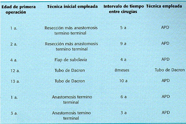
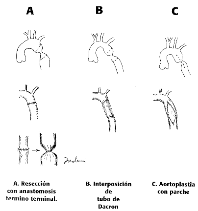
<body><![CDATA[<br> &nbsp; </p>     <p><b><font face="arial"><font size="-1">Introducci&oacute;n</font></font></b> </p>     <p><font face="arial"><font size="-1">La coartaci&oacute;n de la aorta es una malformaci&oacute;n cong&eacute;nita que se presenta cl&iacute;nicamente por lo general a edades tempranas y que produce la muerte si no es tratada (<a href="#4">4</a>,<a href="#14">14</a>). Se ha reportado espor&aacute;dicamente individuos de m&aacute;s de 70 a&ntilde;os. (<a href="#19">19</a>) Representa de un 5 a un 10% de las patolog&iacute;as cong&eacute;nitas cardiovasculares y se observa en 1 de cada 4000 a 5000 necropsias (<a href="#11">11</a>,<a  href="#18">18</a> ) sigue siendo una patolog&iacute;a de suma importancia debido a lo limitante del padecimiento para el enfermo(<a href="#4">4</a>,<a href="#5">5</a>,6,<a  href="#12">12</a>). <b>Cuadro cl&iacute;nico</b> (<a href="#4">4</a>,6) 1. HTA que puede producir al paciente cefalea, visi&oacute;n borrosa, hemorragias retinianas (<a  href="#30">30</a>), muerte s&uacute;bita por ruptura de aneurismas intercostales (<a  href="#20">20</a>) o de la aorta (<a href="#12">12</a>,<a href="#13">13</a> ) y hemorragia subaracnoidea por ruptura de aneurismas del pol&iacute;gono de Willis (<a  href="#29">29</a>). 2. Endarteritis y endocarditis bacteriana por anomal&iacute;as valvulares asociadas (<a href="#25">25</a>). 3. Disminuci&oacute;n de flujo arterial a miembros inferiores con la consecuente claudicaci&oacute;n intermitente(<a href="#4">4</a>,<a href="#10">10</a>). Compromete en sus distintas variedades un estrechamiento del lumen de la aorta mayormente en situaci&oacute;n periductal, pero que puede involucrar el &aacute;rea itsmica y en diferentes sitios del arco a&oacute;rtico inclusive la aorta abdominal (<a href="#27"> 27</a> ). Desde el punto de vista funcional tiene importancia la situaci&oacute;n con respecto al ductus arterioso, reconoci&eacute;ndose una variedad <b>preductal o del reci&eacute;n nacido</b>, con un ductus permeable, una circulaci&oacute;n del hemicuerpo superior arterial y del hemicuerpo inferior venosa que lleva a la insuficiencia card&iacute;aca y muerte tempranamente relacionada a la permeabilidad del ductus (<a href="#11">11</a>,<a href="#25">25</a>), y una variedad <b>postductal o del adulto</b>, con los cambios tr&oacute;ficos, hipertensi&oacute;n arterial y una gran circulaci&oacute;n colateral ( <a href="#4">4</a>,6,<a  href="#25">25</a> ). Por lo general la coartaci&oacute;n est&aacute; en relaci&oacute;n con el ductus arterioso, con una proliferaci&oacute;n de tejido ductal proveniente del desarrollo del sexto arco a&oacute;rtico que ocluye el lumen en forma de membrana incompleta por lo cual se ha denominado <b>coartaci&oacute;n yuxtaductal</b> (<a href="#11">11</a>). El diagn&oacute;stico se reliza generalmente al ex&aacute;men cl&iacute;nico del paciente con desarrollo del hemicuerpo superior y con hipotrofia del hemicuerpo inferior, en pacientes hipertensos con un gradiente brazo - pierna de m&aacute;s de 20 mm hg. Existe un soplo en el espacio interescapulo vertebral y signos radiol&oacute;gicos como la "E invertida " y el de Roessler ( <a href="#25">25</a> ). El diagn&oacute;stico apropiado se realiza con eco transesof&aacute;gico, resonancia magn&eacute;tica nuclear y/o cateterismo (<a href="#20">20</a>,<a href="#21">21</a> ) y el mismo puede realizarse desde estadios fetales in &uacute;tero. (<a  href="#26">26</a>). <b>Tratamiento</b>: La reparaci&oacute;n quir&uacute;rgica fue realizada por vez primera por Crafoord en 1944 ( <a href="#22">22</a> ) con una resecci&oacute;n del segmento coartado y anastomosis t&eacute;rmino terminal . Posteriormente Vosschulte en 1966 (<a href="#23">23</a>) describi&oacute; la aortoplast&iacute;a con parche de dacron dados los &iacute;ndices de reestenosis. En 1966 Waldhaussen (<a href="#8">8</a>) describe la aortoplast&iacute;a con flap de subclavia izquierda. Recientemente se ha descrito la angioplastia con balon y colocaci&oacute;n de stents autoexpandibles con buenos resultados sobre todo en recoartaci&oacute;n (<a href="#24">24</a> ).</font></font>     <br> &nbsp; </p>     <p><b><font face="arial"><font size="-1">Material y M&eacute;todos.</font></font></b> </p>     <p><font face="arial"><font size="-1">Se analizaron retrospectivamente los archivos cl&iacute;nicos de ochenta y un pacientes operados con el diagn&oacute;stico de coartaci&oacute;n de la aorta en el Hospital M&eacute;xico, San Jos&eacute;, Costa Rica, en un per&iacute;odo comprendido entre Enero de 1969 y Diciembre de 1999, anot&aacute;ndose hallazgos cl&iacute;nicos, radiol&oacute;gicos y los estudios hemodin&aacute;micos con aortograma y ecocardiograma. Se utilizaron diferentes t&eacute;cnicas de reparaci&oacute;n a trav&eacute;s de una toracotom&iacute;a posterior izquierda, como son la resecci&oacute;n con anastomosis termino terminal y la interposici&oacute;n de pr&oacute;tesis tubular de dacron. A partir de 1984 se ha usado la aortoplast&iacute;a descrita por Vosschulte modificado con un parche de duramadre.</font></font> </p>     <p><font face="arial"><font size="-1">Postoperatoriamente se determinaron gradientes transa&oacute;rticos y se controlaron valores de PA, adem&aacute;s de complementar con estudios radiol&oacute;gicos y ecogr&aacute;ficos en un per&iacute;odo que oscil&oacute; entre 0.2 y 30 a.</font></font>     <br> &nbsp; </p>     <p><font face="arial"><font size="-1"><b>Hallazgos</b>.</font></font> </p>     <p><font face="arial"><font size="-1">Un total de 81 pacientes fueron operados 56 de ellos hombres y 25 mujeres. El rango de edades oscil&oacute; entre los 0 y los 53 a. con un promedio de 20.3 a. . Las manifestaciones cl&iacute;nicas fueron : 1) En ni&ntilde;os menores de 1 a. : ICC ( 4ptes). 2) De los 2 a los 5 a : Cefalea, ausencia de pulsos femorales y claudicaci&oacute;n.( 4 ptes ) y 3) En el resto de pacientes: HTA y claudicaci&oacute;n intermitente de Ms Is. (73 ptes). Como defectos cong&eacute;nitos asociados se document&oacute; un ductus arterioso permeable en el 31% de los pacientes as&iacute; como una v&aacute;lvula a&oacute;rtica con doble lesi&oacute;n, o insuficiencia mitral en el 19%, siendo estos hallazgos de hasta un 37% en otras series ( 28 ). Con respecto a la t&eacute;cnica quir&uacute;rgica utilizada, se raliz&oacute; (<a href="#fig1">fig. 1</a>) Resecci&oacute;n con anastomosis termino terminal : 29 pacientes (36%), Interposici&oacute;n de tubo de Dacron :1 pte (1.2%), y aortoplast&iacute;a con parche de duramadre 51 ptes ( 62.8%). Reoperaciones fueron practicadas en 7 pacientes, todos operados previamente en otros centros, los cuales fueron tratados con parche de duramadre en 6 casos y con tubo de dacron en uno (<a href="#Cuadro1">cuadro 1</a> ).    ]]></body>
<body><![CDATA[<br> </font></font></p>     <center><a name="fig1"></a><img src="/img/fbpe/rcc/v2n2/0484i1.GIF"  height="432" width="404"></center> <font face="arial"><font size="-1"><b>&nbsp;&nbsp;&nbsp;&nbsp;&nbsp;&nbsp;&nbsp;&nbsp;&nbsp;&nbsp;&nbsp;&nbsp;&nbsp;&nbsp;&nbsp;&nbsp;&nbsp;&nbsp;&nbsp;&nbsp;&nbsp;&nbsp;&nbsp;&nbsp;&nbsp;&nbsp;&nbsp;&nbsp;&nbsp;&nbsp;&nbsp;&nbsp;&nbsp;&nbsp;&nbsp;&nbsp;&nbsp;&nbsp;&nbsp;&nbsp;&nbsp;&nbsp;&nbsp;&nbsp;&nbsp;&nbsp;&nbsp;&nbsp;&nbsp;&nbsp;&nbsp;&nbsp;&nbsp;&nbsp;&nbsp;&nbsp; FIGURA 1:</b> ver explicaci&oacute;n en el texto    
<br>     <br> </font></font>     <center><a name="Cuadro1"></a><b><font face="arial"><font size="-1">Cuadro 1. Reoperaciones por coartaci&oacute;n de la aorta</font></font></b></center>     <center></center>     <center><img src="/img/fbpe/rcc/v2n2/0484i2.JPG" height="408"  width="614"></center> <font face="Arial"><font size="-1">&nbsp;&nbsp;&nbsp;&nbsp;&nbsp;&nbsp;&nbsp;&nbsp;&nbsp;&nbsp;&nbsp;&nbsp;&nbsp;&nbsp;&nbsp;&nbsp;&nbsp; APD: Aortoplastia con Parche de Duramadre.</font></font>    
<br>     <p> </p>     <p><font face="arial"><font size="-1">La morbilidad quir&uacute;rgica fue de un 16%, correspondiendo a neumot&oacute;rax residual, hemot&oacute;rax, arritmias postoperatorias e hipertensi&oacute;n en el postoperatorio inmediato. La mortalidad fue de 2.4% que correspondi&oacute; a dos pacientes, uno de 4 meses de edad con ICC y bronconeumon&iacute;a postoperatoria y uno de 23 a. con una reoperaci&oacute;n con tubo de dacron que sangr&oacute; de la pared costal siendo reintervenido m&uacute;ltiples veces presentando sepsis y fallo multisist&eacute;mico.</font></font> </p>     ]]></body>
<body><![CDATA[<p><font face="arial"><font size="-1">Con respecto a la hipertensi&oacute;n arterial el 84% de los pacientes se tornaron normotensos en el postoperatorio el 10 % obtuvo mejor&iacute;a, siendo controlados con antihipertensivos satisfactoriamente y el 6% permaneci&oacute; sin cambios. El gradiente de presi&oacute;n brazo pierna en reposo fue en todos los pacientes menor de 20 mmHg, con buena evoluci&oacute;n (<a href="#15">15</a>). Durante el seguimiento de los pacientes no se han presentado complicaciones tard&iacute;as derivadas de la cirug&iacute;a como recoartaci&oacute;n, endarteritis o formaci&oacute;n de aneurisma cerca del parche, no habiendo necesidad de reoperar a ning&uacute;n paciente de los originalmente intervenidos en nuestro centro.</font></font>     <br> &nbsp; </p>     <p><b><font face="arial"><font size="-1">Comentario.</font></font></b> </p>     <p><font face="arial"><font size="-1">Desde principios de los 80 , la aortoplast&iacute;a con la variaci&oacute;n del parche de Duramadre descrito por Ross (<a  href="#16">16</a> ) se ha implementado en nuestro Servicio, siendo desde entonces la t&eacute;cnica de elecci&oacute;n por las siguientes ventajas sobre las otras: A) El di&aacute;metro de la aorta permanece amplio a&uacute;n en casos de ni&ntilde;os en crecimiento, cosa que no sucede en todos los casos de anastomosis termino terminal y de interposici&oacute;n de tubo (<a href="#1">1</a>,<a href="#3">3</a>,<a  href="#17">17</a> ) B) Est&aacute; demostrado que la aortoplast&iacute;a con parche es m&aacute;s efectiva que la resecci&oacute;n con anastomosis T-T en la reducci&oacute;n del gradiente transa&oacute;rtico(<a href="#3">3</a>,<a href="#8">8</a>). C) La hemostasia es &oacute;ptima puesto que toda la cirug&iacute;a se realiza en el plano frontal de la aorta y no manipula la cara posterior la cual es de dif&iacute;cil abordaje( <a href="#15">15</a> ) y desde el punto de vista t&eacute;cnico nos resulta m&aacute;s c&oacute;modo de realizar. Finalmente, no utilizamos flap de arteria subclavia por la poblaci&oacute;n adulta que actualmente manejamos ademas del sacrificio de la arteria subclavia que esta conlleva (<a href="#2">2</a>). Es as&iacute; como la aortoplast&iacute;a con parche de duramadre es la t&eacute;cnica preferida por nosotros tanto por ser t&eacute;cnicamente la m&aacute;s sencilla, as&iacute; como por los resultados a largo plazo donde no hemos tenido complicaciones relacionadas a la misma, tales como son la reestenosis o la formaci&oacute;n de aneurismas tard&iacute;os en un per&iacute;odo que lleva 20 a.</font></font>     <br> &nbsp;</p>     <p><b><font face="Arial"><font size="-1">Referencias</font></font></b> </p>     <!-- ref --><p><a name="1"></a><font face="ARIAL"><font size="-1"><b>1</b> Morris G. Et al. Coarctation of the aorta with special emphasis upon improved techniques of surgical repair. J. of Thorac and Cardiovasc. Surgery, Vol 4 , Dec 1960.</font></font> &nbsp;&nbsp;&nbsp;&nbsp;&nbsp;&nbsp;&nbsp;&nbsp;[&#160;<a href="javascript:void(0);" onclick="javascript: window.open('/scielo.php?script=sci_nlinks&ref=725663&pid=S1409-4142200000020000400001&lng=','','width=640,height=500,resizable=yes,scrollbars=1,menubar=yes,');">Links</a>&#160;]<!-- end-ref --><!-- ref --><p><a name="2"></a><font face="ARIAL"><font size="-1"><b>2</b> Williams S.Pierceet al. Late results of the subclavian flap. Procedure in infants with coarctation of the thoracic aorta. Cardiovascular surgery 1977. Circulation suppl. 1 Vol 58, sept 1978</font></font> &nbsp;&nbsp;&nbsp;&nbsp;&nbsp;&nbsp;&nbsp;&nbsp;[&#160;<a href="javascript:void(0);" onclick="javascript: window.open('/scielo.php?script=sci_nlinks&ref=725664&pid=S1409-4142200000020000400002&lng=','','width=640,height=500,resizable=yes,scrollbars=1,menubar=yes,');">Links</a>&#160;]<!-- end-ref --><!-- ref --><p><a name="3"></a><font face="ARIAL"><font size="-1"><b>3</b> Morris R. Sade et al .Aortoplasty compared with aortic resection in young children . Ann of Thor ac. Surg. Vol 28,oct 1979</font></font> &nbsp;&nbsp;&nbsp;&nbsp;&nbsp;&nbsp;&nbsp;&nbsp;[&#160;<a href="javascript:void(0);" onclick="javascript: window.open('/scielo.php?script=sci_nlinks&ref=725665&pid=S1409-4142200000020000400003&lng=','','width=640,height=500,resizable=yes,scrollbars=1,menubar=yes,');">Links</a>&#160;]<!-- end-ref --><!-- ref --><p><a name="4"></a><font face="ARIAL"><font size="-1"><b>4</b> Gibbons ,Spencer .Cirugia toracica . Ed Salvat P 943-45, ed 1981.</font></font> &nbsp;&nbsp;&nbsp;&nbsp;&nbsp;&nbsp;&nbsp;&nbsp;[&#160;<a href="javascript:void(0);" onclick="javascript: window.open('/scielo.php?script=sci_nlinks&ref=725666&pid=S1409-4142200000020000400004&lng=','','width=640,height=500,resizable=yes,scrollbars=1,menubar=yes,');">Links</a>&#160;]<!-- end-ref --><!-- ref --><p><a name="5"></a><font face="ARIAL"><font size="-1"><b>5</b> Cowgill D. Corction of the aorta . Cardiac Surg. Vol.13 Jun 1987.</font></font> &nbsp;&nbsp;&nbsp;&nbsp;&nbsp;&nbsp;&nbsp;&nbsp;[&#160;<a href="javascript:void(0);" onclick="javascript: window.open('/scielo.php?script=sci_nlinks&ref=725667&pid=S1409-4142200000020000400005&lng=','','width=640,height=500,resizable=yes,scrollbars=1,menubar=yes,');">Links</a>&#160;]<!-- end-ref --><!-- ref --><p><font face="ARIAL"><font size="-1">Litwin S.et al . Surgical resection of aortic coarctation of the aorta in infancy. Jour. Ped.Surg. 11:307, 1971</font></font> &nbsp;&nbsp;&nbsp;&nbsp;&nbsp;&nbsp;&nbsp;&nbsp;[&#160;<a href="javascript:void(0);" onclick="javascript: window.open('/scielo.php?script=sci_nlinks&ref=725668&pid=S1409-4142200000020000400006&lng=','','width=640,height=500,resizable=yes,scrollbars=1,menubar=yes,');">Links</a>&#160;]<!-- end-ref --><!-- ref --><p><a name="7"></a><font face="ARIAL"><font size="-1"><b>7</b> Tawes L. Coarctation of the aorta in infants and children. Circulation ,39:173,1969.</font></font> &nbsp;&nbsp;&nbsp;&nbsp;&nbsp;&nbsp;&nbsp;&nbsp;[&#160;<a href="javascript:void(0);" onclick="javascript: window.open('/scielo.php?script=sci_nlinks&ref=725669&pid=S1409-4142200000020000400007&lng=','','width=640,height=500,resizable=yes,scrollbars=1,menubar=yes,');">Links</a>&#160;]<!-- end-ref --><!-- ref --><p><a name="8"></a><font face="ARIAL"><font size="-1"><b>8</b> Waldhaussen J. Repair of the coarctation of the aorta with subclavian flap. J. Thorac.Cardiovasc. Surg51:532,1966.</font></font> &nbsp;&nbsp;&nbsp;&nbsp;&nbsp;&nbsp;&nbsp;&nbsp;[&#160;<a href="javascript:void(0);" onclick="javascript: window.open('/scielo.php?script=sci_nlinks&ref=725670&pid=S1409-4142200000020000400008&lng=','','width=640,height=500,resizable=yes,scrollbars=1,menubar=yes,');">Links</a>&#160;]<!-- end-ref --><!-- ref --><p><a name="9"></a><font face="ARIAL"><font size="-1"><b>9</b> Palatianos G. Changing trends in the surgical teatment of aortic coarctation. Annals of thoracic surgery. Vol 40, jul 1985</font></font> &nbsp;&nbsp;&nbsp;&nbsp;&nbsp;&nbsp;&nbsp;&nbsp;[&#160;<a href="javascript:void(0);" onclick="javascript: window.open('/scielo.php?script=sci_nlinks&ref=725671&pid=S1409-4142200000020000400009&lng=','','width=640,height=500,resizable=yes,scrollbars=1,menubar=yes,');">Links</a>&#160;]<!-- end-ref --><!-- ref --><p><a name="10"></a><font face="ARIAL"><font size="-1"><b>10</b> Morua E. Coartacion de la Aorta. Acta Med. Costarricense Vol.31:n2;1988.</font></font> &nbsp;&nbsp;&nbsp;&nbsp;&nbsp;&nbsp;&nbsp;&nbsp;[&#160;<a href="javascript:void(0);" onclick="javascript: window.open('/scielo.php?script=sci_nlinks&ref=725672&pid=S1409-4142200000020000400010&lng=','','width=640,height=500,resizable=yes,scrollbars=1,menubar=yes,');">Links</a>&#160;]<!-- end-ref --><!-- ref --><p><a name="11"></a><font face="ARIAL"><font size="-1"><b>11</b> Sabiston Textbook of surgery 16 th ed. WBSaunders Co 1996.</font></font> &nbsp;&nbsp;&nbsp;&nbsp;&nbsp;&nbsp;&nbsp;&nbsp;[&#160;<a href="javascript:void(0);" onclick="javascript: window.open('/scielo.php?script=sci_nlinks&ref=725673&pid=S1409-4142200000020000400011&lng=','','width=640,height=500,resizable=yes,scrollbars=1,menubar=yes,');">Links</a>&#160;]<!-- end-ref --><!-- ref --><p><a name="12"></a><font face="ARIAL"><font size="-1"><b>12</b> Findlow D. Congenital Heart disease in adults. Br J Anaesth. 1997;78(4):416-30</font></font> &nbsp;&nbsp;&nbsp;&nbsp;&nbsp;&nbsp;&nbsp;&nbsp;[&#160;<a href="javascript:void(0);" onclick="javascript: window.open('/scielo.php?script=sci_nlinks&ref=725674&pid=S1409-4142200000020000400012&lng=','','width=640,height=500,resizable=yes,scrollbars=1,menubar=yes,');">Links</a>&#160;]<!-- end-ref --><!-- ref --><p><a name="13"></a><font face="ARIAL"><font size="-1"><b>13</b> Heineman NK. Extraanatomic Thoracic aortic by pass grafs. Indications ,Technics and results. Eur.J. Cardiothorac-Surg.1997;11(1):169-75</font></font> &nbsp;&nbsp;&nbsp;&nbsp;&nbsp;&nbsp;&nbsp;&nbsp;[&#160;<a href="javascript:void(0);" onclick="javascript: window.open('/scielo.php?script=sci_nlinks&ref=725675&pid=S1409-4142200000020000400013&lng=','','width=640,height=500,resizable=yes,scrollbars=1,menubar=yes,');">Links</a>&#160;]<!-- end-ref --><!-- ref --><p><a name="14"></a><font face="ARIAL"><font size="-1"><b>14</b> Heger M. Aortic coarctation Long term follow-up in adults (Abstract) Z Kardiol 1997;86(1):50-5</font></font> &nbsp;&nbsp;&nbsp;&nbsp;&nbsp;&nbsp;&nbsp;&nbsp;[&#160;<a href="javascript:void(0);" onclick="javascript: window.open('/scielo.php?script=sci_nlinks&ref=725676&pid=S1409-4142200000020000400014&lng=','','width=640,height=500,resizable=yes,scrollbars=1,menubar=yes,');">Links</a>&#160;]<!-- end-ref --><!-- ref --><p><a name="15"></a><font face="ARIAL"><font size="-1"><b>15</b> Guenthard J. Arm leg pressure gradients on late follow up after aortic coarctation repair. Possible causes and implications. Eur.Heart J. 1996;17(10):1572-75</font></font> &nbsp;&nbsp;&nbsp;&nbsp;&nbsp;&nbsp;&nbsp;&nbsp;[&#160;<a href="javascript:void(0);" onclick="javascript: window.open('/scielo.php?script=sci_nlinks&ref=725677&pid=S1409-4142200000020000400015&lng=','','width=640,height=500,resizable=yes,scrollbars=1,menubar=yes,');">Links</a>&#160;]<!-- end-ref --><!-- ref --><p><a name="16"></a><font face="ARIAL"><font size="-1"><b>16</b> Ross D. comunicacion personal</font></font> &nbsp;&nbsp;&nbsp;&nbsp;&nbsp;&nbsp;&nbsp;&nbsp;[&#160;<a href="javascript:void(0);" onclick="javascript: window.open('/scielo.php?script=sci_nlinks&ref=725678&pid=S1409-4142200000020000400016&lng=','','width=640,height=500,resizable=yes,scrollbars=1,menubar=yes,');">Links</a>&#160;]<!-- end-ref --><!-- ref --><p><a name="17"></a><font face="ARIAL"><font size="-1"><b>17</b> Venturini A. Primary repair of coarctation of the aorta by patch graft aortoplasty. Eur J Cardiiothorac Surg. 1996;10(10):890-6.</font></font> &nbsp;&nbsp;&nbsp;&nbsp;&nbsp;&nbsp;&nbsp;&nbsp;[&#160;<a href="javascript:void(0);" onclick="javascript: window.open('/scielo.php?script=sci_nlinks&ref=725679&pid=S1409-4142200000020000400017&lng=','','width=640,height=500,resizable=yes,scrollbars=1,menubar=yes,');">Links</a>&#160;]<!-- end-ref --><!-- ref --><p><a name="18"></a><font face="ARIAL"><font size="-1"><b>18</b> Mc Connell M. Congenital diseases of the aorta. Rev Esp Cardiol 1996;49 Suppl. 4 :92-9.</font></font> &nbsp;&nbsp;&nbsp;&nbsp;&nbsp;&nbsp;&nbsp;&nbsp;[&#160;<a href="javascript:void(0);" onclick="javascript: window.open('/scielo.php?script=sci_nlinks&ref=725680&pid=S1409-4142200000020000400018&lng=','','width=640,height=500,resizable=yes,scrollbars=1,menubar=yes,');">Links</a>&#160;]<!-- end-ref --><!-- ref --><p><a name="19"></a><font face="ARIAL"><font size="-1"><b>19</b> Convens C. Aortic coarctation : A rare and unexpected cause of secondary arterial arterial hypertension in the elderly. CathetCardiovasc Diagn.1996 ;39(1):71-4.</font></font> &nbsp;&nbsp;&nbsp;&nbsp;&nbsp;&nbsp;&nbsp;&nbsp;[&#160;<a href="javascript:void(0);" onclick="javascript: window.open('/scielo.php?script=sci_nlinks&ref=725681&pid=S1409-4142200000020000400019&lng=','','width=640,height=500,resizable=yes,scrollbars=1,menubar=yes,');">Links</a>&#160;]<!-- end-ref --><!-- ref --><p><a name="20"></a><font face="ARIAL"><font size="-1"><b>20</b> Yamaguchi T. Intercostal artery aneurism associated with coarctation of the aorta: specific finding on MRI. Int J Card Imaging. 1997;13(4):357-8.</font></font> &nbsp;&nbsp;&nbsp;&nbsp;&nbsp;&nbsp;&nbsp;&nbsp;[&#160;<a href="javascript:void(0);" onclick="javascript: window.open('/scielo.php?script=sci_nlinks&ref=725682&pid=S1409-4142200000020000400020&lng=','','width=640,height=500,resizable=yes,scrollbars=1,menubar=yes,');">Links</a>&#160;]<!-- end-ref --><!-- ref --><p><a name="21"></a><font face="ARIAL"><font size="-1"><b>21</b> Linsay J. Diagnosis and treatment of diseases of the aorta. Curr Probl Cardiol. 1997 ;22(10): 485-542.</font></font> &nbsp;&nbsp;&nbsp;&nbsp;&nbsp;&nbsp;&nbsp;&nbsp;[&#160;<a href="javascript:void(0);" onclick="javascript: window.open('/scielo.php?script=sci_nlinks&ref=725683&pid=S1409-4142200000020000400021&lng=','','width=640,height=500,resizable=yes,scrollbars=1,menubar=yes,');">Links</a>&#160;]<!-- end-ref --><!-- ref --><p><a name="22"></a><font face="ARIAL"><font size="-1"><b>22</b> Crafoord S. Congenital coarctation of the aorta and its surgical management. Jthorac Cardivasc Surg, 1945 ; 14:347.</font></font> &nbsp;&nbsp;&nbsp;&nbsp;&nbsp;&nbsp;&nbsp;&nbsp;[&#160;<a href="javascript:void(0);" onclick="javascript: window.open('/scielo.php?script=sci_nlinks&ref=725684&pid=S1409-4142200000020000400022&lng=','','width=640,height=500,resizable=yes,scrollbars=1,menubar=yes,');">Links</a>&#160;]<!-- end-ref --><!-- ref --><p><a name="23"></a><font face="ARIAL"><font size="-1"><b>23</b> Vosschulte K. Surgical correction of coarctation of the aorta by an isthmuplastic operation. Thorax 1961;16: 338.</font></font> &nbsp;&nbsp;&nbsp;&nbsp;&nbsp;&nbsp;&nbsp;&nbsp;[&#160;<a href="javascript:void(0);" onclick="javascript: window.open('/scielo.php?script=sci_nlinks&ref=725685&pid=S1409-4142200000020000400023&lng=','','width=640,height=500,resizable=yes,scrollbars=1,menubar=yes,');">Links</a>&#160;]<!-- end-ref --><!-- ref --><p><a name="24"></a><font face="ARIAL"><font size="-1"><b>24</b> Descalzo-Sonorans A. Ballon percutaneus angioplasty in postoperative aortic coarctation. Rev Esp Cardiol. 1994; 47(4):247-50.</font></font> &nbsp;&nbsp;&nbsp;&nbsp;&nbsp;&nbsp;&nbsp;&nbsp;[&#160;<a href="javascript:void(0);" onclick="javascript: window.open('/scielo.php?script=sci_nlinks&ref=725686&pid=S1409-4142200000020000400024&lng=','','width=640,height=500,resizable=yes,scrollbars=1,menubar=yes,');">Links</a>&#160;]<!-- end-ref --><!-- ref --><p><a name="25"></a><font face="ARIAL"><font size="-1"><b>25</b> Hillis LD. Manual of Clinical problems in Cardiology, third ed. Little Brown, 1989.</font></font> &nbsp;&nbsp;&nbsp;&nbsp;&nbsp;&nbsp;&nbsp;&nbsp;[&#160;<a href="javascript:void(0);" onclick="javascript: window.open('/scielo.php?script=sci_nlinks&ref=725687&pid=S1409-4142200000020000400025&lng=','','width=640,height=500,resizable=yes,scrollbars=1,menubar=yes,');">Links</a>&#160;]<!-- end-ref --><!-- ref --><p><a name="26"></a><font face="ARIAL"><font size="-1"><b>26</b> Hornberger L. Antenatal diagnosis of coarctation of the aorta: a multicenter experience. J Am Coll Cardiol. 1994;23(2):417-23.</font></font> &nbsp;&nbsp;&nbsp;&nbsp;&nbsp;&nbsp;&nbsp;&nbsp;[&#160;<a href="javascript:void(0);" onclick="javascript: window.open('/scielo.php?script=sci_nlinks&ref=725688&pid=S1409-4142200000020000400026&lng=','','width=640,height=500,resizable=yes,scrollbars=1,menubar=yes,');">Links</a>&#160;]<!-- end-ref --><!-- ref --><p><a name="27"></a><font face="ARIAL"><font size="-1"><b>27</b> Janzen J . The disease picture of atypical aortic coarctation Helv.Chir.Acta 1993;60(1-2):291-8.</font></font> &nbsp;&nbsp;&nbsp;&nbsp;&nbsp;&nbsp;&nbsp;&nbsp;[&#160;<a href="javascript:void(0);" onclick="javascript: window.open('/scielo.php?script=sci_nlinks&ref=725689&pid=S1409-4142200000020000400027&lng=','','width=640,height=500,resizable=yes,scrollbars=1,menubar=yes,');">Links</a>&#160;]<!-- end-ref --><!-- ref --><p><a name="28"></a><font face="ARIAL"><font size="-1"><b>28</b> Poliakova N. Aortic coarctation and the bicuspid aortic valve. Ter Arkh. 1996;68(4) : 48-50.</font></font> &nbsp;&nbsp;&nbsp;&nbsp;&nbsp;&nbsp;&nbsp;&nbsp;[&#160;<a href="javascript:void(0);" onclick="javascript: window.open('/scielo.php?script=sci_nlinks&ref=725690&pid=S1409-4142200000020000400028&lng=','','width=640,height=500,resizable=yes,scrollbars=1,menubar=yes,');">Links</a>&#160;]<!-- end-ref --><!-- ref --><p><a name="29"></a><font face="ARIAL"><font size="-1"><b>29</b> Gire C. Rupture of cerebral aneurysm associated with aortic coarctation. Rev Neurol Paris .1997;153(5):357-8.</font></font> &nbsp;&nbsp;&nbsp;&nbsp;&nbsp;&nbsp;&nbsp;&nbsp;[&#160;<a href="javascript:void(0);" onclick="javascript: window.open('/scielo.php?script=sci_nlinks&ref=725691&pid=S1409-4142200000020000400029&lng=','','width=640,height=500,resizable=yes,scrollbars=1,menubar=yes,');">Links</a>&#160;]<!-- end-ref --><!-- ref --><p><a name="30"></a><font face="ARIAL"><font size="-1"><b>30</b> Tabandeh H. Retinal vascular abnormalities in aortic coarctation . Eye . 1996;10 (4) :525-7.</font></font>    &nbsp;&nbsp;&nbsp;&nbsp;&nbsp;&nbsp;&nbsp;&nbsp;[&#160;<a href="javascript:void(0);" onclick="javascript: window.open('/scielo.php?script=sci_nlinks&ref=725692&pid=S1409-4142200000020000400030&lng=','','width=640,height=500,resizable=yes,scrollbars=1,menubar=yes,');">Links</a>&#160;]<!-- end-ref --><br> &nbsp; </p>     <p><a name="*"></a><font face="ARIAL"><font size="-1"><a href="#*a">*</a> Servicio de Cirug&iacute;a Tor&aacute;cica y Cardiovascular Hospital M&eacute;xico,</font></font>     <br> <font face="ARIAL"><font size="-1">San Jos&eacute;, Costa Rica.</font></font>     <br> <font face="ARIAL"><font size="-1">e-mail:<a  href="mailto:eiduni@medicos.sa.cr">eiduni@medicos.sa.cr</a></font></font> </p>     ]]></body>
<body><![CDATA[ ]]></body><back>
<ref-list>
<ref id="B1">
<nlm-citation citation-type="journal">
<person-group person-group-type="author">
<name>
<surname><![CDATA[Morris]]></surname>
<given-names><![CDATA[G]]></given-names>
</name>
</person-group>
<article-title xml:lang="en"><![CDATA[Coarctation of the aorta with special emphasis upon improved techniques of surgical repair]]></article-title>
<source><![CDATA[J. of Thorac and Cardiovasc. Surgery]]></source>
<year>Dec </year>
<month>19</month>
<day>60</day>
<volume>4</volume>
</nlm-citation>
</ref>
<ref id="B2">
<nlm-citation citation-type="journal">
<person-group person-group-type="author">
<name>
<surname><![CDATA[Williams]]></surname>
<given-names><![CDATA[S]]></given-names>
</name>
<name>
<surname><![CDATA[Pierce]]></surname>
</name>
</person-group>
<article-title xml:lang="en"><![CDATA[Late results of the subclavian flap: Procedure in infants with coarctation of the thoracic aorta]]></article-title>
<source><![CDATA[Cardiovascular surgery 1977]]></source>
<year>sept</year>
<month> 1</month>
<day>97</day>
<volume>58</volume>
<numero>^s1</numero>
<issue>^s1</issue>
<supplement>1</supplement>
</nlm-citation>
</ref>
<ref id="B3">
<nlm-citation citation-type="journal">
<person-group person-group-type="author">
<name>
<surname><![CDATA[Morris]]></surname>
<given-names><![CDATA[R]]></given-names>
</name>
<name>
<surname><![CDATA[Sade]]></surname>
</name>
</person-group>
<article-title xml:lang="en"><![CDATA[Aortoplasty compared with aortic resection in young children]]></article-title>
<source><![CDATA[Ann of Thor ac. Surg]]></source>
<year>oct </year>
<month>19</month>
<day>79</day>
<volume>28</volume>
</nlm-citation>
</ref>
<ref id="B4">
<nlm-citation citation-type="book">
<person-group person-group-type="author">
<name>
<surname><![CDATA[Gibbons]]></surname>
<given-names><![CDATA[Spencer]]></given-names>
</name>
</person-group>
<source><![CDATA[Cirugia toracica]]></source>
<year>1981</year>
<page-range>943-45</page-range><publisher-name><![CDATA[Ed Salvat P]]></publisher-name>
</nlm-citation>
</ref>
<ref id="B5">
<nlm-citation citation-type="journal">
<person-group person-group-type="author">
<name>
<surname><![CDATA[Cowgill]]></surname>
<given-names><![CDATA[D]]></given-names>
</name>
</person-group>
<article-title xml:lang="en"><![CDATA[Corction of the aorta]]></article-title>
<source><![CDATA[Cardiac Surg]]></source>
<year>Jun </year>
<month>19</month>
<day>87</day>
<volume>13</volume>
</nlm-citation>
</ref>
<ref id="B6">
<nlm-citation citation-type="journal">
<person-group person-group-type="author">
<name>
<surname><![CDATA[Litwin]]></surname>
<given-names><![CDATA[S]]></given-names>
</name>
</person-group>
<article-title xml:lang="en"><![CDATA[Surgical resection of aortic coarctation of the aorta in infancy]]></article-title>
<source><![CDATA[Jour. Ped.Surg]]></source>
<year>1971</year>
<volume>11</volume>
<page-range>307</page-range></nlm-citation>
</ref>
<ref id="B7">
<nlm-citation citation-type="journal">
<person-group person-group-type="author">
<name>
<surname><![CDATA[Tawes]]></surname>
<given-names><![CDATA[L]]></given-names>
</name>
</person-group>
<article-title xml:lang="en"><![CDATA[Coarctation of the aorta in infants and children]]></article-title>
<source><![CDATA[Circulation]]></source>
<year>1969</year>
<volume>39</volume>
<page-range>173</page-range></nlm-citation>
</ref>
<ref id="B8">
<nlm-citation citation-type="journal">
<person-group person-group-type="author">
<name>
<surname><![CDATA[Waldhaussen]]></surname>
<given-names><![CDATA[J]]></given-names>
</name>
</person-group>
<article-title xml:lang="en"><![CDATA[Repair of the coarctation of the aorta with subclavian flap]]></article-title>
<source><![CDATA[J. Thorac.Cardiovasc. Surg]]></source>
<year>1966</year>
<volume>51</volume>
<page-range>532</page-range></nlm-citation>
</ref>
<ref id="B9">
<nlm-citation citation-type="journal">
<person-group person-group-type="author">
<name>
<surname><![CDATA[Palatianos]]></surname>
<given-names><![CDATA[G]]></given-names>
</name>
</person-group>
<article-title xml:lang="en"><![CDATA[Changing trends in the surgical teatment of aortic coarctation]]></article-title>
<source><![CDATA[Annals of thoracic surgery]]></source>
<year>jul </year>
<month>19</month>
<day>85</day>
<volume>40</volume>
</nlm-citation>
</ref>
<ref id="B10">
<nlm-citation citation-type="journal">
<person-group person-group-type="author">
<name>
<surname><![CDATA[Morua]]></surname>
<given-names><![CDATA[E]]></given-names>
</name>
</person-group>
<article-title xml:lang="es"><![CDATA[Coartacion de la Aorta]]></article-title>
<source><![CDATA[Acta Med. Costarricense]]></source>
<year>1988</year>
<volume>31</volume>
<numero>^s2</numero>
<issue>^s2</issue>
<supplement>2</supplement>
</nlm-citation>
</ref>
<ref id="B11">
<nlm-citation citation-type="book">
<source><![CDATA[Sabiston Textbook of surgery]]></source>
<year>1996</year>
<edition>16</edition>
<publisher-name><![CDATA[WBSaunders Co]]></publisher-name>
</nlm-citation>
</ref>
<ref id="B12">
<nlm-citation citation-type="journal">
<person-group person-group-type="author">
<name>
<surname><![CDATA[Findlow]]></surname>
<given-names><![CDATA[D]]></given-names>
</name>
</person-group>
<article-title xml:lang="en"><![CDATA[Congenital Heart disease in adults]]></article-title>
<source><![CDATA[Br J Anaesth]]></source>
<year>1997</year>
<volume>78</volume>
<numero>^s4</numero>
<issue>^s4</issue>
<supplement>4</supplement>
<page-range>416-30</page-range></nlm-citation>
</ref>
<ref id="B13">
<nlm-citation citation-type="journal">
<person-group person-group-type="author">
<name>
<surname><![CDATA[Heineman]]></surname>
<given-names><![CDATA[NK]]></given-names>
</name>
</person-group>
<article-title xml:lang="en"><![CDATA[Extraanatomic Thoracic aortic by pass grafs: Indications ,Technics and results]]></article-title>
<source><![CDATA[Eur.J. Cardiothorac-Surg]]></source>
<year>1997</year>
<volume>11</volume>
<numero>^s1</numero>
<issue>^s1</issue>
<supplement>1</supplement>
<page-range>169-75</page-range></nlm-citation>
</ref>
<ref id="B14">
<nlm-citation citation-type="journal">
<person-group person-group-type="author">
<name>
<surname><![CDATA[Heger]]></surname>
<given-names><![CDATA[M]]></given-names>
</name>
</person-group>
<article-title xml:lang="en"><![CDATA[Aortic coarctation Long term follow-up in adults]]></article-title>
<source><![CDATA[Z Kardiol]]></source>
<year>1997</year>
<volume>86</volume>
<numero>^s1</numero>
<issue>^s1</issue>
<supplement>1</supplement>
<page-range>50-5</page-range></nlm-citation>
</ref>
<ref id="B15">
<nlm-citation citation-type="journal">
<person-group person-group-type="author">
<name>
<surname><![CDATA[Guenthard]]></surname>
<given-names><![CDATA[J]]></given-names>
</name>
</person-group>
<article-title xml:lang="en"><![CDATA[Arm leg pressure gradients on late follow up after aortic coarctation repair: Possible causes and implications]]></article-title>
<source><![CDATA[Eur.Heart J]]></source>
<year>1996</year>
<volume>17</volume>
<numero>^s10</numero>
<issue>^s10</issue>
<supplement>10</supplement>
<page-range>1572-75</page-range></nlm-citation>
</ref>
<ref id="B16">
<nlm-citation citation-type="">
<person-group person-group-type="author">
<name>
<surname><![CDATA[Ross]]></surname>
<given-names><![CDATA[D]]></given-names>
</name>
</person-group>
<source><![CDATA[comunicacion personal]]></source>
<year></year>
</nlm-citation>
</ref>
<ref id="B17">
<nlm-citation citation-type="journal">
<person-group person-group-type="author">
<name>
<surname><![CDATA[Venturini]]></surname>
<given-names><![CDATA[A]]></given-names>
</name>
</person-group>
<article-title xml:lang="en"><![CDATA[Primary repair of coarctation of the aorta by patch graft aortoplasty]]></article-title>
<source><![CDATA[Eur J Cardiiothorac Surg]]></source>
<year>1996</year>
<volume>10</volume>
<numero>^s10</numero>
<issue>^s10</issue>
<supplement>10</supplement>
<page-range>890-6</page-range></nlm-citation>
</ref>
<ref id="B18">
<nlm-citation citation-type="journal">
<person-group person-group-type="author">
<name>
<surname><![CDATA[Mc Connell]]></surname>
<given-names><![CDATA[M]]></given-names>
</name>
</person-group>
<article-title xml:lang="en"><![CDATA[Congenital diseases of the aorta]]></article-title>
<source><![CDATA[Rev Esp Cardiol]]></source>
<year>1996</year>
<volume>49</volume>
<numero>^s4</numero>
<issue>^s4</issue>
<supplement>4</supplement>
<page-range>92-9</page-range></nlm-citation>
</ref>
<ref id="B19">
<nlm-citation citation-type="journal">
<person-group person-group-type="author">
<name>
<surname><![CDATA[Convens]]></surname>
<given-names><![CDATA[C]]></given-names>
</name>
</person-group>
<article-title xml:lang="en"><![CDATA[Aortic coarctation: A rare and unexpected cause of secondary arterial arterial hypertension in the elderly]]></article-title>
<source><![CDATA[CathetCardiovasc Diagn]]></source>
<year>1996</year>
<volume>39</volume>
<numero>^s1</numero>
<issue>^s1</issue>
<supplement>1</supplement>
<page-range>71-4</page-range></nlm-citation>
</ref>
<ref id="B20">
<nlm-citation citation-type="journal">
<person-group person-group-type="author">
<name>
<surname><![CDATA[Yamaguchi]]></surname>
<given-names><![CDATA[T]]></given-names>
</name>
</person-group>
<article-title xml:lang="en"><![CDATA[Intercostal artery aneurism associated with coarctation of the aorta: specific finding on MRI]]></article-title>
<source><![CDATA[Int J Card Imaging]]></source>
<year>1997</year>
<volume>13</volume>
<numero>^s4</numero>
<issue>^s4</issue>
<supplement>4</supplement>
<page-range>357-8</page-range></nlm-citation>
</ref>
<ref id="B21">
<nlm-citation citation-type="journal">
<person-group person-group-type="author">
<name>
<surname><![CDATA[Linsay]]></surname>
<given-names><![CDATA[J]]></given-names>
</name>
</person-group>
<article-title xml:lang="en"><![CDATA[Diagnosis and treatment of diseases of the aorta]]></article-title>
<source><![CDATA[Curr Probl Cardiol]]></source>
<year>1997</year>
<volume>22</volume>
<numero>^s10</numero>
<issue>^s10</issue>
<supplement>10</supplement>
<page-range>485-542</page-range></nlm-citation>
</ref>
<ref id="B22">
<nlm-citation citation-type="journal">
<person-group person-group-type="author">
<name>
<surname><![CDATA[Crafoord]]></surname>
<given-names><![CDATA[S]]></given-names>
</name>
</person-group>
<article-title xml:lang="en"><![CDATA[Congenital coarctation of the aorta and its surgical management]]></article-title>
<source><![CDATA[Jthorac Cardivasc Surg]]></source>
<year>1945</year>
<volume>14</volume>
<page-range>347</page-range></nlm-citation>
</ref>
<ref id="B23">
<nlm-citation citation-type="journal">
<person-group person-group-type="author">
<name>
<surname><![CDATA[Vosschulte]]></surname>
<given-names><![CDATA[K]]></given-names>
</name>
</person-group>
<article-title xml:lang="en"><![CDATA[Surgical correction of coarctation of the aorta by an isthmuplastic operation]]></article-title>
<source><![CDATA[Thorax]]></source>
<year>1961</year>
<volume>16</volume>
<page-range>338</page-range></nlm-citation>
</ref>
<ref id="B24">
<nlm-citation citation-type="journal">
<person-group person-group-type="author">
<name>
<surname><![CDATA[Descalzo-Sonorans]]></surname>
<given-names><![CDATA[A]]></given-names>
</name>
</person-group>
<article-title xml:lang="en"><![CDATA[Ballon percutaneus angioplasty in postoperative aortic coarctation]]></article-title>
<source><![CDATA[Rev Esp Cardiol]]></source>
<year>1994</year>
<volume>47</volume>
<numero>^s4</numero>
<issue>^s4</issue>
<supplement>4</supplement>
<page-range>247-50</page-range></nlm-citation>
</ref>
<ref id="B25">
<nlm-citation citation-type="book">
<person-group person-group-type="author">
<name>
<surname><![CDATA[Hillis]]></surname>
<given-names><![CDATA[LD]]></given-names>
</name>
</person-group>
<source><![CDATA[Manual of Clinical problems in Cardiology]]></source>
<year>1989</year>
<edition>third</edition>
<publisher-name><![CDATA[Little Brown]]></publisher-name>
</nlm-citation>
</ref>
<ref id="B26">
<nlm-citation citation-type="journal">
<person-group person-group-type="author">
<name>
<surname><![CDATA[Hornberger]]></surname>
<given-names><![CDATA[L]]></given-names>
</name>
</person-group>
<article-title xml:lang="en"><![CDATA[Antenatal diagnosis of coarctation of the aorta: a multicenter experience]]></article-title>
<source><![CDATA[J Am Coll Cardiol]]></source>
<year>1994</year>
<volume>23</volume>
<numero>^s2</numero>
<issue>^s2</issue>
<supplement>2</supplement>
<page-range>417-23</page-range></nlm-citation>
</ref>
<ref id="B27">
<nlm-citation citation-type="journal">
<person-group person-group-type="author">
<name>
<surname><![CDATA[Janzen]]></surname>
<given-names><![CDATA[J]]></given-names>
</name>
</person-group>
<article-title xml:lang="en"><![CDATA[The disease picture of atypical aortic coarctation]]></article-title>
<source><![CDATA[Helv.Chir.Acta]]></source>
<year>1993</year>
<volume>60</volume>
<numero>^s1-2</numero>
<issue>^s1-2</issue>
<supplement>1-2</supplement>
<page-range>291-8</page-range></nlm-citation>
</ref>
<ref id="B28">
<nlm-citation citation-type="journal">
<person-group person-group-type="author">
<name>
<surname><![CDATA[Poliakova]]></surname>
<given-names><![CDATA[N]]></given-names>
</name>
</person-group>
<article-title xml:lang="en"><![CDATA[Aortic coarctation and the bicuspid aortic valve]]></article-title>
<source><![CDATA[Ter Arkh]]></source>
<year>1996</year>
<volume>68</volume>
<numero>^s4</numero>
<issue>^s4</issue>
<supplement>4</supplement>
<page-range>48-50</page-range></nlm-citation>
</ref>
<ref id="B29">
<nlm-citation citation-type="journal">
<person-group person-group-type="author">
<name>
<surname><![CDATA[Gire]]></surname>
<given-names><![CDATA[C]]></given-names>
</name>
</person-group>
<article-title xml:lang="en"><![CDATA[Rupture of cerebral aneurysm associated with aortic coarctation]]></article-title>
<source><![CDATA[Rev Neurol Paris]]></source>
<year>1997</year>
<volume>153</volume>
<numero>^s5</numero>
<issue>^s5</issue>
<supplement>5</supplement>
<page-range>357-8</page-range></nlm-citation>
</ref>
<ref id="B30">
<nlm-citation citation-type="journal">
<person-group person-group-type="author">
<name>
<surname><![CDATA[Tabandeh]]></surname>
<given-names><![CDATA[H]]></given-names>
</name>
</person-group>
<article-title xml:lang="en"><![CDATA[Retinal vascular abnormalities in aortic coarctation]]></article-title>
<source><![CDATA[Eye]]></source>
<year>1996</year>
<volume>10</volume>
<numero>^s4</numero>
<issue>^s4</issue>
<supplement>4</supplement>
<page-range>525-7</page-range></nlm-citation>
</ref>
</ref-list>
</back>
</article>