<?xml version="1.0" encoding="ISO-8859-1"?><article xmlns:mml="http://www.w3.org/1998/Math/MathML" xmlns:xlink="http://www.w3.org/1999/xlink" xmlns:xsi="http://www.w3.org/2001/XMLSchema-instance">
<front>
<journal-meta>
<journal-id>1409-0015</journal-id>
<journal-title><![CDATA[Medicina Legal de Costa Rica]]></journal-title>
<abbrev-journal-title><![CDATA[Med. leg. Costa Rica]]></abbrev-journal-title>
<issn>1409-0015</issn>
<publisher>
<publisher-name><![CDATA[Asociación Costarricense de Medicina Forense]]></publisher-name>
</publisher>
</journal-meta>
<article-meta>
<article-id>S1409-00152001000200006</article-id>
<title-group>
<article-title xml:lang="es"><![CDATA[Lesiones traqueales por intubación prolongada y su relación con la presión del balón del tubo endotraqueal]]></article-title>
</title-group>
<contrib-group>
<contrib contrib-type="author">
<name>
<surname><![CDATA[Miranda]]></surname>
<given-names><![CDATA[J.E.]]></given-names>
</name>
</contrib>
<contrib contrib-type="author">
<name>
<surname><![CDATA[Herrera]]></surname>
<given-names><![CDATA[M.]]></given-names>
</name>
</contrib>
<contrib contrib-type="author">
<name>
<surname><![CDATA[Villalobos]]></surname>
<given-names><![CDATA[M.]]></given-names>
</name>
<xref ref-type="aff" rid="A01"/>
</contrib>
<contrib contrib-type="author">
<name>
<surname><![CDATA[Rodríguez]]></surname>
<given-names><![CDATA[L.]]></given-names>
</name>
</contrib>
<contrib contrib-type="author">
<name>
<surname><![CDATA[Jiménez]]></surname>
<given-names><![CDATA[E.]]></given-names>
</name>
<xref ref-type="aff" rid="A01"/>
</contrib>
</contrib-group>
<aff id="A01">
<institution><![CDATA[,CCSS  ]]></institution>
<addr-line><![CDATA[ ]]></addr-line>
</aff>
<aff id="A02">
<institution><![CDATA[,Hospital San Juan de Dios  ]]></institution>
<addr-line><![CDATA[ ]]></addr-line>
</aff>
<pub-date pub-type="pub">
<day>00</day>
<month>04</month>
<year>2001</year>
</pub-date>
<pub-date pub-type="epub">
<day>00</day>
<month>04</month>
<year>2001</year>
</pub-date>
<volume>18</volume>
<numero>1</numero>
<fpage>23</fpage>
<lpage>28</lpage>
<copyright-statement/>
<copyright-year/>
<self-uri xlink:href="http://www.scielo.sa.cr/scielo.php?script=sci_arttext&amp;pid=S1409-00152001000200006&amp;lng=en&amp;nrm=iso"></self-uri><self-uri xlink:href="http://www.scielo.sa.cr/scielo.php?script=sci_abstract&amp;pid=S1409-00152001000200006&amp;lng=en&amp;nrm=iso"></self-uri><self-uri xlink:href="http://www.scielo.sa.cr/scielo.php?script=sci_pdf&amp;pid=S1409-00152001000200006&amp;lng=en&amp;nrm=iso"></self-uri></article-meta>
</front><body><![CDATA[ <CENTER><B><FONT FACE="Arial,Helvetica">Lesiones traqueales por intubaci&oacute;n prolongada</FONT></B></CENTER>      <CENTER><B><FONT FACE="Arial,Helvetica">y su relaci&oacute;n con la presi&oacute;n del bal&oacute;n del tubo endotraqueal</FONT></B></CENTER> <FONT SIZE=-1>&nbsp;</FONT>     <BR><FONT SIZE=-1>&nbsp;</FONT>     <BR><FONT SIZE=-1>&nbsp;</FONT>     <CENTER><B><FONT FACE="Arial,Helvetica"><FONT SIZE=-1>J.E. Miranda, M. Herrera, M. Villalobos<A NAME="*a"></A><A HREF="#*">*</A> ,&nbsp; L. Rodr&iacute;guez, E. Jim&eacute;nez<A NAME="**a"></A><A HREF="#**">**</A></FONT></FONT></B></CENTER> <B><FONT SIZE=-1>&nbsp;</FONT></B>     <BR><FONT SIZE=-1>&nbsp;</FONT><FONT SIZE=-1></FONT>      <P><FONT SIZE=-1><B><FONT FACE="Arial,Helvetica">Introducci&oacute;n</FONT></B></FONT><FONT SIZE=-1></FONT>      <P><FONT SIZE=-1><FONT FACE="Arial,Helvetica">Desde la d&eacute;cada de los a&ntilde;os 60 y debido al desarrollo de t&eacute;cnicas para el soporte de los pacientes en estado cr&iacute;tico, han aparecido lesiones consecuentes a dicha invasi&oacute;n del cuerpo humano por cat&eacute;teres y drenajes. La traquea no est&aacute; exenta de tal invasi&oacute;n, y el efecto mec&aacute;nico del Tubo Endotraqueal (TET) sobre la mucosa traqueal puede causar lesiones epiteliales debidas a la presi&oacute;n directa (<A HREF="#14.">14</A>,<A HREF="#18.">18</A>) y a la isquemia secundaria generada por la compresi&oacute;n del plexo submucoso (<A HREF="#44.">44</A>,<A HREF="#45.">45</A>,<A HREF="#47.">47</A>). La expresi&oacute;n m&aacute;s grave de este tipo de lesiones es la estenosis traqueal que podr&iacute;a amenazar la vida del paciente ya de por s&iacute; comprometida por las enfermedades que indujeron su situaci&oacute;n cr&iacute;tica. El monitoreo y control de los pacientes es trabajo rutinario en las Unidades de Cuidado Intensivo (UCI); sin embargo en nuestra instituci&oacute;n son manejados, en algunas ocasiones, pacientes intubados en los salones de internamiento que permanecen as&iacute; por largo tiempo debido a diferentes circunstancias, unas inherentes al paciente otras dependientes de la limitada infraestructura y de personal, que inclinan la balanza de la oferta y la demanda de servicios hacia la carencia. Sumado a esto se hall&oacute; la ausencia de lineamientos estandarizados para&nbsp; el manejo de la v&iacute;a a&eacute;rea de tales enfermos. A causa de &eacute;sta situaci&oacute;n llevamos a cabo este trabajo para demostrar las lesiones causadas por el TET y definir claramente los est&aacute;ndares para el manejo de pacientes intubados en nuestro hospital.</FONT></FONT>     <BR><FONT SIZE=-1>&nbsp;</FONT>     <BR><B><FONT FACE="Arial,Helvetica"><FONT SIZE=-1>&nbsp;</FONT></FONT></B>     ]]></body>
<body><![CDATA[<BR><FONT SIZE=-1><B><FONT FACE="Arial,Helvetica">Objetivo</FONT></B></FONT><FONT SIZE=-1></FONT>      <P><FONT SIZE=-1><FONT FACE="Arial,Helvetica">El prop&oacute;sito de este estudio es observar la relaci&oacute;n de las lesiones traqueales, el tiempo de intubaci&oacute;n y la presi&oacute;n del bal&oacute;n del TET y definir el tiempo m&aacute;ximo que se debe esperar para realizar la traqueostom&iacute;a.</FONT></FONT>     <BR><FONT SIZE=-1>&nbsp;</FONT>     <BR><FONT SIZE=-1>&nbsp;</FONT>     <BR><FONT SIZE=-1><B><FONT FACE="Arial,Helvetica">Pacientes y M&eacute;todos</FONT></B></FONT><FONT SIZE=-1></FONT>      <P><FONT SIZE=-1><FONT FACE="Arial,Helvetica">Se tomaron en cuenta dos series de pacientes colectadas entre 1995 y 1997: la primera de 17 casos y la segunda de 18 pacientes, todos los cuales requirieron traqueostom&iacute;a por intubaci&oacute;n prolongada. Al segundo grupo le fue controlada la presi&oacute;n del bal&oacute;n del TET pre y postoperatoriamente. De todos los pacientes fueron tomadas biopsias traqueales de espesor total del 4o. anillo traqueal, justo en el momento de efectuarse la traqueostom&iacute;a. Durante este procedimiento se observ&oacute; y anot&oacute; la posici&oacute;n del bal&oacute;n del TET como sigue: Zona A: &aacute;rea subgl&oacute;tica. Zona B: &aacute;rea &iacute;stmica. Zona C: &aacute;rea infra&iacute;stmica.</FONT></FONT><FONT SIZE=-1></FONT>      <P><FONT SIZE=-1><FONT FACE="Arial,Helvetica">Para la evaluaci&oacute;n de los resultados se tomaron en cuenta como factores de exposici&oacute;n m&aacute;s de 14 d&iacute;as de intubaci&oacute;n y m&aacute;s de 30 cmH2O en la presi&oacute;n del bal&oacute;n del TET.</FONT></FONT><FONT SIZE=-1></FONT>      <P><FONT SIZE=-1><FONT FACE="Arial,Helvetica">Todas las biopsias fueron procesadas&nbsp; en forma estandard, te&ntilde;idas con Hematoxilina-Eosina y analisadas con microscop&iacute;a de luz. Con el fin de obtener un valor cuantitativo de las lesiones traqueales se dise&ntilde;&oacute; la siguiente tabla de calificaci&oacute;n:</FONT></FONT>     <BR><FONT SIZE=-1>&nbsp;</FONT>     <BR><FONT SIZE=-1>&nbsp;</FONT>     ]]></body>
<body><![CDATA[<BR><FONT SIZE=-1>&nbsp;</FONT>     <CENTER><B><FONT FACE="Arial,Helvetica"><FONT SIZE=-1>Lesiones traqueales por TET</FONT></FONT></B></CENTER>      <CENTER><B><FONT FACE="Arial,Helvetica"><FONT SIZE=-1>Tabla de puntuaci&oacute;n de las biopsias de traquea</FONT></FONT></B></CENTER>      <CENTER><B><FONT FACE="Arial,Helvetica"><FONT SIZE=-1>H.S.J.D.</FONT></FONT></B></CENTER>      <CENTER><B><FONT FACE="Arial,Helvetica"><FONT SIZE=-1>1995</FONT></FONT></B></CENTER> <B><FONT SIZE=-1>&nbsp;</FONT></B>     <CENTER><TABLE BORDER=0 CELLSPACING=0 CELLPADDING=0 WIDTH="53%" > <TR> <TD COLSPAN="2"> <HR WIDTH="100%"></TD> </TR>  <TR> <TD><FONT FACE="Arial,Helvetica"><FONT SIZE=-1>&nbsp; <B>CARACTERISTICA</B></FONT></FONT></TD>  <TD>     <CENTER><B><FONT FACE="Arial,Helvetica"><FONT SIZE=-1>PUNTOS</FONT></FONT></B></CENTER> </TD> </TR>  <TR> <TD><B><I><FONT FACE="Arial,Helvetica"><FONT SIZE=-1>Ulceraci&oacute;n</FONT></FONT></I></B></TD>  <TD>     <CENTER><FONT SIZE=-1>&nbsp;</FONT></CENTER> </TD> </TR>  <TR> <TD><FONT FACE="Arial,Helvetica"><FONT SIZE=-1>- del epitelio superficial</FONT></FONT></TD>  <TD>     <CENTER><FONT FACE="Arial,Helvetica"><FONT SIZE=-1>1</FONT></FONT></CENTER> </TD> </TR>  <TR> <TD><FONT FACE="Arial,Helvetica"><FONT SIZE=-1>- de la submucosa</FONT></FONT></TD>  <TD>     <CENTER><FONT FACE="Arial,Helvetica"><FONT SIZE=-1>2</FONT></FONT></CENTER> </TD> </TR>  <TR> <TD><FONT FACE="Arial,Helvetica"><FONT SIZE=-1>- del pericondrio</FONT></FONT></TD>  <TD>     ]]></body>
<body><![CDATA[<CENTER><FONT FACE="Arial,Helvetica"><FONT SIZE=-1>3</FONT></FONT></CENTER> </TD> </TR>  <TR> <TD><FONT FACE="Arial,Helvetica"><FONT SIZE=-1>- ulceraci&oacute;n profunda con exposici&oacute;n del cart&iacute;lago</FONT></FONT></TD>  <TD>     <CENTER><FONT FACE="Arial,Helvetica"><FONT SIZE=-1>4</FONT></FONT></CENTER> </TD> </TR>  <TR> <TD><FONT FACE="Arial,Helvetica"><FONT SIZE=-1>- necrosis o degeneraci&oacute;n del cart&iacute;lago</FONT></FONT></TD>  <TD>     <CENTER><FONT FACE="Arial,Helvetica"><FONT SIZE=-1>5</FONT></FONT></CENTER> </TD> </TR>  <TR> <TD><B><I><FONT FACE="Arial,Helvetica"><FONT SIZE=-1>Epitelio adyacente a la ulceraci&oacute;n</FONT></FONT></I></B></TD>  <TD>     <CENTER><FONT SIZE=-1>&nbsp;</FONT></CENTER> </TD> </TR>  <TR> <TD><FONT FACE="Arial,Helvetica"><FONT SIZE=-1>- regeneraci&oacute;n epitelial m&iacute;nima</FONT></FONT></TD>  <TD>     <CENTER><FONT FACE="Arial,Helvetica"><FONT SIZE=-1>1</FONT></FONT></CENTER> </TD> </TR>  <TR> <TD><FONT FACE="Arial,Helvetica"><FONT SIZE=-1>- metaplasia escamosa focal</FONT></FONT></TD>  <TD>     <CENTER><FONT FACE="Arial,Helvetica"><FONT SIZE=-1>2</FONT></FONT></CENTER> </TD> </TR>  <TR> <TD><FONT FACE="Arial,Helvetica"><FONT SIZE=-1>- metaplasia escamosa extensa</FONT></FONT></TD>  <TD>     <CENTER><FONT FACE="Arial,Helvetica"><FONT SIZE=-1>3</FONT></FONT></CENTER> </TD> </TR>  <TR> <TD><FONT FACE="Arial,Helvetica"><FONT SIZE=-1>- ausencia de epitelio por necrosis extensa</FONT></FONT></TD>  <TD>     <CENTER><FONT FACE="Arial,Helvetica"><FONT SIZE=-1>4</FONT></FONT></CENTER> </TD> </TR>  <TR> <TD><B><I><FONT FACE="Arial,Helvetica"><FONT SIZE=-1>Vasos sangu&iacute;neos</FONT></FONT></I></B></TD>  <TD>     <CENTER><FONT SIZE=-1>&nbsp;</FONT></CENTER> </TD> </TR>  <TR> <TD><FONT FACE="Arial,Helvetica"><FONT SIZE=-1>- vac&iacute;os o dilatados</FONT></FONT></TD>  <TD>     <CENTER><FONT FACE="Arial,Helvetica"><FONT SIZE=-1>1</FONT></FONT></CENTER> </TD> </TR>  <TR> <TD><FONT FACE="Arial,Helvetica"><FONT SIZE=-1>- dilataci&oacute;n con congesti&oacute;n superficial</FONT></FONT></TD>  <TD>     ]]></body>
<body><![CDATA[<CENTER><FONT FACE="Arial,Helvetica"><FONT SIZE=-1>2</FONT></FONT></CENTER> </TD> </TR>  <TR> <TD><FONT FACE="Arial,Helvetica"><FONT SIZE=-1>- dilataci&oacute;n con congesti&oacute;n profunda</FONT></FONT></TD>  <TD>     <CENTER><FONT FACE="Arial,Helvetica"><FONT SIZE=-1>3</FONT></FONT></CENTER> </TD> </TR>  <TR> <TD><FONT FACE="Arial,Helvetica"><FONT SIZE=-1>- trombosis</FONT></FONT></TD>  <TD>     <CENTER><FONT FACE="Arial,Helvetica"><FONT SIZE=-1>4</FONT></FONT></CENTER> </TD> </TR>  <TR> <TD><B><I><FONT FACE="Arial,Helvetica"><FONT SIZE=-1>Estroma</FONT></FONT></I></B></TD>  <TD>     <CENTER><FONT SIZE=-1>&nbsp;</FONT></CENTER> </TD> </TR>  <TR> <TD><FONT FACE="Arial,Helvetica"><FONT SIZE=-1>- edema</FONT></FONT></TD>  <TD>     <CENTER><FONT FACE="Arial,Helvetica"><FONT SIZE=-1>1</FONT></FONT></CENTER> </TD> </TR>  <TR> <TD><FONT FACE="Arial,Helvetica"><FONT SIZE=-1>- necrosis + edema</FONT></FONT></TD>  <TD>     <CENTER><FONT FACE="Arial,Helvetica"><FONT SIZE=-1>2</FONT></FONT></CENTER> </TD> </TR>  <TR> <TD><FONT FACE="Arial,Helvetica"><FONT SIZE=-1>- proliferaci&oacute;n de tejido de granulaci&oacute;n</FONT></FONT></TD>  <TD>     <CENTER><FONT FACE="Arial,Helvetica"><FONT SIZE=-1>3</FONT></FONT></CENTER> </TD> </TR>  <TR> <TD><FONT FACE="Arial,Helvetica"><FONT SIZE=-1>- proliferaci&oacute;n de fibroblastos</FONT></FONT></TD>  <TD>     <CENTER><FONT FACE="Arial,Helvetica"><FONT SIZE=-1>4</FONT></FONT></CENTER> </TD> </TR>  <TR> <TD><B><I><FONT FACE="Arial,Helvetica"><FONT SIZE=-1>Respuesta inflamatoria</FONT></FONT></I></B></TD>  <TD>     <CENTER><FONT SIZE=-1>&nbsp;</FONT></CENTER> </TD> </TR>  <TR> <TD><FONT FACE="Arial,Helvetica"><FONT SIZE=-1>- perivascular</FONT></FONT></TD>  <TD>     <CENTER><FONT FACE="Arial,Helvetica"><FONT SIZE=-1>1</FONT></FONT></CENTER> </TD> </TR>  <TR> <TD><FONT FACE="Arial,Helvetica"><FONT SIZE=-1>- limitada a la submucosa</FONT></FONT></TD>  <TD>     ]]></body>
<body><![CDATA[<CENTER><FONT FACE="Arial,Helvetica"><FONT SIZE=-1>2</FONT></FONT></CENTER> </TD> </TR>  <TR> <TD><FONT FACE="Arial,Helvetica"><FONT SIZE=-1>- submucosa con extensi&oacute;n&nbsp;</FONT></FONT></TD>  <TD>     <CENTER><FONT FACE="Arial,Helvetica"><FONT SIZE=-1>3</FONT></FONT></CENTER> </TD> </TR>  <TR> <TD><FONT FACE="Arial,Helvetica"><FONT SIZE=-1>M&iacute;nima intraepitelial</FONT></FONT></TD>  <TD>     <CENTER><FONT SIZE=-1>&nbsp;</FONT></CENTER> </TD> </TR>  <TR> <TD><FONT FACE="Arial,Helvetica"><FONT SIZE=-1>- submucosa + intraepitelial extensa</FONT></FONT></TD>  <TD>     <CENTER><FONT FACE="Arial,Helvetica"><FONT SIZE=-1>4</FONT></FONT></CENTER> </TD> </TR>  <TR> <TD><B><I><FONT FACE="Arial,Helvetica"><FONT SIZE=-1>Colonizaci&oacute;n por bacterias</FONT></FONT></I></B></TD>  <TD>     <CENTER><FONT SIZE=-1>&nbsp;</FONT></CENTER> </TD> </TR>  <TR> <TD><FONT FACE="Arial,Helvetica"><FONT SIZE=-1>- ausente</FONT></FONT></TD>  <TD>     <CENTER><FONT FACE="Arial,Helvetica"><FONT SIZE=-1>0</FONT></FONT></CENTER> </TD> </TR>  <TR> <TD><FONT FACE="Arial,Helvetica"><FONT SIZE=-1>- presente</FONT></FONT></TD>  <TD>     <CENTER><FONT FACE="Arial,Helvetica"><FONT SIZE=-1>1</FONT></FONT></CENTER> </TD> </TR>  <TR> <TD><B><I><FONT FACE="Arial,Helvetica"><FONT SIZE=-1>Colonizaci&oacute;n por hongos</FONT></FONT></I></B></TD>  <TD>     <CENTER><FONT SIZE=-1>&nbsp;</FONT></CENTER> </TD> </TR>  <TR> <TD><FONT FACE="Arial,Helvetica"><FONT SIZE=-1>- ausente</FONT></FONT></TD>  <TD>     <CENTER><FONT FACE="Arial,Helvetica"><FONT SIZE=-1>0</FONT></FONT></CENTER> </TD> </TR>  <TR> <TD><FONT FACE="Arial,Helvetica"><FONT SIZE=-1>- presente</FONT></FONT></TD>  <TD>     <CENTER><FONT FACE="Arial,Helvetica"><FONT SIZE=-1>1</FONT></FONT></CENTER> </TD> </TR>  <TR> <TD><FONT FACE="Arial,Helvetica"><FONT SIZE=-1>Puntaje m&iacute;nimo posible: 5 - Puntaje m&aacute;ximo posible:</FONT></FONT></TD>  <TD>     ]]></body>
<body><![CDATA[<CENTER><FONT FACE="Arial,Helvetica"><FONT SIZE=-1>23</FONT></FONT></CENTER> </TD> </TR>  <TR> <TD COLSPAN="2"> <HR WIDTH="100%"></TD> </TR> </TABLE></CENTER> <FONT SIZE=-1>&nbsp;</FONT>     <CENTER><FONT FACE="Arial,Helvetica"><FONT SIZE=-1>&nbsp;Fuente: 44,45 reproducido con permiso.</FONT></FONT></CENTER> <FONT SIZE=-1>&nbsp;</FONT><FONT SIZE=-1></FONT>      <P><FONT SIZE=-1><FONT FACE="Arial,Helvetica">Debido a que el rango de lesiones de todos los pacientes estuvo entre 7 y 19 puntos con una media de 13; se tom&oacute; valor representativo de lesi&oacute;n histol&oacute;gica signigficativa un grado de lesi&oacute;n igual o mayor de 13. Los resultados fueron analizados usando el programa estad&iacute;stico de Epi-info.</FONT></FONT>     <BR><FONT SIZE=-1>&nbsp;</FONT><FONT SIZE=-1></FONT>      <P><B><FONT FACE="Arial,Helvetica"><FONT SIZE=-1>Resultados</FONT></FONT></B><FONT SIZE=-1></FONT>      <P><FONT SIZE=-1><FONT FACE="Arial,Helvetica">
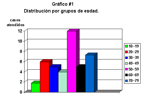
El total de la muestra de 35 pacientes tuvo&nbsp; representaci&oacute;n etaria en todos los grupos, con una predominancia del grupo de 50 a 59 a&ntilde;os; con un promedio de edad de 50,45 a&ntilde;os, con una m&iacute;nima de 14 y m&aacute;xima de 76 a&ntilde;os, con una mediana de 53 y moda de 54 a&ntilde;os. En general predominaron los pacientes mayores de 50 a&ntilde;os, como se ve en el <A HREF="#grafico 1">gr&aacute;fico #1</A>.</FONT></FONT>     <BR><FONT SIZE=-1>&nbsp;</FONT>     <BR><FONT SIZE=-1>&nbsp;</FONT>     <CENTER><A NAME="grafico 1"></A><IMG SRC="/img/fbpe/mlcr/v18n1/0928i01.JPG" HEIGHT=334 WIDTH=505></CENTER>      
<CENTER><B><FONT FACE="Arial,Helvetica"><FONT SIZE=-1>Grupos de edad</FONT></FONT></B></CENTER>      ]]></body>
<body><![CDATA[<CENTER><FONT SIZE=-1>&nbsp;</FONT></CENTER> <FONT SIZE=-1><FONT FACE="Arial,Helvetica">&nbsp;</FONT></FONT>     <BR><FONT SIZE=-1><FONT FACE="Arial,Helvetica">La&nbsp; distribuci&oacute;n por sexo mostr&oacute; que 16 pacientes fueron femeninos y 19 masculinos, para una relaci&oacute;n porcentual de 45,7% y 54,3% respectivamente.</FONT></FONT>     <BR><FONT SIZE=-1><FONT FACE="Arial,Helvetica">&nbsp;</FONT></FONT>     <CENTER><IMG SRC="/img/fbpe/mlcr/v18n1/0928i02.JPG" HEIGHT=231 WIDTH=325></CENTER> <FONT SIZE=-1>&nbsp;</FONT><FONT SIZE=-1></FONT>      
<P><FONT SIZE=-1><FONT FACE="Arial,Helvetica">Todos los pacientes estuvieron intubados por periodos significativos con un rango de 5 a 55 d&iacute;as, con un promedio de 17,34 d&iacute;as y una mediana de 14 d&iacute;as. La posici&oacute;n del bal&oacute;n del tubo endotraqueal se observ&oacute; as&iacute;: 6 pacientes lo ten&iacute;an localizado en el &aacute;rea A, 7 en el &aacute;rea&nbsp; B y 22 lo tuvieron en el C.</FONT></FONT><FONT SIZE=-1></FONT>      <P><FONT SIZE=-1><FONT FACE="Arial,Helvetica">Los 18 pacientes de la segunda serie a quienes se les midi&oacute; la presi&oacute;n del bal&oacute;n del tubo endotraqueal tuvieron un rango de 25 a 34 cm de presi&oacute;n de agua (cmH2O), con un promedio de 28,67cmH2O y una moda de 25,0cmH2O.
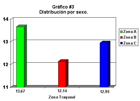
</FONT></FONT><FONT SIZE=-1></FONT>      <P><FONT SIZE=-1><FONT FACE="Arial,Helvetica">El grado de lesi&oacute;n promedio en los pacientes que ten&iacute;an localizado el bal&oacute;n del tubo en la zona subgl&oacute;tica (zona A) fu&eacute; de 13,67, en los que estuvo ubicado en el &aacute;rea istmica (zona B, de la biopsia y traqueostom&iacute;a) de 12,14 y en los que se ubic&oacute; en el &aacute;rea infraistmica (zona C) fu&eacute; de 12,95.</FONT></FONT>     <BR><FONT SIZE=-1>&nbsp;</FONT>     <BR><FONT SIZE=-1>&nbsp;</FONT>     <CENTER><IMG SRC="/img/fbpe/mlcr/v18n1/0928i03.JPG" BORDER=0 HEIGHT=356 WIDTH=491></CENTER> <FONT SIZE=-1>&nbsp;</FONT><FONT SIZE=-1></FONT>      
]]></body>
<body><![CDATA[<P><FONT SIZE=-1><FONT FACE="Arial,Helvetica">El grado de lesi&oacute;n histol&oacute;gica, cuantificado en puntos seg&uacute;n la tabla arriba expuesta, tuvo un rango en el grupo de los 35 pacientes entre&nbsp; 7 y 19, con una mediana de 13 y un promedio de 12,91.</FONT></FONT><FONT SIZE=-1></FONT>      <P><FONT SIZE=-1><FONT FACE="Arial,Helvetica">La relaci&oacute;n entre los d&iacute;as de intubaci&oacute;n y el grado de lesi&oacute;n histol&oacute;gica muestra que a mayor periodo de intubaci&oacute;n la lesi&oacute;n tiende a ser mayor. Cuando se analiza los enfermos expuestos al riesgo de permanecer m&aacute;s de 14 d&iacute;as intubados con el hecho de tener una lesi&oacute;n histol&oacute;gica significativa (mayor o igual a 13 puntos), se encuentra un riesgo relativo de 1,89 ( L&iacute;mite de Confianza&nbsp; 95% - serie&nbsp; de&nbsp; Taylor -); con un valor Chi=3,35 y valor p= 0,067.</FONT></FONT><FONT SIZE=-1></FONT>      <P><FONT SIZE=-1><FONT FACE="Arial,Helvetica">La relaci&oacute;n de los pacientes expuestos a presiones en el bal&oacute;n del TET iguales o mayores de 30 cmH2O, se encontr&oacute; que el riesgo relativo de &eacute;stos de tener una lesi&oacute;n histol&oacute;gica significativa (> 13 puntos) es de 3,93 (L&iacute;mite de Confianza 95% -serie de Taylor); con un valor de Chi de 4,82 y p=0,028.</FONT></FONT><FONT SIZE=-1></FONT>      <P><FONT SIZE=-1><FONT FACE="Arial,Helvetica">El riesgo relativo de los pacientes que no tuvieron control de la presi&oacute;n del bal&oacute;n de tener una lesi&oacute;n significativa es de 1,66 (L&iacute;mite de Confianza 95%-serie de Taylor), con un valor Chi=2,27 y p=0,132.</FONT></FONT>     <BR><FONT SIZE=-1>&nbsp;</FONT><FONT SIZE=-1></FONT>      <P><B><FONT FACE="Arial,Helvetica"><FONT SIZE=-1>Discusi&oacute;n</FONT></FONT></B><FONT SIZE=-1></FONT>      <P><FONT SIZE=-1><FONT FACE="Arial,Helvetica">
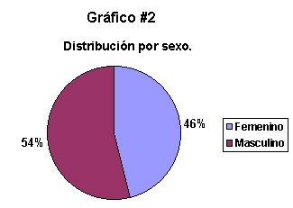
El predominio de un grupo de edad mayor de 50 a&ntilde;os refleja en cierta forma la tendencia de la pir&aacute;mide poblacional de nuestra sociedad, que con el pasar de los a&ntilde;os se invierte y&nbsp; hace necesario que las pol&iacute;ticas de salud del sistema se adec&uacute;en a tal situaci&oacute;n; y la distribuci&oacute;n de los pacientes por sexo indica que ambos grupos enfrentan riesgos y requieren de atenci&oacute;n intensiva en proporciones pr&aacute;cticamente iguales,</FONT></FONT><FONT SIZE=-1></FONT>      <P><FONT SIZE=-1><FONT FACE="Arial,Helvetica">A pesar de las recomendaciones dadas en 1995(<A HREF="#45.">45</A>), persisten manteniendose los pacientes intubados por largos periodos (17,34 d&iacute;as en promedio); &eacute;sto aunado a la evidencia estad&iacute;stica obtenida en este estudio, de que el hecho de permanecer intubado m&aacute;s de 14 d&iacute;as representa para el paciente un riesgo real de padecer una lesi&oacute;n traqueal secundaria al tubo endotraqueal, hace necesario proponer y recomendar de nuevo pautas en el manejo de la v&iacute;a a&eacute;rea de los pacientes cr&iacute;ticamente enfermos pues el riesgo relativo es pr&aacute;cticamente de 2:1.</FONT></FONT><FONT SIZE=-1></FONT>      <P><FONT SIZE=-1><FONT FACE="Arial,Helvetica">Existe evidencia anatomopatol&oacute;gica de lesiones traqueales significativas en &aacute;reas de la traquea que no est&aacute;n en contacto directo con el bal&oacute;n del TET, hecho puesto en evidencia por 22 casos en que el bal&oacute;n se hall&oacute; en la regi&oacute;n infra&iacute;stmica-distal al sitio de toma de la biopsia- los cuales tuvieron un promedio de lesi&oacute;n de 12,95; lo que habla de que no solamente el bal&oacute;n del tubo produce lesi&oacute;n traqueal sino que tambi&eacute;n el resto del tubo por s&iacute;; adem&aacute;s, los hallasgos histol&oacute;gicos informan que gran parte de las lesiones son de origen isqu&eacute;mico por la compresi&oacute;n que ejerce el bal&oacute;n sobre el plexo vascular submucoso de la traquea. Al mismo tiempo llama la atenci&oacute;n que cuando el bal&oacute;n se encontr&oacute; situado en las zonas A y C (proximal y distal) de la traquea los promedios de lesi&oacute;n son notablemente m&aacute;s altos que cuando el bal&oacute;n se sit&uacute;a en la regi&oacute;n media de la traquea, lo que hace pensar que en &eacute;stas ubicaciones el bal&oacute;n compromete la irrigaci&oacute;n proporcionada por las arterias m&aacute;s importantes de la Arteria Longitudinal Lateral&nbsp; (Ped&iacute;culo de Grillo) (Salassa-Pearson 1977) provenientes de la Arteria Tiroidea Inferior y de las Arterias Bronquiales.</FONT></FONT><FONT SIZE=-1></FONT>      <P><FONT SIZE=-1><FONT FACE="Arial,Helvetica">Los hallazgos relativos a la relaci&oacute;n existente entre la presi&oacute;n del bal&oacute;n del TET y la lesi&oacute;n traqueal comprueban en primer lugar que no controlar la presi&oacute;n del bal&oacute;n es exponer al paciente a un riesgo 1,66 veces mayor de sufrir una lesi&oacute;n traqueal significativa secundaria al&nbsp; TET;&nbsp; en segundo lugar que a mayor presi&oacute;n del bal&oacute;n, mayor es la lesi&oacute;n traqueal y que cuando la presi&oacute;n del bal&oacute;n alcanza y supera los 30 cmH2O el riesgo relativo es de 3,93:1, con una significancia estad&iacute;stica importante (Chi=4,82 y p=0,028). Esto indica que este control debiera establecerse en con caracter obligatorio y rutinario</FONT></FONT>     ]]></body>
<body><![CDATA[<BR><FONT SIZE=-1>&nbsp;</FONT><FONT SIZE=-1></FONT>      <P><B><FONT FACE="Arial,Helvetica"><FONT SIZE=-1>Conclusiones y recomendaciones</FONT></FONT></B><FONT SIZE=-1></FONT>      <P><FONT SIZE=-1><FONT FACE="Arial,Helvetica">1.-Existe evidencia anatomopatol&oacute;gica de lesiones traqueales significativas en zonas que no est&aacute;n en contacto con el bal&oacute;n del tubo endotraqueal (TET); probablemente relacionadas con el efecto mec&aacute;nico del TET en s&iacute; y con la compresi&oacute;n del plexo vascular submucoso de la traquea por parte del bal&oacute;n.</FONT></FONT><FONT SIZE=-1></FONT>      <P><FONT SIZE=-1><FONT FACE="Arial,Helvetica">2.-Permitir que un paciente pase intubado m&aacute;s de 14 d&iacute;as, sin realizarle traqueostom&iacute;a, expone al paciente a un riesgo 1,89 veces mayor de sufrir una lesi&oacute;n traqueal significativa secundaria al TET.</FONT></FONT><FONT SIZE=-1></FONT>      <P><FONT SIZE=-1><FONT FACE="Arial,Helvetica">3.-El hecho de no cuantificar ni controlar la presi&oacute;n del bal&oacute;n del TET expone al paciente a un riesgo relativo de 1,66:1 de tener una lesi&oacute;n traqueal significativa con respecto a aquellos pacientes a quienes s&iacute; se les controla.</FONT></FONT><FONT SIZE=-1></FONT>      <P><FONT SIZE=-1><FONT FACE="Arial,Helvetica">4.-A mayor presi&oacute;n del bal&oacute;n del TET mayor es la lesi&oacute;n traqueal desarrollada por el paciente.</FONT></FONT><FONT SIZE=-1></FONT>      <P><FONT SIZE=-1><FONT FACE="Arial,Helvetica">5.-Cuando la presi&oacute;n del bal&oacute;n excede los 30 cmH20 el riesgo de desarrollar una lesi&oacute;n traqueal significativa es 3,93:1.</FONT></FONT><FONT SIZE=-1></FONT>      <P><FONT SIZE=-1><FONT FACE="Arial,Helvetica">6.-Se recomienda adoptar las siguientes medidas con respecto al manejo de la v&iacute;a a&eacute;rea en los pacientes cr&iacute;ticos con ventilaci&oacute;n mec&aacute;nica:</FONT></FONT><FONT SIZE=-1></FONT>      <P><FONT SIZE=-1><FONT FACE="Arial,Helvetica">&nbsp;a.-Se puede mantener un paciente intubado sin problema por 7 d&iacute;as mientras se logra estabilizar su situaci&oacute;n cr&iacute;tica. Si la causa primaria de su fallo respiratorio indica que va a permanecer intubado por largo tiempo debe realizarse la traqueostom&iacute;a en &eacute;sta primera semana;(ej. Lesi&oacute;n neurol&oacute;gica severa).</FONT></FONT><FONT FACE="Arial,Helvetica"><FONT SIZE=-1></FONT></FONT>      <P><FONT SIZE=-1><FONT FACE="Arial,Helvetica">&nbsp;b.-Durante los siguientes 7 d&iacute;as los pacientes que a&uacute;n permanecen intubados deber&aacute;n evaluarse diariamente por la posibilidad de ser extubados; pero cuando la evidencia cl&iacute;nica indique que va a sobrepasar los 14 d&iacute;as de intubaci&oacute;n, deber&aacute; realizarsele traqueostom&iacute;a. Ning&uacute;n paciente deber&iacute;a permanecer con tubo endotraqueal m&aacute;s de 14 d&iacute;as.</FONT></FONT><FONT FACE="Arial,Helvetica"><FONT SIZE=-1></FONT></FONT>      ]]></body>
<body><![CDATA[<P><FONT SIZE=-1><FONT FACE="Arial,Helvetica">&nbsp;c.-Debe establecerse que la medici&oacute;n de la presi&oacute;n del bal&oacute;n del tubo endotraqueal sea obligatoria en los pacientes intubados al menos una vez al d&iacute;a (idealmente una vez en cada turno), y registrada en la tabla de cuidados respiratorios; ya sea que fuese medida por el m&eacute;dico a cargo, por la enfermera del servicio o por el t&eacute;cnico de terapia respiratoria.</FONT></FONT><FONT FACE="Arial,Helvetica"><FONT SIZE=-1></FONT></FONT>      <P><FONT SIZE=-1><FONT FACE="Arial,Helvetica">&nbsp;d.-La presi&oacute;n del bal&oacute;n deber&aacute; manejarse en un rango entre los 20 a los 29 cm de presi&oacute;n de agua. Cuando el paciente se encuentre hipotenso el l&iacute;mite superior deber&aacute; reducirse a 25 cmH2O.</FONT></FONT>     <BR><FONT SIZE=-1>&nbsp;</FONT><FONT SIZE=-1></FONT>      <P><B><FONT FACE="Arial,Helvetica"><FONT SIZE=-1>Bibliograf&iacute;a</FONT></FONT></B><FONT SIZE=-1></FONT>      <!-- ref --><P><A NAME="1."></A><FONT SIZE=-1><FONT FACE="Arial,Helvetica">1.-Arnold, W. Iatrogenic changes of the trachea. Diseases of head and&nbsp; neck, an&nbsp; atlas of histopathology. Cap 9.</FONT></FONT><FONT SIZE=-1></FONT>    &nbsp;&nbsp;&nbsp;&nbsp;&nbsp;&nbsp;&nbsp;&nbsp;[&#160;<a href="javascript:void(0);" onclick="javascript: window.open('/scielo.php?script=sci_nlinks&ref=599491&pid=S1409-0015200100020000600001&lng=','','width=640,height=500,resizable=yes,scrollbars=1,menubar=yes,');">Links</a>&#160;]<!-- end-ref --><!-- ref --><P><A NAME="2."></A><FONT SIZE=-1><FONT FACE="Arial,Helvetica">2.-Astrachan,D. Prolonged intubation vs. tracheotomy: complications,&nbsp; practical, and psycological considerations. Laringoscope. 1988;98:1165-9</FONT></FONT><FONT SIZE=-1></FONT>    &nbsp;&nbsp;&nbsp;&nbsp;&nbsp;&nbsp;&nbsp;&nbsp;[&#160;<a href="javascript:void(0);" onclick="javascript: window.open('/scielo.php?script=sci_nlinks&ref=599492&pid=S1409-0015200100020000600002&lng=','','width=640,height=500,resizable=yes,scrollbars=1,menubar=yes,');">Links</a>&#160;]<!-- end-ref --><!-- ref --><P><A NAME="3."></A><FONT SIZE=-1><FONT FACE="Arial,Helvetica">3.-Ayes,F- J.Zafra. Estenosis traqueal. Experiencia de 15 a&ntilde;os en el&nbsp; Hospital&nbsp; San Juan de Dios. CENDEISSS. No publicado</FONT></FONT><FONT SIZE=-1></FONT>    &nbsp;&nbsp;&nbsp;&nbsp;&nbsp;&nbsp;&nbsp;&nbsp;[&#160;<a href="javascript:void(0);" onclick="javascript: window.open('/scielo.php?script=sci_nlinks&ref=599493&pid=S1409-0015200100020000600003&lng=','','width=640,height=500,resizable=yes,scrollbars=1,menubar=yes,');">Links</a>&#160;]<!-- end-ref --><!-- ref --><P><A NAME="4."></A><FONT SIZE=-1><FONT FACE="Arial,Helvetica">4.-Benizhak,O. Necrotizing squamous metaplasia in herpetic&nbsp; tracheitis&nbsp; following prolonged intubation: a lesion similar to&nbsp; necrotizing&nbsp; sialometaplasia.&nbsp; Histopathology. 1993;22:265- 269</FONT></FONT><FONT SIZE=-1></FONT>    &nbsp;&nbsp;&nbsp;&nbsp;&nbsp;&nbsp;&nbsp;&nbsp;[&#160;<a href="javascript:void(0);" onclick="javascript: window.open('/scielo.php?script=sci_nlinks&ref=599494&pid=S1409-0015200100020000600004&lng=','','width=640,height=500,resizable=yes,scrollbars=1,menubar=yes,');">Links</a>&#160;]<!-- end-ref --><!-- ref --><P><A NAME="5."></A><FONT SIZE=-1><FONT FACE="Arial,Helvetica">5.-Bishop, M. Mechanisms of laryngotracheal injury following&nbsp; prolonged&nbsp; tracheal intubation. Chest. 1989;96:185-90</FONT></FONT><FONT SIZE=-1></FONT>    &nbsp;&nbsp;&nbsp;&nbsp;&nbsp;&nbsp;&nbsp;&nbsp;[&#160;<a href="javascript:void(0);" onclick="javascript: window.open('/scielo.php?script=sci_nlinks&ref=599495&pid=S1409-0015200100020000600005&lng=','','width=640,height=500,resizable=yes,scrollbars=1,menubar=yes,');">Links</a>&#160;]<!-- end-ref --><!-- ref --><P><A NAME="6."></A><FONT SIZE=-1><FONT FACE="Arial,Helvetica">6.-Bunegin,N. Canine tracheal blood flow after endotracheal&nbsp; tube&nbsp; cuff&nbsp; inflation&nbsp; during normotension and hypotension. Anesth.- Analg.&nbsp; 1993;76(5):1083-90</FONT></FONT><FONT SIZE=-1></FONT>    &nbsp;&nbsp;&nbsp;&nbsp;&nbsp;&nbsp;&nbsp;&nbsp;[&#160;<a href="javascript:void(0);" onclick="javascript: window.open('/scielo.php?script=sci_nlinks&ref=599496&pid=S1409-0015200100020000600006&lng=','','width=640,height=500,resizable=yes,scrollbars=1,menubar=yes,');">Links</a>&#160;]<!-- end-ref --><!-- ref --><P><A NAME="7."></A><FONT SIZE=-1><FONT FACE="Arial,Helvetica">7.-Coleman,J. Traumatic lesion of the trachea. Otolaryng.Clin.N.A. August,1995</FONT></FONT><FONT SIZE=-1></FONT>    &nbsp;&nbsp;&nbsp;&nbsp;&nbsp;&nbsp;&nbsp;&nbsp;[&#160;<a href="javascript:void(0);" onclick="javascript: window.open('/scielo.php?script=sci_nlinks&ref=599497&pid=S1409-0015200100020000600007&lng=','','width=640,height=500,resizable=yes,scrollbars=1,menubar=yes,');">Links</a>&#160;]<!-- end-ref --><!-- ref --><P><A NAME="8."></A><FONT SIZE=-1><FONT FACE="Arial,Helvetica">8.-Colice,G. Laryngeal complications of prolonged intubation. Chest.&nbsp; 1989;96:877-83</FONT></FONT><FONT SIZE=-1></FONT>    &nbsp;&nbsp;&nbsp;&nbsp;&nbsp;&nbsp;&nbsp;&nbsp;[&#160;<a href="javascript:void(0);" onclick="javascript: window.open('/scielo.php?script=sci_nlinks&ref=599498&pid=S1409-0015200100020000600008&lng=','','width=640,height=500,resizable=yes,scrollbars=1,menubar=yes,');">Links</a>&#160;]<!-- end-ref --><!-- ref --><P><A NAME="9."></A><FONT SIZE=-1><FONT FACE="Arial,Helvetica">9.-Colice,G. Resolution of laryngeal injury following translaryngeal&nbsp; intubation. Am.Rev.Resp.Dis. 1992;145:361-364</FONT></FONT><FONT SIZE=-1></FONT>    &nbsp;&nbsp;&nbsp;&nbsp;&nbsp;&nbsp;&nbsp;&nbsp;[&#160;<a href="javascript:void(0);" onclick="javascript: window.open('/scielo.php?script=sci_nlinks&ref=599499&pid=S1409-0015200100020000600009&lng=','','width=640,height=500,resizable=yes,scrollbars=1,menubar=yes,');">Links</a>&#160;]<!-- end-ref --><!-- ref --><P><A NAME="10."></A><FONT SIZE=-1><FONT FACE="Arial,Helvetica">10.-Coppolo,DP. Self extubation; a 12 month experience. Chest.&nbsp; 1990;98:165-9</FONT></FONT><FONT SIZE=-1></FONT>    &nbsp;&nbsp;&nbsp;&nbsp;&nbsp;&nbsp;&nbsp;&nbsp;[&#160;<a href="javascript:void(0);" onclick="javascript: window.open('/scielo.php?script=sci_nlinks&ref=599500&pid=S1409-0015200100020000600010&lng=','','width=640,height=500,resizable=yes,scrollbars=1,menubar=yes,');">Links</a>&#160;]<!-- end-ref --><!-- ref --><P><A NAME="11."></A><FONT SIZE=-1><FONT FACE="Arial,Helvetica">11.-Chuzonagaishi,MD. Functional anatomy and hystology of the&nbsp; lung. Public&nbsp; University.Park Press. Baltimore,1972 pp:5-12</FONT></FONT><FONT SIZE=-1></FONT>    &nbsp;&nbsp;&nbsp;&nbsp;&nbsp;&nbsp;&nbsp;&nbsp;[&#160;<a href="javascript:void(0);" onclick="javascript: window.open('/scielo.php?script=sci_nlinks&ref=599501&pid=S1409-0015200100020000600011&lng=','','width=640,height=500,resizable=yes,scrollbars=1,menubar=yes,');">Links</a>&#160;]<!-- end-ref --><!-- ref --><P><A NAME="12."></A><FONT SIZE=-1><FONT FACE="Arial,Helvetica">12.-Delgado, A. Tracheal reconstruction. Rev.&nbsp; Laryngol.Otol.Rhinol.Board. 1993;114(1):21-4</FONT></FONT><FONT SIZE=-1></FONT>    &nbsp;&nbsp;&nbsp;&nbsp;&nbsp;&nbsp;&nbsp;&nbsp;[&#160;<a href="javascript:void(0);" onclick="javascript: window.open('/scielo.php?script=sci_nlinks&ref=599502&pid=S1409-0015200100020000600012&lng=','','width=640,height=500,resizable=yes,scrollbars=1,menubar=yes,');">Links</a>&#160;]<!-- end-ref --><!-- ref --><P><A NAME="13."></A><FONT SIZE=-1><FONT FACE="Arial,Helvetica">13.-Donnelly,W. Laryngotracheal damage after prolonged use of&nbsp; orotracheal&nbsp; tubes in adults. JAMA. 1969;207(4):703-708</FONT></FONT><FONT SIZE=-1></FONT>    &nbsp;&nbsp;&nbsp;&nbsp;&nbsp;&nbsp;&nbsp;&nbsp;[&#160;<a href="javascript:void(0);" onclick="javascript: window.open('/scielo.php?script=sci_nlinks&ref=599503&pid=S1409-0015200100020000600013&lng=','','width=640,height=500,resizable=yes,scrollbars=1,menubar=yes,');">Links</a>&#160;]<!-- end-ref --><!-- ref --><P><A NAME="14."></A><FONT SIZE=-1><FONT FACE="Arial,Helvetica">14.-Donnelly,W. Histopathology of endotracheal intubation. Arch.&nbsp; Pathol.1969;88</FONT></FONT><FONT SIZE=-1></FONT>    &nbsp;&nbsp;&nbsp;&nbsp;&nbsp;&nbsp;&nbsp;&nbsp;[&#160;<a href="javascript:void(0);" onclick="javascript: window.open('/scielo.php?script=sci_nlinks&ref=599504&pid=S1409-0015200100020000600014&lng=','','width=640,height=500,resizable=yes,scrollbars=1,menubar=yes,');">Links</a>&#160;]<!-- end-ref --><!-- ref --><P><A NAME="15."></A><FONT SIZE=-1><FONT FACE="Arial,Helvetica">15.-Dunham,M. Prolonged intubation in the trauma patient.&nbsp; J.Trauma.&nbsp; 1984;24(2):120-24</FONT></FONT><FONT SIZE=-1></FONT>    &nbsp;&nbsp;&nbsp;&nbsp;&nbsp;&nbsp;&nbsp;&nbsp;[&#160;<a href="javascript:void(0);" onclick="javascript: window.open('/scielo.php?script=sci_nlinks&ref=599505&pid=S1409-0015200100020000600015&lng=','','width=640,height=500,resizable=yes,scrollbars=1,menubar=yes,');">Links</a>&#160;]<!-- end-ref --><!-- ref --><P><A NAME="16."></A><FONT SIZE=-1><FONT FACE="Arial,Helvetica">16.-Eliachar,I. Imaging evaluation of laryngotracheal stenosis. J.&nbsp; Otolaryngol. 1993;22(4):265-77</FONT></FONT><FONT SIZE=-1></FONT>    &nbsp;&nbsp;&nbsp;&nbsp;&nbsp;&nbsp;&nbsp;&nbsp;[&#160;<a href="javascript:void(0);" onclick="javascript: window.open('/scielo.php?script=sci_nlinks&ref=599506&pid=S1409-0015200100020000600016&lng=','','width=640,height=500,resizable=yes,scrollbars=1,menubar=yes,');">Links</a>&#160;]<!-- end-ref --><!-- ref --><P><A NAME="17."></A><FONT SIZE=-1><FONT FACE="Arial,Helvetica">17.-Faria, V. The complicatios of tracheal intubation, a new&nbsp; classification&nbsp; with a review of literature. Anesth.- Analg.1974;53(2)</FONT></FONT><FONT SIZE=-1></FONT>    &nbsp;&nbsp;&nbsp;&nbsp;&nbsp;&nbsp;&nbsp;&nbsp;[&#160;<a href="javascript:void(0);" onclick="javascript: window.open('/scielo.php?script=sci_nlinks&ref=599507&pid=S1409-0015200100020000600017&lng=','','width=640,height=500,resizable=yes,scrollbars=1,menubar=yes,');">Links</a>&#160;]<!-- end-ref --><!-- ref --><P><A NAME="18."></A><FONT SIZE=-1><FONT FACE="Arial,Helvetica">18.-Grillo,H. - Cooper. The evolution of tracheal injury due to ventilatory&nbsp; assistance trough cuffed tubes: a pathologic study.Ann.Surg.&nbsp; 1969;169(3):334-48</FONT></FONT><FONT SIZE=-1></FONT>    &nbsp;&nbsp;&nbsp;&nbsp;&nbsp;&nbsp;&nbsp;&nbsp;[&#160;<a href="javascript:void(0);" onclick="javascript: window.open('/scielo.php?script=sci_nlinks&ref=599508&pid=S1409-0015200100020000600018&lng=','','width=640,height=500,resizable=yes,scrollbars=1,menubar=yes,');">Links</a>&#160;]<!-- end-ref --><!-- ref --><P><A NAME="19."></A><FONT SIZE=-1><FONT FACE="Arial,Helvetica">19.-Grillo,H- Cooper. Experimental production and prevention of&nbsp; injury due to&nbsp; cuffed tracheal tubes. Surg.Gynecol.Obstet.&nbsp; 1969;129:1235</FONT></FONT><FONT SIZE=-1></FONT>    &nbsp;&nbsp;&nbsp;&nbsp;&nbsp;&nbsp;&nbsp;&nbsp;[&#160;<a href="javascript:void(0);" onclick="javascript: window.open('/scielo.php?script=sci_nlinks&ref=599509&pid=S1409-0015200100020000600019&lng=','','width=640,height=500,resizable=yes,scrollbars=1,menubar=yes,');">Links</a>&#160;]<!-- end-ref --><!-- ref --><P><A NAME="20."></A><FONT SIZE=-1><FONT FACE="Arial,Helvetica">20.-Grillo,H-Cooper. A low pressure cuff for tracheostomy to minimize&nbsp; tracheal&nbsp; injury-A comparative clinical trial.&nbsp; J.Thorac.Cardiovasc.Surg.&nbsp; 1971;62:898</FONT></FONT><FONT SIZE=-1></FONT>    &nbsp;&nbsp;&nbsp;&nbsp;&nbsp;&nbsp;&nbsp;&nbsp;[&#160;<a href="javascript:void(0);" onclick="javascript: window.open('/scielo.php?script=sci_nlinks&ref=599510&pid=S1409-0015200100020000600020&lng=','','width=640,height=500,resizable=yes,scrollbars=1,menubar=yes,');">Links</a>&#160;]<!-- end-ref --><!-- ref --><P><A NAME="21."></A><FONT SIZE=-1><FONT FACE="Arial,Helvetica">21.-Grillo,H. Surgical treatment of postintubation tracheal injuries.&nbsp; J.Cardiovasc.Surg. 1979;78(6)</FONT></FONT><FONT SIZE=-1></FONT>    &nbsp;&nbsp;&nbsp;&nbsp;&nbsp;&nbsp;&nbsp;&nbsp;[&#160;<a href="javascript:void(0);" onclick="javascript: window.open('/scielo.php?script=sci_nlinks&ref=599511&pid=S1409-0015200100020000600021&lng=','','width=640,height=500,resizable=yes,scrollbars=1,menubar=yes,');">Links</a>&#160;]<!-- end-ref --><!-- ref --><P><A NAME="22."></A><FONT SIZE=-1><FONT FACE="Arial,Helvetica">22.-Grillo,H. Upper airway Compromise after inhalation injury.&nbsp; Complex&nbsp; Strictures of larinx and trachea and their&nbsp; management.&nbsp;&nbsp; Ann.Surg.1993;218(5):672-8</FONT></FONT><FONT SIZE=-1></FONT>    &nbsp;&nbsp;&nbsp;&nbsp;&nbsp;&nbsp;&nbsp;&nbsp;[&#160;<a href="javascript:void(0);" onclick="javascript: window.open('/scielo.php?script=sci_nlinks&ref=599512&pid=S1409-0015200100020000600022&lng=','','width=640,height=500,resizable=yes,scrollbars=1,menubar=yes,');">Links</a>&#160;]<!-- end-ref --><!-- ref --><P><A NAME="23."></A><FONT SIZE=-1><FONT FACE="Arial,Helvetica">23.-Grillo,H. Idiopathic laryngotracheal stenosis and its management.&nbsp; Ann.Thor.Surg. 1993;56(1):80-7</FONT></FONT><FONT SIZE=-1></FONT>    &nbsp;&nbsp;&nbsp;&nbsp;&nbsp;&nbsp;&nbsp;&nbsp;[&#160;<a href="javascript:void(0);" onclick="javascript: window.open('/scielo.php?script=sci_nlinks&ref=599513&pid=S1409-0015200100020000600023&lng=','','width=640,height=500,resizable=yes,scrollbars=1,menubar=yes,');">Links</a>&#160;]<!-- end-ref --><!-- ref --><P><A NAME="24."></A><FONT SIZE=-1><FONT FACE="Arial,Helvetica">24.-Heffner,J. Timing the tracheostomy in mechanically ventilated&nbsp; patients.&nbsp; Am.Rev.Resp.Dis. 1993;147(3):768-71</FONT></FONT><FONT SIZE=-1></FONT>    &nbsp;&nbsp;&nbsp;&nbsp;&nbsp;&nbsp;&nbsp;&nbsp;[&#160;<a href="javascript:void(0);" onclick="javascript: window.open('/scielo.php?script=sci_nlinks&ref=599514&pid=S1409-0015200100020000600024&lng=','','width=640,height=500,resizable=yes,scrollbars=1,menubar=yes,');">Links</a>&#160;]<!-- end-ref --><!-- ref --><P><A NAME="25."></A><FONT SIZE=-1><FONT FACE="Arial,Helvetica">25.-Heffner, J. Tracheostomy en the Intensive Care Unit. Chest.&nbsp; 1986;90(3)</FONT></FONT><FONT SIZE=-1></FONT>    &nbsp;&nbsp;&nbsp;&nbsp;&nbsp;&nbsp;&nbsp;&nbsp;[&#160;<a href="javascript:void(0);" onclick="javascript: window.open('/scielo.php?script=sci_nlinks&ref=599515&pid=S1409-0015200100020000600025&lng=','','width=640,height=500,resizable=yes,scrollbars=1,menubar=yes,');">Links</a>&#160;]<!-- end-ref --><!-- ref --><P><A NAME="26."></A><FONT SIZE=-1><FONT FACE="Arial,Helvetica">26.-Holgaard,H. Complications and late sequelae following&nbsp; nasotracheal&nbsp; intubation. Acta Anaesth.Scand.&nbsp; 1993;37(5):475-80</FONT></FONT><FONT SIZE=-1></FONT>    &nbsp;&nbsp;&nbsp;&nbsp;&nbsp;&nbsp;&nbsp;&nbsp;[&#160;<a href="javascript:void(0);" onclick="javascript: window.open('/scielo.php?script=sci_nlinks&ref=599516&pid=S1409-0015200100020000600026&lng=','','width=640,height=500,resizable=yes,scrollbars=1,menubar=yes,');">Links</a>&#160;]<!-- end-ref --><!-- ref --><P><A NAME="27."></A><FONT SIZE=-1><FONT FACE="Arial,Helvetica">27.-Lester,B. Bacterial colonization profile with tracheal intubation&nbsp; and&nbsp; mechanical ventilation. Arch. Surg. 1972;104:647</FONT></FONT><FONT SIZE=-1></FONT>    &nbsp;&nbsp;&nbsp;&nbsp;&nbsp;&nbsp;&nbsp;&nbsp;[&#160;<a href="javascript:void(0);" onclick="javascript: window.open('/scielo.php?script=sci_nlinks&ref=599517&pid=S1409-0015200100020000600027&lng=','','width=640,height=500,resizable=yes,scrollbars=1,menubar=yes,');">Links</a>&#160;]<!-- end-ref --><!-- ref --><P><A NAME="28."></A><FONT SIZE=-1><FONT FACE="Arial,Helvetica">28.-LoCicero,J. Tracheo-carotid&nbsp; artery erosion following endotracheal&nbsp; intubation. J. Trauma. 1984;24:907-09</FONT></FONT><FONT SIZE=-1></FONT>    &nbsp;&nbsp;&nbsp;&nbsp;&nbsp;&nbsp;&nbsp;&nbsp;[&#160;<a href="javascript:void(0);" onclick="javascript: window.open('/scielo.php?script=sci_nlinks&ref=599518&pid=S1409-0015200100020000600028&lng=','','width=640,height=500,resizable=yes,scrollbars=1,menubar=yes,');">Links</a>&#160;]<!-- end-ref --><!-- ref --><P><A NAME="29."></A><FONT SIZE=-1><FONT FACE="Arial,Helvetica">29.-Mansour,K.Tracheal resections:lessons learned. Ann.Thorac.Surg. 1994;57(5):1120-4</FONT></FONT><FONT SIZE=-1></FONT>    &nbsp;&nbsp;&nbsp;&nbsp;&nbsp;&nbsp;&nbsp;&nbsp;[&#160;<a href="javascript:void(0);" onclick="javascript: window.open('/scielo.php?script=sci_nlinks&ref=599519&pid=S1409-0015200100020000600029&lng=','','width=640,height=500,resizable=yes,scrollbars=1,menubar=yes,');">Links</a>&#160;]<!-- end-ref --><!-- ref --><P><A NAME="30."></A><FONT SIZE=-1><FONT FACE="Arial,Helvetica">30.-McCafrey,T. Management of laryngotracheal stenosis on the basis&nbsp; of site&nbsp; and severity. Otolarygol.Head and Neck Surg.&nbsp; 1993;109(3):468-73</FONT></FONT><FONT SIZE=-1></FONT>    &nbsp;&nbsp;&nbsp;&nbsp;&nbsp;&nbsp;&nbsp;&nbsp;[&#160;<a href="javascript:void(0);" onclick="javascript: window.open('/scielo.php?script=sci_nlinks&ref=599520&pid=S1409-0015200100020000600030&lng=','','width=640,height=500,resizable=yes,scrollbars=1,menubar=yes,');">Links</a>&#160;]<!-- end-ref --><!-- ref --><P><A NAME="31."></A><FONT SIZE=-1><FONT FACE="Arial,Helvetica">31.-McGinnis, G. An enginnering analysis of intratracheal tube cuff.&nbsp; Anesth- Analg. 1971;50(4)</FONT></FONT><FONT SIZE=-1></FONT>    &nbsp;&nbsp;&nbsp;&nbsp;&nbsp;&nbsp;&nbsp;&nbsp;[&#160;<a href="javascript:void(0);" onclick="javascript: window.open('/scielo.php?script=sci_nlinks&ref=599521&pid=S1409-0015200100020000600031&lng=','','width=640,height=500,resizable=yes,scrollbars=1,menubar=yes,');">Links</a>&#160;]<!-- end-ref --><!-- ref --><P><A NAME="32."></A><FONT SIZE=-1><FONT FACE="Arial,Helvetica">32.-Miller,D. Tracheal stenosis following prolonged cuffed intubation.&nbsp; Ann.&nbsp; Surg. 1970;171(283)</FONT></FONT><FONT SIZE=-1></FONT>    &nbsp;&nbsp;&nbsp;&nbsp;&nbsp;&nbsp;&nbsp;&nbsp;[&#160;<a href="javascript:void(0);" onclick="javascript: window.open('/scielo.php?script=sci_nlinks&ref=599522&pid=S1409-0015200100020000600032&lng=','','width=640,height=500,resizable=yes,scrollbars=1,menubar=yes,');">Links</a>&#160;]<!-- end-ref --><!-- ref --><P><A NAME="33."></A><FONT SIZE=-1><FONT FACE="Arial,Helvetica">33.-Paegle, R. Rapid tracheal injury by cuffed airway and healing&nbsp; with loss of&nbsp; ciliate epithelium. Arch.Surg. 1973;106</FONT></FONT><FONT SIZE=-1></FONT>    &nbsp;&nbsp;&nbsp;&nbsp;&nbsp;&nbsp;&nbsp;&nbsp;[&#160;<a href="javascript:void(0);" onclick="javascript: window.open('/scielo.php?script=sci_nlinks&ref=599523&pid=S1409-0015200100020000600033&lng=','','width=640,height=500,resizable=yes,scrollbars=1,menubar=yes,');">Links</a>&#160;]<!-- end-ref --><!-- ref --><P><A NAME="34."></A><FONT SIZE=-1><FONT FACE="Arial,Helvetica">34.-Pearson,F. Incidence and pathogenesis of tracheal injury&nbsp; following&nbsp; cuffed&nbsp; tube tracheostomy with asisted ventilation.&nbsp; Ann. Surg.&nbsp; 1971;173(2)249</FONT></FONT><FONT SIZE=-1></FONT>    &nbsp;&nbsp;&nbsp;&nbsp;&nbsp;&nbsp;&nbsp;&nbsp;[&#160;<a href="javascript:void(0);" onclick="javascript: window.open('/scielo.php?script=sci_nlinks&ref=599524&pid=S1409-0015200100020000600034&lng=','','width=640,height=500,resizable=yes,scrollbars=1,menubar=yes,');">Links</a>&#160;]<!-- end-ref --><!-- ref --><P><A NAME="35."></A><FONT SIZE=-1><FONT FACE="Arial,Helvetica">35.-Pearson, F. Thoracic Surgery,Churchill-Livingstone. New&nbsp; York.1995</FONT></FONT><FONT SIZE=-1></FONT>    &nbsp;&nbsp;&nbsp;&nbsp;&nbsp;&nbsp;&nbsp;&nbsp;[&#160;<a href="javascript:void(0);" onclick="javascript: window.open('/scielo.php?script=sci_nlinks&ref=599525&pid=S1409-0015200100020000600035&lng=','','width=640,height=500,resizable=yes,scrollbars=1,menubar=yes,');">Links</a>&#160;]<!-- end-ref --><!-- ref --><P><A NAME="36."></A><FONT SIZE=-1><FONT FACE="Arial,Helvetica">36.-Staufer,J. Complications and consequences of endotracheal&nbsp; intubation and&nbsp; tracheostomy. Am.J.Med. 1981;70:65-76</FONT></FONT><FONT SIZE=-1></FONT>    &nbsp;&nbsp;&nbsp;&nbsp;&nbsp;&nbsp;&nbsp;&nbsp;[&#160;<a href="javascript:void(0);" onclick="javascript: window.open('/scielo.php?script=sci_nlinks&ref=599526&pid=S1409-0015200100020000600036&lng=','','width=640,height=500,resizable=yes,scrollbars=1,menubar=yes,');">Links</a>&#160;]<!-- end-ref --><!-- ref --><P><A NAME="37."></A><FONT SIZE=-1><FONT FACE="Arial,Helvetica">37.-Shields,T. General Thoracic Surgery. Williams &amp;&nbsp; Wilkins.Baltimore.&nbsp; 1994. 4th.Ed.</FONT></FONT><FONT SIZE=-1></FONT>    &nbsp;&nbsp;&nbsp;&nbsp;&nbsp;&nbsp;&nbsp;&nbsp;[&#160;<a href="javascript:void(0);" onclick="javascript: window.open('/scielo.php?script=sci_nlinks&ref=599527&pid=S1409-0015200100020000600037&lng=','','width=640,height=500,resizable=yes,scrollbars=1,menubar=yes,');">Links</a>&#160;]<!-- end-ref --><!-- ref --><P><A NAME="38."></A><FONT SIZE=-1><FONT FACE="Arial,Helvetica">38.-Schmidt,W. Inmediate mucosal effects of short-term soft cuff&nbsp; endotracheal&nbsp; intubation. Arch.Pathol.Lab.Med. 1979;103</FONT></FONT><FONT SIZE=-1></FONT>    &nbsp;&nbsp;&nbsp;&nbsp;&nbsp;&nbsp;&nbsp;&nbsp;[&#160;<a href="javascript:void(0);" onclick="javascript: window.open('/scielo.php?script=sci_nlinks&ref=599528&pid=S1409-0015200100020000600038&lng=','','width=640,height=500,resizable=yes,scrollbars=1,menubar=yes,');">Links</a>&#160;]<!-- end-ref --><!-- ref --><P><A NAME="39."></A><FONT SIZE=-1><FONT FACE="Arial,Helvetica">39.-Stockh. Structure of the tracheal wall after different durations of&nbsp; intubation.Acta Otolaryng.Suppl.1977;245:1-71</FONT></FONT><FONT SIZE=-1></FONT>    &nbsp;&nbsp;&nbsp;&nbsp;&nbsp;&nbsp;&nbsp;&nbsp;[&#160;<a href="javascript:void(0);" onclick="javascript: window.open('/scielo.php?script=sci_nlinks&ref=599529&pid=S1409-0015200100020000600039&lng=','','width=640,height=500,resizable=yes,scrollbars=1,menubar=yes,');">Links</a>&#160;]<!-- end-ref --><!-- ref --><P><A NAME="40."></A><FONT SIZE=-1><FONT FACE="Arial,Helvetica">40.-Tayal,V. Tracheostomies. Emerg.Clin.N.A. 1994;12(3):707</FONT></FONT><FONT SIZE=-1></FONT>    &nbsp;&nbsp;&nbsp;&nbsp;&nbsp;&nbsp;&nbsp;&nbsp;[&#160;<a href="javascript:void(0);" onclick="javascript: window.open('/scielo.php?script=sci_nlinks&ref=599530&pid=S1409-0015200100020000600040&lng=','','width=640,height=500,resizable=yes,scrollbars=1,menubar=yes,');">Links</a>&#160;]<!-- end-ref --><!-- ref --><P><A NAME="41."></A><FONT SIZE=-1><FONT FACE="Arial,Helvetica">41.-Whited, R. A prospective study of laryngotracheal sequelae in&nbsp; long term&nbsp; intubation. Laryngoscope. 1983;94:367-77</FONT></FONT><FONT SIZE=-1></FONT>    &nbsp;&nbsp;&nbsp;&nbsp;&nbsp;&nbsp;&nbsp;&nbsp;[&#160;<a href="javascript:void(0);" onclick="javascript: window.open('/scielo.php?script=sci_nlinks&ref=599531&pid=S1409-0015200100020000600041&lng=','','width=640,height=500,resizable=yes,scrollbars=1,menubar=yes,');">Links</a>&#160;]<!-- end-ref --><!-- ref --><P><A NAME="42."></A><FONT SIZE=-1><FONT FACE="Arial,Helvetica">42.-Weymuller, E. Quatification of intralaryngeal pressure exerted by&nbsp; endotracheal tubes. Ann.Otol.Rhinol.Laryngol. 1983;92:444-46</FONT></FONT><FONT SIZE=-1></FONT>    &nbsp;&nbsp;&nbsp;&nbsp;&nbsp;&nbsp;&nbsp;&nbsp;[&#160;<a href="javascript:void(0);" onclick="javascript: window.open('/scielo.php?script=sci_nlinks&ref=599532&pid=S1409-0015200100020000600042&lng=','','width=640,height=500,resizable=yes,scrollbars=1,menubar=yes,');">Links</a>&#160;]<!-- end-ref --><!-- ref --><P><A NAME="43."></A><FONT SIZE=-1><FONT FACE="Arial,Helvetica">43.-Wright, P. In vitro vs. in vivo comparison of endotracheal tube&nbsp; airflow&nbsp; resistence. Am.Rev.Resp.Dis.1989;140:10-6</FONT></FONT><FONT SIZE=-1></FONT>    &nbsp;&nbsp;&nbsp;&nbsp;&nbsp;&nbsp;&nbsp;&nbsp;[&#160;<a href="javascript:void(0);" onclick="javascript: window.open('/scielo.php?script=sci_nlinks&ref=599533&pid=S1409-0015200100020000600043&lng=','','width=640,height=500,resizable=yes,scrollbars=1,menubar=yes,');">Links</a>&#160;]<!-- end-ref --><!-- ref --><P><A NAME="44."></A><FONT SIZE=-1><FONT FACE="Arial,Helvetica">44.-Rodr&iacute;guez,L. Estudio histol&oacute;gico de las lesiones traqueales en pacientes con&nbsp; intubaci&oacute;n prolongada. Tesis de Graduaci&oacute;n de Postgrado en Anatom&iacute;a&nbsp; Patol&oacute;gica. CENDEISSS-UCR.1998</FONT></FONT><FONT SIZE=-1></FONT>    &nbsp;&nbsp;&nbsp;&nbsp;&nbsp;&nbsp;&nbsp;&nbsp;[&#160;<a href="javascript:void(0);" onclick="javascript: window.open('/scielo.php?script=sci_nlinks&ref=599534&pid=S1409-0015200100020000600044&lng=','','width=640,height=500,resizable=yes,scrollbars=1,menubar=yes,');">Links</a>&#160;]<!-- end-ref --><!-- ref --><P><A NAME="45."></A><FONT SIZE=-1><FONT FACE="Arial,Helvetica">45.-Miranda, J. Estudio cl&iacute;nico-patol&oacute;gico de las lesiones traqueales en pacientes&nbsp; con intubaci&oacute;n prolongada. Tesis de Graduaci&oacute;n&nbsp; de Postgrado en Cirug&iacute;a General. CENDEISSS-UCR.1995</FONT></FONT><FONT SIZE=-1></FONT>    &nbsp;&nbsp;&nbsp;&nbsp;&nbsp;&nbsp;&nbsp;&nbsp;[&#160;<a href="javascript:void(0);" onclick="javascript: window.open('/scielo.php?script=sci_nlinks&ref=599535&pid=S1409-0015200100020000600045&lng=','','width=640,height=500,resizable=yes,scrollbars=1,menubar=yes,');">Links</a>&#160;]<!-- end-ref --><!-- ref --><P><A NAME="46."></A><FONT SIZE=-1><FONT FACE="Arial,Helvetica">46.-Salassa JR, Pearson BW et.al. Gross and Microscopical blood supply of the&nbsp; trachea. Ann Thorac Surg 24:100-107,1977</FONT></FONT><FONT SIZE=-1></FONT>    &nbsp;&nbsp;&nbsp;&nbsp;&nbsp;&nbsp;&nbsp;&nbsp;[&#160;<a href="javascript:void(0);" onclick="javascript: window.open('/scielo.php?script=sci_nlinks&ref=599536&pid=S1409-0015200100020000600046&lng=','','width=640,height=500,resizable=yes,scrollbars=1,menubar=yes,');">Links</a>&#160;]<!-- end-ref --><!-- ref --><P><A NAME="47."></A><FONT SIZE=-1><FONT FACE="Arial,Helvetica">47.-Allen,M. Surgical Anatomy of the Trachea.Chest Surgery Clinics of N.A.&nbsp; 6(4) November,1996</FONT></FONT>    &nbsp;&nbsp;&nbsp;&nbsp;&nbsp;&nbsp;&nbsp;&nbsp;[&#160;<a href="javascript:void(0);" onclick="javascript: window.open('/scielo.php?script=sci_nlinks&ref=599537&pid=S1409-0015200100020000600047&lng=','','width=640,height=500,resizable=yes,scrollbars=1,menubar=yes,');">Links</a>&#160;]<!-- end-ref --><BR><FONT SIZE=-1>&nbsp;</FONT>     <BR><FONT SIZE=-1>&nbsp;</FONT>     <BR><A NAME="*"></A><FONT SIZE=-1><FONT FACE="Arial,Helvetica"><A HREF="#*a">*</A>Servicio de Cirug&iacute;a 2 y T&oacute;rax,</FONT></FONT>     <BR><FONT SIZE=-1>&nbsp;</FONT>     <BR><A NAME="**"></A><FONT SIZE=-1><FONT FACE="Arial,Helvetica"><A HREF="#**a">**</A>Servicio de Anatom&iacute;a Patol&oacute;gica</FONT></FONT>     <BR><FONT SIZE=-1><FONT FACE="Arial,Helvetica">&nbsp;Hospital San Juan de Dios. C.C.S.S.</FONT></FONT>      ]]></body><back>
<ref-list>
<ref id="B1">
<nlm-citation citation-type="">
<person-group person-group-type="author">
<name>
<surname><![CDATA[Arnold]]></surname>
<given-names><![CDATA[W]]></given-names>
</name>
</person-group>
<source><![CDATA[Iatrogenic changes of the trachea. Diseases of head and neck, an atlas of histopathology]]></source>
<year></year>
<volume>9</volume>
</nlm-citation>
</ref>
<ref id="B2">
<nlm-citation citation-type="journal">
<person-group person-group-type="author">
<name>
<surname><![CDATA[Astrachan]]></surname>
<given-names><![CDATA[D]]></given-names>
</name>
</person-group>
<article-title xml:lang="en"><![CDATA[Prolonged intubation vs. tracheotomy: complications, practical, and psycological considerations]]></article-title>
<source><![CDATA[Laringoscope]]></source>
<year>1988</year>
<volume>98</volume>
<page-range>1165-9</page-range></nlm-citation>
</ref>
<ref id="B3">
<nlm-citation citation-type="book">
<person-group person-group-type="author">
<name>
<surname><![CDATA[Ayes]]></surname>
<given-names><![CDATA[F]]></given-names>
</name>
<name>
<surname><![CDATA[Zafra]]></surname>
<given-names><![CDATA[J]]></given-names>
</name>
</person-group>
<source><![CDATA[Estenosis traqueal: Experiencia de 15 años en el Hospital San Juan de Dios]]></source>
<year></year>
<publisher-name><![CDATA[CENDEISSS]]></publisher-name>
</nlm-citation>
</ref>
<ref id="B4">
<nlm-citation citation-type="journal">
<person-group person-group-type="author">
<name>
<surname><![CDATA[Benizhak]]></surname>
<given-names><![CDATA[O]]></given-names>
</name>
</person-group>
<article-title xml:lang="en"><![CDATA[Necrotizing squamous metaplasia in herpetic tracheitis following prolonged intubation: a lesion similar to necrotizing sialometaplasia]]></article-title>
<source><![CDATA[Histopathology]]></source>
<year>1993</year>
<volume>22</volume>
<page-range>265- 269</page-range></nlm-citation>
</ref>
<ref id="B5">
<nlm-citation citation-type="journal">
<person-group person-group-type="author">
<name>
<surname><![CDATA[Bishop]]></surname>
<given-names><![CDATA[M]]></given-names>
</name>
</person-group>
<article-title xml:lang="en"><![CDATA[Mechanisms of laryngotracheal injury following prolonged tracheal intubation]]></article-title>
<source><![CDATA[Chest]]></source>
<year>1989</year>
<volume>96</volume>
<page-range>185-90</page-range></nlm-citation>
</ref>
<ref id="B6">
<nlm-citation citation-type="journal">
<person-group person-group-type="author">
<name>
<surname><![CDATA[Bunegin]]></surname>
<given-names><![CDATA[N]]></given-names>
</name>
</person-group>
<article-title xml:lang="en"><![CDATA[Canine tracheal blood flow after endotracheal tube cuff inflation during normotension and hypotension]]></article-title>
<source><![CDATA[Anesth.- Analg]]></source>
<year>1993</year>
<volume>76</volume>
<numero>^s5</numero>
<issue>^s5</issue>
<supplement>5</supplement>
<page-range>1083-90</page-range></nlm-citation>
</ref>
<ref id="B7">
<nlm-citation citation-type="journal">
<person-group person-group-type="author">
<name>
<surname><![CDATA[Coleman]]></surname>
<given-names><![CDATA[J]]></given-names>
</name>
</person-group>
<article-title xml:lang="en"><![CDATA[Traumatic lesion of the trachea]]></article-title>
<source><![CDATA[Otolaryng.Clin.N.A]]></source>
<year>Augu</year>
<month>st</month>
<day>,1</day>
</nlm-citation>
</ref>
<ref id="B8">
<nlm-citation citation-type="journal">
<person-group person-group-type="author">
<name>
<surname><![CDATA[Colice]]></surname>
<given-names><![CDATA[G]]></given-names>
</name>
</person-group>
<article-title xml:lang="en"><![CDATA[Laryngeal complications of prolonged intubation]]></article-title>
<source><![CDATA[Chest]]></source>
<year>1989</year>
<volume>96</volume>
<page-range>877-83</page-range></nlm-citation>
</ref>
<ref id="B9">
<nlm-citation citation-type="journal">
<person-group person-group-type="author">
<name>
<surname><![CDATA[Colice]]></surname>
<given-names><![CDATA[G]]></given-names>
</name>
</person-group>
<article-title xml:lang="en"><![CDATA[Resolution of laryngeal injury following translaryngeal intubation]]></article-title>
<source><![CDATA[Am.Rev.Resp.Dis]]></source>
<year>1992</year>
<volume>145</volume>
<page-range>361-364</page-range></nlm-citation>
</ref>
<ref id="B10">
<nlm-citation citation-type="journal">
<person-group person-group-type="author">
<name>
<surname><![CDATA[Coppolo]]></surname>
<given-names><![CDATA[DP]]></given-names>
</name>
</person-group>
<article-title xml:lang="en"><![CDATA[Self extubation: a 12 month experience]]></article-title>
<source><![CDATA[Chest]]></source>
<year>1990</year>
<volume>98</volume>
<page-range>165-9</page-range></nlm-citation>
</ref>
<ref id="B11">
<nlm-citation citation-type="book">
<person-group person-group-type="author">
<name>
<surname><![CDATA[Chuzonagaishi]]></surname>
<given-names><![CDATA[MD]]></given-names>
</name>
</person-group>
<source><![CDATA[Functional anatomy and hystology of the lung]]></source>
<year>1972</year>
<page-range>5-12</page-range><publisher-loc><![CDATA[Baltimore ]]></publisher-loc>
<publisher-name><![CDATA[Public University. Park Press]]></publisher-name>
</nlm-citation>
</ref>
<ref id="B12">
<nlm-citation citation-type="journal">
<person-group person-group-type="author">
<name>
<surname><![CDATA[Delgado]]></surname>
<given-names><![CDATA[A]]></given-names>
</name>
</person-group>
<article-title xml:lang="en"><![CDATA[Tracheal reconstruction]]></article-title>
<source><![CDATA[Rev. Laryngol.Otol.Rhinol.Board]]></source>
<year>1993</year>
<volume>114</volume>
<numero>^s1</numero>
<issue>^s1</issue>
<supplement>1</supplement>
<page-range>21-4</page-range></nlm-citation>
</ref>
<ref id="B13">
<nlm-citation citation-type="journal">
<person-group person-group-type="author">
<name>
<surname><![CDATA[Donnelly]]></surname>
<given-names><![CDATA[W]]></given-names>
</name>
</person-group>
<article-title xml:lang="en"><![CDATA[Laryngotracheal damage after prolonged use of orotracheal tubes in adults]]></article-title>
<source><![CDATA[JAMA]]></source>
<year>1969</year>
<volume>207</volume>
<numero>^s4</numero>
<issue>^s4</issue>
<supplement>4</supplement>
<page-range>703-708</page-range></nlm-citation>
</ref>
<ref id="B14">
<nlm-citation citation-type="journal">
<person-group person-group-type="author">
<name>
<surname><![CDATA[Donnelly]]></surname>
<given-names><![CDATA[W]]></given-names>
</name>
</person-group>
<article-title xml:lang="en"><![CDATA[Histopathology of endotracheal intubation]]></article-title>
<source><![CDATA[Arch. Pathol]]></source>
<year>1969</year>
<volume>88</volume>
</nlm-citation>
</ref>
<ref id="B15">
<nlm-citation citation-type="journal">
<person-group person-group-type="author">
<name>
<surname><![CDATA[Dunham]]></surname>
<given-names><![CDATA[M]]></given-names>
</name>
</person-group>
<article-title xml:lang="en"><![CDATA[Prolonged intubation in the trauma patient]]></article-title>
<source><![CDATA[J.Trauma]]></source>
<year>1984</year>
<volume>24</volume>
<numero>^s2</numero>
<issue>^s2</issue>
<supplement>2</supplement>
<page-range>120-24</page-range></nlm-citation>
</ref>
<ref id="B16">
<nlm-citation citation-type="journal">
<person-group person-group-type="author">
<name>
<surname><![CDATA[Eliachar]]></surname>
<given-names><![CDATA[I]]></given-names>
</name>
</person-group>
<article-title xml:lang="en"><![CDATA[Imaging evaluation of laryngotracheal stenosis]]></article-title>
<source><![CDATA[J. Otolaryngol]]></source>
<year>1993</year>
<volume>22</volume>
<numero>^s4</numero>
<issue>^s4</issue>
<supplement>4</supplement>
<page-range>265-77</page-range></nlm-citation>
</ref>
<ref id="B17">
<nlm-citation citation-type="journal">
<person-group person-group-type="author">
<name>
<surname><![CDATA[Faria]]></surname>
<given-names><![CDATA[V]]></given-names>
</name>
</person-group>
<article-title xml:lang="en"><![CDATA[The complicatios of tracheal intubation, a new classification with a review of literature]]></article-title>
<source><![CDATA[Anesth.- Analg]]></source>
<year>1974</year>
<volume>53</volume>
<numero>^s2</numero>
<issue>^s2</issue>
<supplement>2</supplement>
</nlm-citation>
</ref>
<ref id="B18">
<nlm-citation citation-type="journal">
<person-group person-group-type="author">
<name>
<surname><![CDATA[Grillo]]></surname>
<given-names><![CDATA[H. - Cooper]]></given-names>
</name>
</person-group>
<article-title xml:lang="en"><![CDATA[The evolution of tracheal injury due to ventilatory assistance trough cuffed tubes: a pathologic study]]></article-title>
<source><![CDATA[Ann.Surg]]></source>
<year>1969</year>
<volume>169</volume>
<numero>^s3</numero>
<issue>^s3</issue>
<supplement>3</supplement>
<page-range>334-48</page-range></nlm-citation>
</ref>
<ref id="B19">
<nlm-citation citation-type="journal">
<person-group person-group-type="author">
<name>
<surname><![CDATA[Grillo]]></surname>
<given-names><![CDATA[H- Cooper]]></given-names>
</name>
</person-group>
<article-title xml:lang="en"><![CDATA[Experimental production and prevention of injury due to cuffed tracheal tubes]]></article-title>
<source><![CDATA[Surg.Gynecol.Obstet]]></source>
<year>1969</year>
<volume>129</volume>
<page-range>1235</page-range></nlm-citation>
</ref>
<ref id="B20">
<nlm-citation citation-type="journal">
<person-group person-group-type="author">
<name>
<surname><![CDATA[Grillo]]></surname>
<given-names><![CDATA[H-Cooper]]></given-names>
</name>
</person-group>
<article-title xml:lang="en"><![CDATA[A low pressure cuff for tracheostomy to minimize tracheal injury-A comparative clinical trial]]></article-title>
<source><![CDATA[J.Thorac.Cardiovasc.Surg]]></source>
<year>1971</year>
<volume>62</volume>
<page-range>898</page-range></nlm-citation>
</ref>
<ref id="B21">
<nlm-citation citation-type="journal">
<person-group person-group-type="author">
<name>
<surname><![CDATA[Grillo]]></surname>
<given-names><![CDATA[H]]></given-names>
</name>
</person-group>
<article-title xml:lang="en"><![CDATA[Surgical treatment of postintubation tracheal injuries]]></article-title>
<source><![CDATA[J.Cardiovasc.Surg]]></source>
<year>1979</year>
<volume>78</volume>
<numero>^s6</numero>
<issue>^s6</issue>
<supplement>6</supplement>
</nlm-citation>
</ref>
<ref id="B22">
<nlm-citation citation-type="journal">
<person-group person-group-type="author">
<name>
<surname><![CDATA[Grillo]]></surname>
<given-names><![CDATA[H]]></given-names>
</name>
</person-group>
<article-title xml:lang="en"><![CDATA[Upper airway Compromise after inhalation injury: Complex Strictures of larinx and trachea and their management]]></article-title>
<source><![CDATA[Ann.Surg]]></source>
<year>1993</year>
<volume>218</volume>
<numero>^s5</numero>
<issue>^s5</issue>
<supplement>5</supplement>
<page-range>672-8</page-range></nlm-citation>
</ref>
<ref id="B23">
<nlm-citation citation-type="journal">
<person-group person-group-type="author">
<name>
<surname><![CDATA[Grillo]]></surname>
<given-names><![CDATA[H]]></given-names>
</name>
</person-group>
<article-title xml:lang="en"><![CDATA[Idiopathic laryngotracheal stenosis and its management]]></article-title>
<source><![CDATA[Ann.Thor.Surg]]></source>
<year>1993</year>
<volume>56</volume>
<numero>^s1</numero>
<issue>^s1</issue>
<supplement>1</supplement>
<page-range>80-7</page-range></nlm-citation>
</ref>
<ref id="B24">
<nlm-citation citation-type="journal">
<person-group person-group-type="author">
<name>
<surname><![CDATA[Heffner]]></surname>
<given-names><![CDATA[J]]></given-names>
</name>
</person-group>
<article-title xml:lang="en"><![CDATA[Timing the tracheostomy in mechanically ventilated patients]]></article-title>
<source><![CDATA[Am.Rev.Resp.Dis]]></source>
<year>1993</year>
<volume>147</volume>
<numero>^s3</numero>
<issue>^s3</issue>
<supplement>3</supplement>
<page-range>768-71</page-range></nlm-citation>
</ref>
<ref id="B25">
<nlm-citation citation-type="journal">
<person-group person-group-type="author">
<name>
<surname><![CDATA[Heffner]]></surname>
<given-names><![CDATA[J]]></given-names>
</name>
</person-group>
<article-title xml:lang="en"><![CDATA[Tracheostomy en the Intensive Care Unit]]></article-title>
<source><![CDATA[Chest]]></source>
<year>1986</year>
<volume>90</volume>
<numero>^s3</numero>
<issue>^s3</issue>
<supplement>3</supplement>
</nlm-citation>
</ref>
<ref id="B26">
<nlm-citation citation-type="journal">
<person-group person-group-type="author">
<name>
<surname><![CDATA[Holgaard]]></surname>
<given-names><![CDATA[H]]></given-names>
</name>
</person-group>
<article-title xml:lang="en"><![CDATA[Complications and late sequelae following nasotracheal intubation]]></article-title>
<source><![CDATA[Acta Anaesth.Scand]]></source>
<year>1993</year>
<volume>37</volume>
<numero>^s5</numero>
<issue>^s5</issue>
<supplement>5</supplement>
<page-range>475-80</page-range></nlm-citation>
</ref>
<ref id="B27">
<nlm-citation citation-type="journal">
<person-group person-group-type="author">
<name>
<surname><![CDATA[Lester]]></surname>
<given-names><![CDATA[B]]></given-names>
</name>
</person-group>
<article-title xml:lang="en"><![CDATA[Bacterial colonization profile with tracheal intubation and mechanical ventilation]]></article-title>
<source><![CDATA[Arch. Surg]]></source>
<year>1972</year>
<volume>104</volume>
<page-range>647</page-range></nlm-citation>
</ref>
<ref id="B28">
<nlm-citation citation-type="journal">
<person-group person-group-type="author">
<name>
<surname><![CDATA[LoCicero]]></surname>
<given-names><![CDATA[J]]></given-names>
</name>
</person-group>
<article-title xml:lang="en"><![CDATA[Tracheo-carotid artery erosion following endotracheal intubation]]></article-title>
<source><![CDATA[J. Trauma]]></source>
<year>1984</year>
<volume>24</volume>
<page-range>907-09</page-range></nlm-citation>
</ref>
<ref id="B29">
<nlm-citation citation-type="journal">
<person-group person-group-type="author">
<name>
<surname><![CDATA[Mansour]]></surname>
<given-names><![CDATA[K]]></given-names>
</name>
</person-group>
<article-title xml:lang="en"><![CDATA[Tracheal resections:lessons learned]]></article-title>
<source><![CDATA[Ann.Thorac.Surg]]></source>
<year>1994</year>
<volume>57</volume>
<numero>^s5</numero>
<issue>^s5</issue>
<supplement>5</supplement>
<page-range>1120-4</page-range></nlm-citation>
</ref>
<ref id="B30">
<nlm-citation citation-type="journal">
<person-group person-group-type="author">
<name>
<surname><![CDATA[McCafrey]]></surname>
<given-names><![CDATA[T]]></given-names>
</name>
</person-group>
<article-title xml:lang="en"><![CDATA[Management of laryngotracheal stenosis on the basis of site and severity]]></article-title>
<source><![CDATA[Otolarygol.Head and Neck Surg]]></source>
<year>1993</year>
<volume>109</volume>
<numero>^s3</numero>
<issue>^s3</issue>
<supplement>3</supplement>
<page-range>468-73</page-range></nlm-citation>
</ref>
<ref id="B31">
<nlm-citation citation-type="journal">
<person-group person-group-type="author">
<name>
<surname><![CDATA[McGinnis]]></surname>
<given-names><![CDATA[G]]></given-names>
</name>
</person-group>
<article-title xml:lang="en"><![CDATA[An enginnering analysis of intratracheal tube cuff]]></article-title>
<source><![CDATA[Anesth- Analg]]></source>
<year>1971</year>
<volume>50</volume>
<numero>^s4</numero>
<issue>^s4</issue>
<supplement>4</supplement>
</nlm-citation>
</ref>
<ref id="B32">
<nlm-citation citation-type="journal">
<person-group person-group-type="author">
<name>
<surname><![CDATA[Miller]]></surname>
<given-names><![CDATA[D]]></given-names>
</name>
</person-group>
<article-title xml:lang="en"><![CDATA[Tracheal stenosis following prolonged cuffed intubation]]></article-title>
<source><![CDATA[Ann. Surg]]></source>
<year>1970</year>
<volume>171</volume>
<numero>^s283</numero>
<issue>^s283</issue>
<supplement>283</supplement>
</nlm-citation>
</ref>
<ref id="B33">
<nlm-citation citation-type="journal">
<person-group person-group-type="author">
<name>
<surname><![CDATA[Paegle]]></surname>
<given-names><![CDATA[R]]></given-names>
</name>
</person-group>
<article-title xml:lang="en"><![CDATA[Rapid tracheal injury by cuffed airway and healing with loss of ciliate epithelium]]></article-title>
<source><![CDATA[Arch.Surg]]></source>
<year>1973</year>
<page-range>106</page-range></nlm-citation>
</ref>
<ref id="B34">
<nlm-citation citation-type="journal">
<person-group person-group-type="author">
<name>
<surname><![CDATA[Pearson]]></surname>
<given-names><![CDATA[F]]></given-names>
</name>
</person-group>
<article-title xml:lang="en"><![CDATA[Incidence and pathogenesis of tracheal injury following cuffed tube tracheostomy with asisted ventilation]]></article-title>
<source><![CDATA[Ann. Surg]]></source>
<year>1971</year>
<volume>173</volume>
<numero>^s2</numero>
<issue>^s2</issue>
<supplement>2</supplement>
<page-range>249</page-range></nlm-citation>
</ref>
<ref id="B35">
<nlm-citation citation-type="book">
<person-group person-group-type="author">
<name>
<surname><![CDATA[Pearson]]></surname>
<given-names><![CDATA[F]]></given-names>
</name>
</person-group>
<source><![CDATA[Thoracic Surgery]]></source>
<year>1995</year>
<publisher-loc><![CDATA[New York ]]></publisher-loc>
<publisher-name><![CDATA[Churchill-Livingstone]]></publisher-name>
</nlm-citation>
</ref>
<ref id="B36">
<nlm-citation citation-type="journal">
<person-group person-group-type="author">
<name>
<surname><![CDATA[Staufer]]></surname>
<given-names><![CDATA[J]]></given-names>
</name>
</person-group>
<article-title xml:lang="en"><![CDATA[Complications and consequences of endotracheal intubation and tracheostomy]]></article-title>
<source><![CDATA[Am.J.Med]]></source>
<year>1981</year>
<volume>70</volume>
<page-range>65-76</page-range></nlm-citation>
</ref>
<ref id="B37">
<nlm-citation citation-type="book">
<person-group person-group-type="author">
<name>
<surname><![CDATA[Shields]]></surname>
<given-names><![CDATA[T]]></given-names>
</name>
</person-group>
<source><![CDATA[General Thoracic Surgery]]></source>
<year>1994</year>
<edition>4th</edition>
<publisher-name><![CDATA[Williams & WilkinsBaltimore]]></publisher-name>
</nlm-citation>
</ref>
<ref id="B38">
<nlm-citation citation-type="journal">
<person-group person-group-type="author">
<name>
<surname><![CDATA[Schmidt]]></surname>
<given-names><![CDATA[W]]></given-names>
</name>
</person-group>
<article-title xml:lang="en"><![CDATA[Inmediate mucosal effects of short-term soft cuff endotracheal intubation]]></article-title>
<source><![CDATA[Arch.Pathol.Lab.Med]]></source>
<year>1979</year>
<page-range>103</page-range></nlm-citation>
</ref>
<ref id="B39">
<nlm-citation citation-type="journal">
<person-group person-group-type="author">
<name>
<surname><![CDATA[Stockh]]></surname>
</name>
</person-group>
<article-title xml:lang="en"><![CDATA[Structure of the tracheal wall after different durations of intubation]]></article-title>
<source><![CDATA[Acta Otolaryng.Suppl]]></source>
<year>1977</year>
<volume>245</volume>
<page-range>1-71</page-range></nlm-citation>
</ref>
<ref id="B40">
<nlm-citation citation-type="journal">
<person-group person-group-type="author">
<name>
<surname><![CDATA[Tayal]]></surname>
<given-names><![CDATA[V]]></given-names>
</name>
</person-group>
<article-title xml:lang="en"><![CDATA[Tracheostomies]]></article-title>
<source><![CDATA[Emerg.Clin.N.A]]></source>
<year>1994</year>
<volume>12</volume>
<numero>^s3</numero>
<issue>^s3</issue>
<supplement>3</supplement>
<page-range>707</page-range></nlm-citation>
</ref>
<ref id="B41">
<nlm-citation citation-type="journal">
<person-group person-group-type="author">
<name>
<surname><![CDATA[Whited]]></surname>
<given-names><![CDATA[R]]></given-names>
</name>
</person-group>
<article-title xml:lang="en"><![CDATA[A prospective study of laryngotracheal sequelae in long term intubation]]></article-title>
<source><![CDATA[Laryngoscope]]></source>
<year>1983</year>
<volume>94</volume>
<page-range>367-77</page-range></nlm-citation>
</ref>
<ref id="B42">
<nlm-citation citation-type="journal">
<person-group person-group-type="author">
<name>
<surname><![CDATA[Weymuller]]></surname>
<given-names><![CDATA[E]]></given-names>
</name>
</person-group>
<article-title xml:lang="en"><![CDATA[Quatification of intralaryngeal pressure exerted by endotracheal tubes]]></article-title>
<source><![CDATA[Ann.Otol.Rhinol.Laryngol]]></source>
<year>1983</year>
<volume>92</volume>
<page-range>444-46</page-range></nlm-citation>
</ref>
<ref id="B43">
<nlm-citation citation-type="journal">
<person-group person-group-type="author">
<name>
<surname><![CDATA[Wright]]></surname>
<given-names><![CDATA[P]]></given-names>
</name>
</person-group>
<article-title xml:lang="en"><![CDATA[In vitro vs. in vivo comparison of endotracheal tube airflow resistence]]></article-title>
<source><![CDATA[Am.Rev.Resp.Dis]]></source>
<year>1989</year>
<volume>140</volume>
<page-range>10-6</page-range></nlm-citation>
</ref>
<ref id="B44">
<nlm-citation citation-type="">
<person-group person-group-type="author">
<name>
<surname><![CDATA[Rodríguez]]></surname>
<given-names><![CDATA[L]]></given-names>
</name>
</person-group>
<source><![CDATA[Estudio histológico de las lesiones traqueales en pacientes con intubación prolongada]]></source>
<year></year>
</nlm-citation>
</ref>
<ref id="B45">
<nlm-citation citation-type="">
<person-group person-group-type="author">
<name>
<surname><![CDATA[Miranda]]></surname>
<given-names><![CDATA[J]]></given-names>
</name>
</person-group>
<source><![CDATA[Estudio clínico-patológico de las lesiones traqueales en pacientes con intubación prolongada]]></source>
<year></year>
</nlm-citation>
</ref>
<ref id="B46">
<nlm-citation citation-type="journal">
<person-group person-group-type="author">
<name>
<surname><![CDATA[Salassa]]></surname>
<given-names><![CDATA[JR]]></given-names>
</name>
<name>
<surname><![CDATA[Pearson]]></surname>
<given-names><![CDATA[BW]]></given-names>
</name>
</person-group>
<article-title xml:lang="en"><![CDATA[Gross and Microscopical blood supply of the trachea]]></article-title>
<source><![CDATA[Ann Thorac Surg]]></source>
<year>1977</year>
<volume>24</volume>
<page-range>100-107</page-range></nlm-citation>
</ref>
<ref id="B47">
<nlm-citation citation-type="journal">
<person-group person-group-type="author">
<name>
<surname><![CDATA[Allen]]></surname>
<given-names><![CDATA[M]]></given-names>
</name>
</person-group>
<article-title xml:lang="en"><![CDATA[Surgical Anatomy of the Trachea]]></article-title>
<source><![CDATA[Chest Surgery Clinics of N.A]]></source>
<year>Nove</year>
<month>mb</month>
<day>er</day>
<volume>6</volume>
<numero>^s4</numero>
<issue>^s4</issue>
<supplement>4</supplement>
</nlm-citation>
</ref>
</ref-list>
</back>
</article>
