<?xml version="1.0" encoding="ISO-8859-1"?><article xmlns:mml="http://www.w3.org/1998/Math/MathML" xmlns:xlink="http://www.w3.org/1999/xlink" xmlns:xsi="http://www.w3.org/2001/XMLSchema-instance">
<front>
<journal-meta>
<journal-id>0253-2948</journal-id>
<journal-title><![CDATA[Revista Costarricense de Ciencias Médicas]]></journal-title>
<abbrev-journal-title><![CDATA[Rev. costarric. cienc. méd]]></abbrev-journal-title>
<issn>0253-2948</issn>
<publisher>
<publisher-name><![CDATA[Editorial Nacional de Salud y Seguridad Social]]></publisher-name>
</publisher>
</journal-meta>
<article-meta>
<article-id>S0253-29482001000200006</article-id>
<title-group>
<article-title xml:lang="es"><![CDATA[Síndrome de Touraine-Solente-Goulé (Paquidermoperiostosis primaria: breve revisión y reporte del primer caso en Costa Rica)]]></article-title>
</title-group>
<contrib-group>
<contrib contrib-type="author">
<name>
<surname><![CDATA[Hidalgo-Matlock]]></surname>
<given-names><![CDATA[Benjamín]]></given-names>
</name>
<xref ref-type="aff" rid="A01"/>
</contrib>
<contrib contrib-type="author">
<name>
<surname><![CDATA[Hines-Jackson]]></surname>
<given-names><![CDATA[Norman]]></given-names>
</name>
<xref ref-type="aff" rid="A02"/>
</contrib>
<contrib contrib-type="author">
<name>
<surname><![CDATA[Gei-Guardia]]></surname>
<given-names><![CDATA[Orlando]]></given-names>
</name>
<xref ref-type="aff" rid="A02"/>
</contrib>
<contrib contrib-type="author">
<name>
<surname><![CDATA[Jaramillo-Antillón]]></surname>
<given-names><![CDATA[Orlando]]></given-names>
</name>
<xref ref-type="aff" rid="A01"/>
</contrib>
</contrib-group>
<aff id="A01">
<institution><![CDATA[,Caja Costarricense de Seguro Social Hospital México Servicio Dermatología-Alergología]]></institution>
<addr-line><![CDATA[San José ]]></addr-line>
<country>Costa Rica</country>
</aff>
<aff id="A02">
<institution><![CDATA[,Caja Costarricense de Seguro Social Hospital México Servicio Endocrinología]]></institution>
<addr-line><![CDATA[San José ]]></addr-line>
<country>Costa Rica</country>
</aff>
<pub-date pub-type="pub">
<day>00</day>
<month>12</month>
<year>2001</year>
</pub-date>
<pub-date pub-type="epub">
<day>00</day>
<month>12</month>
<year>2001</year>
</pub-date>
<volume>22</volume>
<numero>3-4</numero>
<fpage>163</fpage>
<lpage>170</lpage>
<copyright-statement/>
<copyright-year/>
<self-uri xlink:href="http://www.scielo.sa.cr/scielo.php?script=sci_arttext&amp;pid=S0253-29482001000200006&amp;lng=en&amp;nrm=iso"></self-uri><self-uri xlink:href="http://www.scielo.sa.cr/scielo.php?script=sci_abstract&amp;pid=S0253-29482001000200006&amp;lng=en&amp;nrm=iso"></self-uri><self-uri xlink:href="http://www.scielo.sa.cr/scielo.php?script=sci_pdf&amp;pid=S0253-29482001000200006&amp;lng=en&amp;nrm=iso"></self-uri><abstract abstract-type="short" xml:lang="es"><p><![CDATA[La paquidermoperiostosis es una enfermedad poco frecuente que se caracteriza por un engrosamiento de la piel en el cuero cabelludo, frente y párpados, también ocurre en la piel de manos y pies, con un engrosamiento concomitante de las estructuras óseas de las extremidades. El crecimiento acral hace sospechar acromegalia. Su presentación puede ser idiopática, con inicio en la adolescencia y estabilización en la tercera y cuarta década, es de origen genético, de transmisión autosómico dominante y penetrancia variable, o secundaria a enfermedades tumorales, principalmente pulmonares o intratorácicas de inicio en la cuarta y quinta décadas de la vida. El tratamiento para sus dos variedades es quirúrgico, ya sea reconstructivo tratándose de la primaria, con mejoría estética; o excisional en el caso de la secundaria, con regresión total o parcial a la normalidad. Se presenta el primer caso conocido en el ámbito de la medicina costarricense, a fin de dar a conocer esta patología al cuerpo científico y facilitar la detección de otros posibles casos. Esta es una enfermedad poco frecuente en el mundo y hasta ahora desconocida en nuestro país.]]></p></abstract>
<abstract abstract-type="short" xml:lang="en"><p><![CDATA[The disease known as pachydermoperiostosis is of rare occurrence and is characterized by thickening of the skin folds on the forehead and eyelids and in the extremities. It is also accompanied by a gross thickening of the underlying the bony structures of hands and feet, raising the diagnostic possibility of acromegaly. It has two presentations, idiopathic, which is transmitted in an autosomic and dominant fashion with variability in its penetrance and starts in the teen years and halts progression in the third and fourth decades of life, the secondary type starts in the fourth and fifth decade and its secondary to a pulmonary or intrathoracic tumoral lesion. Treatment consists in either reconstructive surgery for the primary type, with cosmetic acceptable results and surgical excision in the secondary type with partial or complete regression of the disease. We present to our knowledge what is the first reported case in Costa Rica with the purpose of informing the scientific body in our country of the existence of the disease and to help detect any other possible cases.]]></p></abstract>
<kwd-group>
<kwd lng="es"><![CDATA[Paquidermoperiostosis primaria]]></kwd>
<kwd lng="en"><![CDATA[Primary pachydermoperiostosis]]></kwd>
</kwd-group>
</article-meta>
</front><body><![CDATA[  <B><FONT FACE="Arial,Helvetica">Casos Cl&iacute;nicos</FONT></B>     <CENTER></CENTER>      <CENTER><B><FONT FACE="Arial,Helvetica">S&iacute;ndrome de Touraine-Solente-Goul&eacute; (Paquidermoperiostosis Primaria:</FONT></B></CENTER>      <CENTER><B><FONT FACE="Arial,Helvetica">Breve Revisi&oacute;n y Reporte del primer caso en Costa Rica)</FONT></B></CENTER>      <CENTER></CENTER>      <CENTER><FONT FACE="Arial,Helvetica"><B><FONT SIZE=-1>Benjam&iacute;n Hidalgo-Matlock<A NAME="1"></A></FONT></B><SUP><A HREF="#1a"><FONT SIZE=-2>1</FONT><B><FONT SIZE=-1>&nbsp;</FONT></B></A><B><FONT SIZE=-1>&nbsp; </FONT></B></SUP><B><FONT SIZE=-1>Norman Hines-Jackson<A NAME="2"></A></FONT></B><SUP><FONT SIZE=-2><A HREF="#2b">2</A></FONT><B><FONT SIZE=-1>&nbsp;&nbsp;&nbsp;&nbsp;&nbsp;&nbsp;</FONT></B></SUP><B><FONT SIZE=-1> Orlando Gei-Guardia<A NAME="2.1"></A></FONT></B><SUP><FONT SIZE=-2><A HREF="#2b">2</A></FONT></SUP></FONT></CENTER>      <CENTER><FONT FACE="Arial,Helvetica"><B><FONT SIZE=-1>Orlando Jaramillo-Antill&oacute;n<A NAME="1.1"></A></FONT></B><SUP><FONT SIZE=-2><A HREF="#1a">1</A></FONT></SUP></FONT></CENTER>       <P><B><FONT FACE="Arial,Helvetica"><FONT SIZE=-1>Resumen</FONT></FONT></B>      <P><FONT FACE="Arial,Helvetica"><FONT SIZE=-1>La paquidermoperiostosis es una enfermedad poco frecuente que se caracteriza por un engrosamiento de la piel en el cuero cabelludo, frente y p&aacute;rpados, tambi&eacute;n ocurre en la piel de manos y pies, con un engrosamiento concomitante de las estructuras &oacute;seas de las extremidades. El crecimiento acral hace sospechar acromegalia. Su presentaci&oacute;n puede ser idiop&aacute;tica, con inicio en la adolescencia y estabilizaci&oacute;n en la tercera y cuarta d&eacute;cada, es de origen gen&eacute;tico, de transmisi&oacute;n autos&oacute;mico dominante y penetrancia variable, o secundaria a enfermedades tumorales, principalmente pulmonares o intrator&aacute;cicas de inicio en la cuarta y quinta d&eacute;cadas de la vida.</FONT></FONT>      <P><FONT FACE="Arial,Helvetica"><FONT SIZE=-1>El tratamiento para sus dos variedades es quir&uacute;rgico, ya sea reconstructivo trat&aacute;ndose de la primaria, con mejor&iacute;a est&eacute;tica; o excisional en el caso de la secundaria, con regresi&oacute;n total o parcial a la normalidad.</FONT></FONT>      ]]></body>
<body><![CDATA[<P><FONT FACE="Arial,Helvetica"><FONT SIZE=-1>Se presenta el primer caso conocido en el &aacute;mbito de la medicina costarricense, a fin de dar a conocer esta patolog&iacute;a al cuerpo cient&iacute;fico y facilitar la detecci&oacute;n de otros posibles casos. Esta es una enfermedad poco frecuente en el mundo y hasta ahora desconocida en nuestro pa&iacute;s.</FONT></FONT>     <BR>&nbsp;      <P><B><FONT FACE="Arial,Helvetica"><FONT SIZE=-1>Palabra Clave</FONT></FONT></B>      <P><FONT FACE="Arial,Helvetica"><FONT SIZE=-1>Paquidermoperiostosis primaria.</FONT></FONT>     <BR>&nbsp;      <P><B><FONT FACE="Arial,Helvetica"><FONT SIZE=-1>Abstract</FONT></FONT></B>      <P><FONT FACE="Arial,Helvetica"><FONT SIZE=-1>The disease known as pachydermoperiostosis is of rare occurrence and is characterized by thickening of the skin folds on the forehead and eyelids and in the extremities. It is also accompanied by a gross thickening of the underlying the bony structures of hands and feet, raising the diagnostic possibility of acromegaly. It has two presentations, idiopathic, which is transmitted in an autosomic and dominant fashion with variability in its penetrance and starts in the teen years and halts progression in the third and fourth decades of life, the secondary type starts in the fourth and fifth decade and its secondary to a pulmonary or intrathoracic tumoral lesion.</FONT></FONT>      <P><FONT FACE="Arial,Helvetica"><FONT SIZE=-1>Treatment consists in either reconstructive surgery for the primary type, with cosmetic acceptable results and surgical excision in the secondary type with partial or complete regression of the disease.</FONT></FONT>      <P><FONT FACE="Arial,Helvetica"><FONT SIZE=-1>We present to our knowledge what is the first reported case in Costa Rica with the purpose of informing the scientific body in our country of the existence of the disease and to help detect any other possible cases.</FONT></FONT>     <BR>&nbsp;      ]]></body>
<body><![CDATA[<P><B><FONT FACE="Arial,Helvetica"><FONT SIZE=-1>Key Word</FONT></FONT></B>      <P><FONT FACE="Arial,Helvetica"><FONT SIZE=-1>Primary pachydermoperiostosis.</FONT></FONT>     <BR>&nbsp;      <P><B><FONT FACE="Arial,Helvetica"><FONT SIZE=-1>Intorducci&oacute;n</FONT></FONT></B>      <P><FONT FACE="Arial,Helvetica"><FONT SIZE=-1>La paquidermoperiostosis primaria o idiop&aacute;tica es una genodermat&oacute;sis poco frecuente, definida como una osteoartropat&iacute;a hipertr&oacute;fica, de origen gen&eacute;tico, es de car&aacute;cter autos&oacute;mico dominante con penetraci&oacute;n variable de predominio en hombres, aunque no se ha encontrado el cromosoma responsable<SUP> </SUP><A HREF="#1autor">(1</A><A HREF="#3autor">-3)</A>.</FONT></FONT>      <P><FONT FACE="Arial,Helvetica"><FONT SIZE=-1>Existe la paquidermoperiostosis secundaria, cuya etiolog&iacute;a es debida a enfermedades pulmonares como: adenocarcinoma y carcinoma epidermoide bronquial, mesotelioma pleural, inclusive abscesos pulmonares. Otras causas son carcinomas g&aacute;stricos, es&oacute;fago y timo <A HREF="#15autor">(15)</A>.</FONT></FONT>      <P><FONT FACE="Arial,Helvetica"><FONT SIZE=-1>Su presentaci&oacute;n cl&iacute;nica se inicia en la pubertad, con cambios faciales, con engrosamiento de la dermis en un patr&oacute;n caracter&iacute;stico, descrito como "<I>cutis verticis gyrata</I> ", que es una presentaci&oacute;n cerebriforme de los pliegues de la piel, presente en otras enfermedades como la acromegalia, mixedema, cretinismo y nevus. La piel de manos y pies tambi&eacute;n se ve comprometida. Hay hiperactividad de las gl&aacute;ndulas seb&aacute;ceas de cara y cuero cabelludo con hiperhidrosis de manos y pies.</FONT></FONT>      <P><FONT FACE="Arial,Helvetica"><FONT SIZE=-1>Ocurre una periostitis proliferativa en la di&aacute;fisis &oacute;sea, con engrosamiento de los huesos de la tibia, radio, ulna y las falanges de pies y manos con ocasional compromiso de los ligamentos y tendones y acro-osteolisis de las falanges. Cl&iacute;nicamente se observa un engrosamiento cil&iacute;ndrico de dedos de pies y manos con aspecto hipocr&aacute;tico. Estos pacientes tambi&eacute;n tienen una alta frecuencia de queloides.</FONT></FONT>      <P><FONT FACE="Arial,Helvetica"><FONT SIZE=-1>Inclusive se ha asociado con hipertrofia g&aacute;strica y el desarrollo de &uacute;lcera g&aacute;strica conocido como enfermedad de Men&eacute;trier <A HREF="#4.autor">(4)</A>.</FONT></FONT>      <P><FONT FACE="Arial,Helvetica"><FONT SIZE=-1>Existen informes de casos primarios asociados a acromegalia<SUP> </SUP><A HREF="#4.autor">(4)</A>, ductus arteriosus patente<SUP> </SUP><A HREF="#5.autor">(5)</A>, y p&eacute;nfigo paraneopl&aacute;sico en caso de secundario<SUP> </SUP><A HREF="#6autor">(6)</A>.</FONT></FONT>      ]]></body>
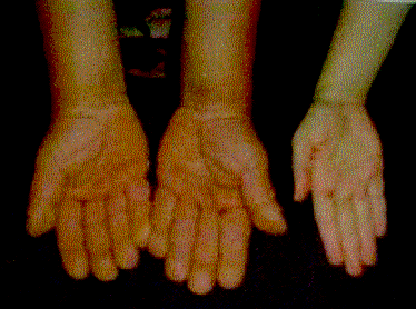
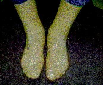
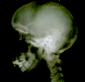
<body><![CDATA[<P><FONT FACE="Arial,Helvetica"><FONT SIZE=-1>Los cambios que inician en la pubertad se extienden por un lapso de 5 a 10 a&ntilde;os y luego no presentan cambios por el resto de la vida, con presencia de comedones, con poco vello facial y pelo p&uacute;bico y la presencia de ginecomastia.</FONT></FONT>      <P><FONT FACE="Arial,Helvetica"><FONT SIZE=-1>Algunos pacientes presentan oligofrenia, que se asocia a una disminuci&oacute;n en la expectancia de vida.</FONT></FONT>      <P><FONT FACE="Arial,Helvetica"><FONT SIZE=-1>A la histolog&iacute;a se observa hipertrofia de fibras col&aacute;genas en dermis, epidermis y ap&eacute;ndices cut&aacute;neos, con incremento en el contenido de mucopolisac&aacute;ridos y mucinosis papular.</FONT></FONT>      <P><FONT FACE="Arial,Helvetica"><FONT SIZE=-1>La diferencia principal entre la forma primaria y secundaria, es la edad de presentaci&oacute;n, donde la primaria es en la adolescencia, mientras que la secundaria es m&aacute;s frecuente entre los 30 y 50 a&ntilde;os de edad. Adicionalmente, la velocidad de crecimiento de las estructuras &oacute;seas es mucho mayor en la presentaci&oacute;n secundaria y causa, importante dolor al paciente. Otra patolog&iacute;a con la que se hace diagn&oacute;stico diferencial es con la acromegalia, pero en esta el compromiso es de aumento de volumen de todo el macizo facial y crecimiento desproporcionado del paciente. En el caso de la acropaquia tiroidea, el crecimiento se da en las manos y pies asociado a exoftalmos, mixedema pretibial y con otros datos de hipertiroidismo.</FONT></FONT>      <P><FONT FACE="Arial,Helvetica"><FONT SIZE=-1>El tratamiento va dirigido hacia le resoluci&oacute;n est&eacute;tica del problema<SUP> </SUP><A HREF="#7autor">(7</A><A HREF="#9autor">-9)</A>. Existen reportes en la literatura del tratamiento sintom&aacute;tico de las manifestaciones reumatol&oacute;gicas <A HREF="#10autor">(10</A>-<A HREF="#12autor">12)</A> con el uso de bifosfonatos <A HREF="#13autor">(13)</A>, de la paquidermia con isotretinoina<SUP> </SUP><A HREF="#14autor">(14)</A>, y en el caso de los pacientes con problema secundario se ha observado una regresi&oacute;n casi completa tras la excisi&oacute;n completa del tumor <A HREF="#15autor">(15)</A>.</FONT></FONT>      <P><B><FONT FACE="Arial,Helvetica"><FONT SIZE=-1>Caso</FONT></FONT></B>      <P><FONT FACE="Arial,Helvetica"><FONT SIZE=-1>Paciente masculino de 24 a&ntilde;os de edad vecino de Alajuela, Costa Rica, agricultor. Como antecedentes heredo-familiares: diabetes melitus y asma en los abuelos paternos, hipertensi&oacute;n y enfermedad arterial coronaria en abuela materna. El paciente carec&iacute;a de antecedentes personales no patol&oacute;gicos, quir&uacute;rgicos y como &uacute;nico antecedente personal patol&oacute;gico, una enfermedad de Gilbert.</FONT></FONT>      <P><FONT FACE="Arial,Helvetica"><FONT SIZE=-1>El paciente consulta por cuadro que inicia a los 15 a&ntilde;os de edad con aumento del tama&ntilde;o de sus manos, pies y la piel sobre la frente <A HREF="#img1">(Fig. 1</A><A HREF="#img3">-3)</A>, cuero cabelludo y manos. Asocia leve intolerancia al calor. Refiere el paciente que el crecimiento se detuvo hace unos a&ntilde;os, ya que not&oacute; que no cambia el tama&ntilde;o de sus zapatos (talla 42).</FONT></FONT>      <P><FONT FACE="Arial,Helvetica"><FONT SIZE=-1>Al examen f&iacute;sico se observan como datos positivos &uacute;nicamente un bocio difuso, con un aumento importante de los pliegues a nivel del cuero cabelludo, frente y p&aacute;rpados, con crecimiento acral muy sugestivo de acromegalia. Aumento de volumen de manos, pies y de los dedos mismos. Se observan u&ntilde;as en vidrio de reloj.</FONT></FONT>      <P><FONT FACE="Arial,Helvetica"><FONT SIZE=-1>Los estudios de laboratorio y gabinete se orientan a descartar como primera posibilidad diagn&oacute;stica una acromegalia. Pruebas de funci&oacute;n tiroidea, LH y FSH normales, cortisol urinario 46,4 g/24 H normal, testosterona 16,6 pg/ml normal, estradiol y PTH normales, Calcio 9,9 mmol/L, f&oacute;sforo 3,2 mmol/L. Hemograma completo normal, pruebas de funci&oacute;n renal normal, pruebas de funci&oacute;n hep&aacute;tica normales. Pruebas de coagulaci&oacute;n normal. Un examen general de orina normal. Se realizan tambi&eacute;n estudios inmunol&oacute;gicos con un factor antin&uacute;cleo negativo, una electroforesis de prote&iacute;nas que se informa como normal. Una tomograf&iacute;a axial computarizada para estudio de silla turca que no muestra alteraciones, una radiograf&iacute;a de t&oacute;rax con hiperclaridad de los campos pulmonares, en las radiograf&iacute;as de pies, manos y cr&aacute;neo se observa un engrosamiento del periostio <A HREF="#img4">(Fig. 4)</A>. Un examen oftalmol&oacute;gico normal.</FONT></FONT>      ]]></body>
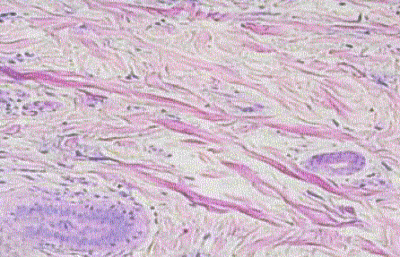
<body><![CDATA[<P><FONT FACE="Arial,Helvetica"><FONT SIZE=-1>Biopsia de piel del cuero cabelludo muestra: un epitelio sin lesiones pero se aprecia hipertrofia de los complejos piloseb&aacute;ceos y aumento de la mucina d&eacute;rmica, con diagn&oacute;stico de hipertrofia de los complejos piloseb&aacute;ceos y mucinosis d&eacute;rmica <A HREF="#img5">(Fig. 5)</A>.</FONT></FONT>      <P><FONT FACE="Arial,Helvetica"><FONT SIZE=-1>La curva de tolerancia a la glucosa con niveles concomitantes de hormona de crecimiento descart&oacute; acromegalia. <A HREF="#tab1">Cuadro 1</A>.</FONT></FONT>      <P>&nbsp;     <CENTER><A NAME="tab1"></A><B><FONT FACE="Arial,Helvetica"><FONT SIZE=-1>CUADRO 1</FONT></FONT></B></CENTER>      <CENTER><B><FONT FACE="Arial,Helvetica"><FONT SIZE=-1>CURVA DE TOLERANCIA DE LA GLUCOSA ASOCIADA A NIVELES DE OTRAS HORMONAS,</FONT></FONT></B></CENTER>      <CENTER><B><FONT FACE="Arial,Helvetica"><FONT SIZE=-1>PARA EL ESTUDIO DE LA ACROMEGALIA</FONT></FONT></B></CENTER> &nbsp;     <CENTER><TABLE BORDER CELLSPACING=0 CELLPADDING=7 WIDTH="590" > <TR> <TD VALIGN=TOP WIDTH="20%">     <CENTER><B><FONT FACE="Arial,Helvetica"><FONT SIZE=-1>Tiempo</FONT></FONT></B></CENTER> </TD>  <TD VALIGN=TOP WIDTH="20%">     <CENTER><B><FONT FACE="Arial,Helvetica"><FONT SIZE=-1>HET uIU/d</FONT></FONT></B></CENTER> </TD>  <TD VALIGN=TOP WIDTH="16%">     <CENTER><B><FONT FACE="Arial,Helvetica"><FONT SIZE=-1>PRL ng/dl</FONT></FONT></B></CENTER> </TD>  <TD VALIGN=TOP WIDTH="22%">     ]]></body>
<body><![CDATA[<CENTER><B><FONT FACE="Arial,Helvetica"><FONT SIZE=-1>Glicemia mg/dl</FONT></FONT></B></CENTER> </TD>  <TD VALIGN=TOP WIDTH="22%">     <CENTER><B><FONT FACE="Arial,Helvetica"><FONT SIZE=-1>HC ng/dl</FONT></FONT></B></CENTER> </TD> </TR>  <TR> <TD VALIGN=TOP WIDTH="20%">     <CENTER><FONT FACE="Arial,Helvetica"><FONT SIZE=-1>0’</FONT></FONT></CENTER> </TD>  <TD VALIGN=TOP WIDTH="20%">     <CENTER><FONT FACE="Arial,Helvetica"><FONT SIZE=-1>1,1</FONT></FONT></CENTER> </TD>  <TD VALIGN=TOP WIDTH="16%">     <CENTER><FONT FACE="Arial,Helvetica"><FONT SIZE=-1>3,1</FONT></FONT></CENTER> </TD>  <TD VALIGN=TOP WIDTH="22%">     <CENTER><FONT FACE="Arial,Helvetica"><FONT SIZE=-1>77</FONT></FONT></CENTER> </TD>  <TD VALIGN=TOP WIDTH="22%">     <CENTER><FONT FACE="Arial,Helvetica"><FONT SIZE=-1>No detectado.</FONT></FONT></CENTER> </TD> </TR>  <TR> <TD VALIGN=TOP WIDTH="20%">     <CENTER><FONT FACE="Arial,Helvetica"><FONT SIZE=-1>30’</FONT></FONT></CENTER> </TD>  <TD VALIGN=TOP WIDTH="20%">     <CENTER><FONT FACE="Arial,Helvetica"><FONT SIZE=-1>10,8</FONT></FONT></CENTER> </TD>  <TD VALIGN=TOP WIDTH="16%">     <CENTER><FONT FACE="Arial,Helvetica"><FONT SIZE=-1>30,3</FONT></FONT></CENTER> </TD>  <TD VALIGN=TOP WIDTH="22%">     ]]></body>
<body><![CDATA[<CENTER><FONT FACE="Arial,Helvetica"><FONT SIZE=-1>117</FONT></FONT></CENTER> </TD>  <TD VALIGN=TOP WIDTH="22%">     <CENTER><FONT FACE="Arial,Helvetica"><FONT SIZE=-1>"</FONT></FONT></CENTER> </TD> </TR>  <TR> <TD VALIGN=TOP WIDTH="20%">     <CENTER><FONT FACE="Arial,Helvetica"><FONT SIZE=-1>60’</FONT></FONT></CENTER> </TD>  <TD VALIGN=TOP WIDTH="20%">     <CENTER><FONT FACE="Arial,Helvetica"><FONT SIZE=-1>8,2</FONT></FONT></CENTER> </TD>  <TD VALIGN=TOP WIDTH="16%">     <CENTER><FONT FACE="Arial,Helvetica"><FONT SIZE=-1>17,2</FONT></FONT></CENTER> </TD>  <TD VALIGN=TOP WIDTH="22%">     <CENTER><FONT FACE="Arial,Helvetica"><FONT SIZE=-1>68</FONT></FONT></CENTER> </TD>  <TD VALIGN=TOP WIDTH="22%">     <CENTER><FONT FACE="Arial,Helvetica"><FONT SIZE=-1>"</FONT></FONT></CENTER> </TD> </TR>  <TR> <TD VALIGN=TOP WIDTH="20%">     <CENTER><FONT FACE="Arial,Helvetica"><FONT SIZE=-1>120’</FONT></FONT></CENTER> </TD>  <TD VALIGN=TOP WIDTH="20%"><FONT FACE="Arial,Helvetica"><FONT SIZE=-1>&nbsp;</FONT></FONT></TD>  <TD VALIGN=TOP WIDTH="16%"><FONT FACE="Arial,Helvetica"><FONT SIZE=-1>&nbsp;</FONT></FONT></TD>  <TD VALIGN=TOP WIDTH="22%">     <CENTER><FONT FACE="Arial,Helvetica"><FONT SIZE=-1>65</FONT></FONT></CENTER> </TD>  <TD VALIGN=TOP WIDTH="22%">     <CENTER><FONT FACE="Arial,Helvetica"><FONT SIZE=-1>"</FONT></FONT></CENTER> </TD> </TR> </TABLE></CENTER>      ]]></body>
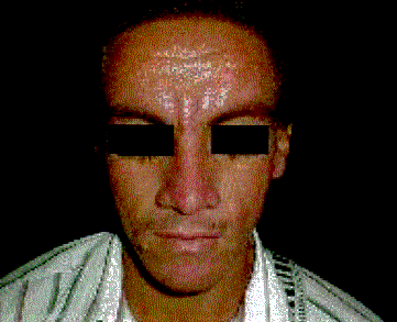
<body><![CDATA[<CENTER><FONT FACE="Arial,Helvetica"><FONT SIZE=-1>HET: hormona estimuladora de tiroides, PRL: prolactina, HC: hormona de crecimiento.</FONT></FONT></CENTER> &nbsp;      <P><B><FONT FACE="Arial,Helvetica"><FONT SIZE=-1>Discusi&oacute;n</FONT></FONT></B>      <P><FONT FACE="Arial,Helvetica"><FONT SIZE=-1>El presente caso, se enfoco inicialmente, como si se tratase de un paciente con acromegalia. Debido a las caracter&iacute;sticas f&iacute;sicas del paciente, como eran: el aumento acral de las extremidades, el aumento en el volumen de las manos, pies y los dedos, el engrosamiento facial prominente y la alteraci&oacute;n a nivel de la piel de la cara y las extremidades. Sin embargo, se presentan caracter&iacute;sticas que hac&iacute;an discrepar de ese diagn&oacute;stico inicial.</FONT></FONT>      <P><FONT FACE="Arial,Helvetica"><FONT SIZE=-1>Como la no afecci&oacute;n del macizo facial espec&iacute;ficamente no compromet&iacute;a el hueso mandibular ni el zigom&aacute;tico, como ocurre en la acromegalia. Se procedi&oacute; a realizar una curva de tolerancia a la glucosa asociada a la hormona de crecimiento, que se considera diagn&oacute;stica en el estudio de la acromegalia. Como se observ&oacute; en la <A HREF="#tab1">Cuadro 1</A>, la hormona de crecimiento no fue detectable, lo que descarta el diagn&oacute;stico previo. En la tomograf&iacute;a axial computadorizada no se encontr&oacute; afecci&oacute;n de la silla turca, la biometr&iacute;a hem&aacute;tica no estaba alterada y no hab&iacute;a alteraciones en los niveles hormonales estudiados. La radiograf&iacute;a de t&oacute;rax no evidenciaba la presencia de masas como para hacernos pensar en la posibilidad de una lesi&oacute;n tumoral, la cual nos dar&iacute;a el cuadro secundario. Los hallazgos en la biopsia de piel, eran compatibles con las descripciones en la literatura para este s&iacute;ndrome <A HREF="#17.autor">(17)</A>. La serie de estudios realizados y la revisi&oacute;n de la literatura confirman el diagn&oacute;stico de una paquidermoperiostosis primaria.</FONT></FONT>      <P><FONT FACE="Arial,Helvetica"><FONT SIZE=-1>Otros diagn&oacute;sticos posibles que se descartan cl&iacute;nicamente son: el s&iacute;ndrome Rosenthal-Kloepfer caracterizado por un aumento en el macizo facial, e inclusive la presencia de cutis verticis gyrata pero sin afecci&oacute;n de la silla turca ni afecci&oacute;n endocrina, es de origen gen&eacute;tico y es auton&oacute;mico dominante la acropaquia tiroidea, que tambi&eacute;n presenta engrosamiento de pies y manos y no en el macizo facial, pero tambi&eacute;n asocia exoftalmos, mixedema pretibial y alteraciones a nivel de las hormonas tiroideas, compatibles con hipertiroidismo, y por ultimo tenemos el gigantismo cerebral o s&iacute;ndrome de Soto, el cual es tambi&eacute;n una genodermatosis extremadamente rara, que se caracteriza por un aumento en peso y talla, con facciones faciales acromeg&aacute;licas, engrosamiento de pies y manos y retardo mental<A HREF="#15autor"> (15)</A>.</FONT></FONT>     <CENTER></CENTER>      <CENTER><A NAME="img1"></A><IMG SRC="/img/fbpe/rccm/v22n3-4/1080i1.GIF" HEIGHT=246 WIDTH=324></CENTER>      
<CENTER><FONT FACE="Arial,Helvetica"><FONT SIZE=-2>Figura 1. Se observa la frente del paciente con engrosamiento de los</FONT></FONT></CENTER>      <CENTER><FONT FACE="Arial,Helvetica"><FONT SIZE=-2>Pliegues con aspecto cerebriforme (cutis verticis gyrata).</FONT></FONT></CENTER>      <CENTER></CENTER>      ]]></body>
<body><![CDATA[<CENTER><A NAME="img2"></A><IMG SRC="/img/fbpe/rccm/v22n3-4/1080i2.GIF" HEIGHT=261 WIDTH=330></CENTER>      
<CENTER><FONT FACE="Arial,Helvetica"><FONT SIZE=-2>Figura 2. Las manos presentan un engrosamiento acral a nivel</FONT></FONT></CENTER>      <CENTER><FONT FACE="Arial,Helvetica"><FONT SIZE=-2>&oacute;seo y subcut&aacute;neo, la apariencia cl&iacute;nica lleva a pensar en acromegalia</FONT></FONT></CENTER>      <CENTER><FONT FACE="Arial,Helvetica"><FONT SIZE=-2>en este caso comparativo.</FONT></FONT></CENTER>      <CENTER></CENTER>      <CENTER><A NAME="img3"></A><IMG SRC="/img/fbpe/rccm/v22n3-4/1080i3.GIF" HEIGHT=267 WIDTH=327></CENTER>      
<CENTER><FONT FACE="Arial,Helvetica"><FONT SIZE=-2>Figura 3. El engrosamiento tambi&eacute;n ocurre a nivel de los pies con</FONT></FONT></CENTER>      <CENTER><FONT FACE="Arial,Helvetica"><FONT SIZE=-2>dedos en palillo de tambor y u&ntilde;as en vidrio de reloj.</FONT></FONT></CENTER>      <CENTER>&nbsp;</CENTER>      <CENTER></CENTER>      ]]></body>
<body><![CDATA[<CENTER><A NAME="img4"></A><IMG SRC="/img/fbpe/rccm/v22n3-4/1080i4.GIF" HEIGHT=265 WIDTH=310></CENTER>      
<CENTER><FONT FACE="Arial,Helvetica"><FONT SIZE=-2>Figura 4. A nivel &oacute;seo se observa engrosamiento leve del periostio,</FONT></FONT></CENTER>      <CENTER><FONT FACE="Arial,Helvetica"><FONT SIZE=-2>especialmente en regi&oacute;n frontal.</FONT></FONT></CENTER>      <CENTER></CENTER>      <CENTER><A NAME="img5"></A><IMG SRC="/img/fbpe/rccm/v22n3-4/1080i5.GIF" HEIGHT=247 WIDTH=360></CENTER>      
<CENTER><FONT FACE="Arial,Helvetica"><FONT SIZE=-2>Figura 5. Corte Histol&oacute;gico donde se observa aumento de las bandas de</FONT></FONT></CENTER>      <CENTER><FONT FACE="Arial,Helvetica"><FONT SIZE=-2>col&aacute;geno en la dermis, con aumento en el n&uacute;mero de fibroblastos y en la</FONT></FONT></CENTER>      <CENTER><FONT FACE="Arial,Helvetica"><FONT SIZE=-2>cantidad de sustancia esencial a expensas de mucina y &aacute;cido hialur&oacute;nico.</FONT></FONT></CENTER> &nbsp;     <BR>&nbsp;      <P><B><FONT FACE="Arial,Helvetica"><FONT SIZE=-1>Referencias</FONT></FONT></B>      ]]></body>
<body><![CDATA[<!-- ref --><P><A NAME="1autor"></A><FONT FACE="Arial,Helvetica"><FONT SIZE=-1>1. Touraine A., Solente G, y Goul&eacute; L. Un s&iacute;ndrome ost&eacute;odermopathique: la pachydermie plicatur&eacute;e avec pachyp&eacute;riostose des extr&eacute;mit&eacute;s. <I>Presse m&eacute;d</I> 43: 1820-1824, 1935.</FONT></FONT>    &nbsp;&nbsp;&nbsp;&nbsp;&nbsp;&nbsp;&nbsp;&nbsp;[&#160;<a href="javascript:void(0);" onclick="javascript: window.open('/scielo.php?script=sci_nlinks&ref=779637&pid=S0253-2948200100020000600001&lng=','','width=640,height=500,resizable=yes,scrollbars=1,menubar=yes,');">Links</a>&#160;]<!-- end-ref --><!-- ref --><P><A NAME="2.autor"></A><FONT FACE="Arial,Helvetica"><FONT SIZE=-1>2. Vogl A., and Goldfischer S. Pachydermoperiostosis: primary or idiopathic hypertrophic osteoarthropathy. <I>Am J Med</I> 1962; 33:166-187.</FONT></FONT>    &nbsp;&nbsp;&nbsp;&nbsp;&nbsp;&nbsp;&nbsp;&nbsp;[&#160;<a href="javascript:void(0);" onclick="javascript: window.open('/scielo.php?script=sci_nlinks&ref=779638&pid=S0253-2948200100020000600002&lng=','','width=640,height=500,resizable=yes,scrollbars=1,menubar=yes,');">Links</a>&#160;]<!-- end-ref --><!-- ref --><P><A NAME="3autor"></A><FONT FACE="Arial,Helvetica"><FONT SIZE=-1>3. Rimoin DL. Pachydermoperiostosis (idiopathic clubbing and periostosis): Genetic and physiologic considerations. <I>The New Engl J of Med</I> 1965; 272: 923-931.</FONT></FONT>    &nbsp;&nbsp;&nbsp;&nbsp;&nbsp;&nbsp;&nbsp;&nbsp;[&#160;<a href="javascript:void(0);" onclick="javascript: window.open('/scielo.php?script=sci_nlinks&ref=779639&pid=S0253-2948200100020000600003&lng=','','width=640,height=500,resizable=yes,scrollbars=1,menubar=yes,');">Links</a>&#160;]<!-- end-ref --><!-- ref --><P><A NAME="4.autor"></A><FONT FACE="Arial,Helvetica"><FONT SIZE=-1>4. Shimizu C., et al. A rare case of acromegaly associated with pachydermoperiostosis. <I>J Endocrinol. Invest </I>1999;<I> </I>22: 386-9.</FONT></FONT>    &nbsp;&nbsp;&nbsp;&nbsp;&nbsp;&nbsp;&nbsp;&nbsp;[&#160;<a href="javascript:void(0);" onclick="javascript: window.open('/scielo.php?script=sci_nlinks&ref=779640&pid=S0253-2948200100020000600004&lng=','','width=640,height=500,resizable=yes,scrollbars=1,menubar=yes,');">Links</a>&#160;]<!-- end-ref --><!-- ref --><P><A NAME="5.autor"></A><FONT FACE="Arial,Helvetica"><FONT SIZE=-1>5. Mart&iacute;nez-Lavin M. y cols. Primary hypertrophic osteoarthropathy: another disorder associated with patent ductus arteriosus. <I>Pediatr Cardiol</I> 1993; 14:181-2.</FONT></FONT>    &nbsp;&nbsp;&nbsp;&nbsp;&nbsp;&nbsp;&nbsp;&nbsp;[&#160;<a href="javascript:void(0);" onclick="javascript: window.open('/scielo.php?script=sci_nlinks&ref=779641&pid=S0253-2948200100020000600005&lng=','','width=640,height=500,resizable=yes,scrollbars=1,menubar=yes,');">Links</a>&#160;]<!-- end-ref --><!-- ref --><P><A NAME="6autor"></A><FONT FACE="Arial,Helvetica"><FONT SIZE=-1>6. Lam S., Stone MS., et al. Paraneoplastic pemphigus, cicatricial conjunctivitis, and acanthosis nigricans with pachydermatoglyphy in a patient with squamous cell carcinoma. <I>Ophthalmology</I> 1992; 99: 108-13.</FONT></FONT>    &nbsp;&nbsp;&nbsp;&nbsp;&nbsp;&nbsp;&nbsp;&nbsp;[&#160;<a href="javascript:void(0);" onclick="javascript: window.open('/scielo.php?script=sci_nlinks&ref=779642&pid=S0253-2948200100020000600006&lng=','','width=640,height=500,resizable=yes,scrollbars=1,menubar=yes,');">Links</a>&#160;]<!-- end-ref --><!-- ref --><P><A NAME="7autor"></A><FONT FACE="Arial,Helvetica"><FONT SIZE=-1>7. Gonz&aacute;lez Saldivar G. y cols. S&iacute;ndrome de Touraine-Solente-Goul&eacute;: Presentaci&oacute;n de un caso cl&iacute;nico. <I>Cir Pl&aacute;st </I>1997; 7: 9-11.</FONT></FONT>    &nbsp;&nbsp;&nbsp;&nbsp;&nbsp;&nbsp;&nbsp;&nbsp;[&#160;<a href="javascript:void(0);" onclick="javascript: window.open('/scielo.php?script=sci_nlinks&ref=779643&pid=S0253-2948200100020000600007&lng=','','width=640,height=500,resizable=yes,scrollbars=1,menubar=yes,');">Links</a>&#160;]<!-- end-ref --><!-- ref --><P><A NAME="8autor"></A><FONT FACE="Arial,Helvetica"><FONT SIZE=-1>8. Ohtsuka M., Takayanagi S. Eyelid reconstruction in pahydermoperiostosis. <I>Plast Reconstr Surg</I> 1988; 81: 88-90.</FONT></FONT>    &nbsp;&nbsp;&nbsp;&nbsp;&nbsp;&nbsp;&nbsp;&nbsp;[&#160;<a href="javascript:void(0);" onclick="javascript: window.open('/scielo.php?script=sci_nlinks&ref=779644&pid=S0253-2948200100020000600008&lng=','','width=640,height=500,resizable=yes,scrollbars=1,menubar=yes,');">Links</a>&#160;]<!-- end-ref --><!-- ref --><P><A NAME="9autor"></A><FONT FACE="Arial,Helvetica"><FONT SIZE=-1>9. Friedhofer H, Salles AG, et al. Correction of eyelid anomalies in pachydermoperiostosis. <I>Ophthal Plast Reconstr Surg</I> 1999; 15: 137-8.</FONT></FONT>    &nbsp;&nbsp;&nbsp;&nbsp;&nbsp;&nbsp;&nbsp;&nbsp;[&#160;<a href="javascript:void(0);" onclick="javascript: window.open('/scielo.php?script=sci_nlinks&ref=779645&pid=S0253-2948200100020000600009&lng=','','width=640,height=500,resizable=yes,scrollbars=1,menubar=yes,');">Links</a>&#160;]<!-- end-ref --><!-- ref --><P><A NAME="10autor"></A><FONT FACE="Arial,Helvetica"><FONT SIZE=-1>10. Heddayati H, Barmada R, Skosey JL. Acrolysis in pachydermoperiostosis. Primary or idiopathic hypertrophic osteoarthropathy. <I>Arch Intern Med</I> 1980; 140:1087-8.</FONT></FONT>    &nbsp;&nbsp;&nbsp;&nbsp;&nbsp;&nbsp;&nbsp;&nbsp;[&#160;<a href="javascript:void(0);" onclick="javascript: window.open('/scielo.php?script=sci_nlinks&ref=779646&pid=S0253-2948200100020000600010&lng=','','width=640,height=500,resizable=yes,scrollbars=1,menubar=yes,');">Links</a>&#160;]<!-- end-ref --><!-- ref --><P><A NAME="11autor"></A><FONT FACE="Arial,Helvetica"><FONT SIZE=-1>11. Schumacher HR Jr. Hypertrophic osteoarthropathy: rheumatologic manifestations. <I>Clin Exp Rheumatol</I> Suppl 1992; 7: 35-40.</FONT></FONT>    &nbsp;&nbsp;&nbsp;&nbsp;&nbsp;&nbsp;&nbsp;&nbsp;[&#160;<a href="javascript:void(0);" onclick="javascript: window.open('/scielo.php?script=sci_nlinks&ref=779647&pid=S0253-2948200100020000600011&lng=','','width=640,height=500,resizable=yes,scrollbars=1,menubar=yes,');">Links</a>&#160;]<!-- end-ref --><!-- ref --><P><A NAME="12autor"></A><FONT FACE="Arial,Helvetica"><FONT SIZE=-1>12. Ansell BM. Hypertrophic osteoarthropathy in the paediatric age. <I>Clin Exp Rheumatol</I> Suppl 1992; 7: 15-8.</FONT></FONT>    &nbsp;&nbsp;&nbsp;&nbsp;&nbsp;&nbsp;&nbsp;&nbsp;[&#160;<a href="javascript:void(0);" onclick="javascript: window.open('/scielo.php?script=sci_nlinks&ref=779648&pid=S0253-2948200100020000600012&lng=','','width=640,height=500,resizable=yes,scrollbars=1,menubar=yes,');">Links</a>&#160;]<!-- end-ref --><!-- ref --><P><A NAME="13autor"></A><FONT FACE="Arial,Helvetica"><FONT SIZE=-1>13. Guyot-Drouot MH., et al. Rheumatologic manifestations of pachydermoperiostosis and preliminary experience with biphosphonates. <I>J Rheumatol </I>2000; 27: 2418-23.</FONT></FONT>    &nbsp;&nbsp;&nbsp;&nbsp;&nbsp;&nbsp;&nbsp;&nbsp;[&#160;<a href="javascript:void(0);" onclick="javascript: window.open('/scielo.php?script=sci_nlinks&ref=779649&pid=S0253-2948200100020000600013&lng=','','width=640,height=500,resizable=yes,scrollbars=1,menubar=yes,');">Links</a>&#160;]<!-- end-ref --><!-- ref --><P><A NAME="14autor"></A><FONT FACE="Arial,Helvetica"><FONT SIZE=-1>14. Beauregard S. Cutis verticis gyrata and pachydermoperiostosis. Several cases in a same family. Initial results of the treatment of pachyderma with isotretinoin. <I>Ann</I> <I>Dermatol Venereol</I> 1994; 121: 134-7.</FONT></FONT>    &nbsp;&nbsp;&nbsp;&nbsp;&nbsp;&nbsp;&nbsp;&nbsp;[&#160;<a href="javascript:void(0);" onclick="javascript: window.open('/scielo.php?script=sci_nlinks&ref=779650&pid=S0253-2948200100020000600014&lng=','','width=640,height=500,resizable=yes,scrollbars=1,menubar=yes,');">Links</a>&#160;]<!-- end-ref --><!-- ref --><P><A NAME="15autor"></A><FONT FACE="Arial,Helvetica"><FONT SIZE=-1>15. Champion RH, et al. <I>Rook/Williams/Ebling: Textbook of Dermatology</I>. Blackwell Science 6</FONT><SUP><FONT SIZE=-2>th</FONT></SUP><FONT SIZE=-1> Ed CD-ROM Version 1.2.0 Software &copy; Gentian I.T. Consultants. 1998 Ch. 12 (pahydermoperiostosis).</FONT></FONT>    &nbsp;&nbsp;&nbsp;&nbsp;&nbsp;&nbsp;&nbsp;&nbsp;[&#160;<a href="javascript:void(0);" onclick="javascript: window.open('/scielo.php?script=sci_nlinks&ref=779651&pid=S0253-2948200100020000600015&lng=','','width=640,height=500,resizable=yes,scrollbars=1,menubar=yes,');">Links</a>&#160;]<!-- end-ref --><!-- ref --><P><A NAME="16autor"></A><FONT FACE="Arial,Helvetica"><FONT SIZE=-1>16. Matucci-Cerinic M., Cinti S., Morroni M, et al. Pachydermoperiostosis (primary hypertrophic osteoarthropathy): report of a case with evidence of endothelial and connective tissue involvement. <I>Ann Rheum Dis</I> 1989; 48: 240-6</FONT></FONT>    &nbsp;&nbsp;&nbsp;&nbsp;&nbsp;&nbsp;&nbsp;&nbsp;[&#160;<a href="javascript:void(0);" onclick="javascript: window.open('/scielo.php?script=sci_nlinks&ref=779652&pid=S0253-2948200100020000600016&lng=','','width=640,height=500,resizable=yes,scrollbars=1,menubar=yes,');">Links</a>&#160;]<!-- end-ref --><!-- ref --><P><A NAME="17.autor"></A><FONT FACE="Arial,Helvetica"><FONT SIZE=-1>17. Elder D., et al. <I>Lever’s Histopathology of the Skin</I>. Lippincot-Raven 8</FONT><SUP><FONT SIZE=-2>th</FONT></SUP><FONT SIZE=-1> Ed. 1997. p. 141.</FONT></FONT>    &nbsp;&nbsp;&nbsp;&nbsp;&nbsp;&nbsp;&nbsp;&nbsp;[&#160;<a href="javascript:void(0);" onclick="javascript: window.open('/scielo.php?script=sci_nlinks&ref=779653&pid=S0253-2948200100020000600017&lng=','','width=640,height=500,resizable=yes,scrollbars=1,menubar=yes,');">Links</a>&#160;]<!-- end-ref --><P><A NAME="1a"></A><FONT FACE="Arial,Helvetica"><SUP><FONT SIZE=-2><A HREF="#1">1</A></FONT></SUP><FONT SIZE=-1>Servicio Dermatolog&iacute;a-Alergolog&iacute;a Hospital M&eacute;xico. Caja Costarricense de Seguro Social. San Jos&eacute; Costa Rica.</FONT></FONT>     <BR><A NAME="2b"></A><FONT FACE="Arial,Helvetica"><SUP><FONT SIZE=-2><A HREF="#2">2</A></FONT></SUP><FONT SIZE=-1>Servicio Endocrinolog&iacute;a Hospital M&eacute;xico. Caja Costarricense de Seguro Social. San Jos&eacute; Costa Rica.</FONT></FONT>      ]]></body><back>
<ref-list>
<ref id="B1">
<nlm-citation citation-type="journal">
<person-group person-group-type="author">
<name>
<surname><![CDATA[Touraine]]></surname>
<given-names><![CDATA[A.]]></given-names>
</name>
<name>
<surname><![CDATA[Solente]]></surname>
<given-names><![CDATA[G]]></given-names>
</name>
<name>
<surname><![CDATA[Goulé]]></surname>
<given-names><![CDATA[L]]></given-names>
</name>
</person-group>
<article-title xml:lang="es"><![CDATA[Un síndrome ostéodermopathique: la pachydermie plicaturée avec pachypériostose des extrémités]]></article-title>
<source><![CDATA[Presse méd]]></source>
<year></year>
<volume>43</volume>
<page-range>1820-1824, 1935</page-range></nlm-citation>
</ref>
<ref id="B2">
<nlm-citation citation-type="journal">
<person-group person-group-type="author">
<name>
<surname><![CDATA[Vogl]]></surname>
<given-names><![CDATA[A]]></given-names>
</name>
<name>
<surname><![CDATA[Goldfischer]]></surname>
<given-names><![CDATA[S.]]></given-names>
</name>
</person-group>
<article-title xml:lang="en"><![CDATA[Pachydermoperiostosis: primary or idiopathic hypertrophic osteoarthropathy]]></article-title>
<source><![CDATA[Am J Med]]></source>
<year>1962</year>
<volume>33</volume>
<page-range>166-187</page-range></nlm-citation>
</ref>
<ref id="B3">
<nlm-citation citation-type="journal">
<person-group person-group-type="author">
<name>
<surname><![CDATA[Rimoin]]></surname>
<given-names><![CDATA[DL]]></given-names>
</name>
</person-group>
<article-title xml:lang="en"><![CDATA[Pachydermoperiostosis (idiopathic clubbing and periostosis): Genetic and physiologic considerations]]></article-title>
<source><![CDATA[The New Engl J of Med]]></source>
<year>1965</year>
<volume>272</volume>
<page-range>923-931</page-range></nlm-citation>
</ref>
<ref id="B4">
<nlm-citation citation-type="journal">
<person-group person-group-type="author">
<name>
<surname><![CDATA[Shimizu]]></surname>
<given-names><![CDATA[C.]]></given-names>
</name>
</person-group>
<article-title xml:lang="en"><![CDATA[A rare case of acromegaly associated with pachydermoperiostosis]]></article-title>
<source><![CDATA[J Endocrinol. Invest]]></source>
<year>1999</year>
<volume>22</volume>
<page-range>386-9</page-range></nlm-citation>
</ref>
<ref id="B5">
<nlm-citation citation-type="journal">
<person-group person-group-type="author">
<name>
<surname><![CDATA[Martínez-Lavin]]></surname>
<given-names><![CDATA[M.]]></given-names>
</name>
</person-group>
<article-title xml:lang="en"><![CDATA[Primary hypertrophic osteoarthropathy: another disorder associated with patent ductus arteriosus]]></article-title>
<source><![CDATA[Pediatr Cardiol]]></source>
<year>1993</year>
<volume>14</volume>
<page-range>181-2</page-range></nlm-citation>
</ref>
<ref id="B6">
<nlm-citation citation-type="journal">
<person-group person-group-type="author">
<name>
<surname><![CDATA[Lam]]></surname>
<given-names><![CDATA[S.]]></given-names>
</name>
<name>
<surname><![CDATA[Stone]]></surname>
<given-names><![CDATA[MS]]></given-names>
</name>
</person-group>
<article-title xml:lang="en"><![CDATA[Paraneoplastic pemphigus, cicatricial conjunctivitis, and acanthosis nigricans with pachydermatoglyphy in a patient with squamous cell carcinoma]]></article-title>
<source><![CDATA[Ophthalmology]]></source>
<year>1992</year>
<volume>99</volume>
<page-range>108-13</page-range></nlm-citation>
</ref>
<ref id="B7">
<nlm-citation citation-type="journal">
<person-group person-group-type="author">
<name>
<surname><![CDATA[González Saldivar]]></surname>
<given-names><![CDATA[G]]></given-names>
</name>
</person-group>
<article-title xml:lang="es"><![CDATA[Síndrome de Touraine-Solente-Goulé: Presentación de un caso clínico]]></article-title>
<source><![CDATA[Cir Plást]]></source>
<year>1997</year>
<volume>7</volume>
<page-range>9-11</page-range></nlm-citation>
</ref>
<ref id="B8">
<nlm-citation citation-type="journal">
<person-group person-group-type="author">
<name>
<surname><![CDATA[Ohtsuka]]></surname>
<given-names><![CDATA[M.]]></given-names>
</name>
<name>
<surname><![CDATA[Takayanagi]]></surname>
<given-names><![CDATA[S.]]></given-names>
</name>
</person-group>
<article-title xml:lang="en"><![CDATA[Eyelid reconstruction in pahydermoperiostosis]]></article-title>
<source><![CDATA[Plast Reconstr Surg]]></source>
<year>1988</year>
<volume>81</volume>
<page-range>88-90</page-range></nlm-citation>
</ref>
<ref id="B9">
<nlm-citation citation-type="journal">
<person-group person-group-type="author">
<name>
<surname><![CDATA[Friedhofer]]></surname>
<given-names><![CDATA[H]]></given-names>
</name>
<name>
<surname><![CDATA[Salles]]></surname>
<given-names><![CDATA[AG]]></given-names>
</name>
</person-group>
<article-title xml:lang="en"><![CDATA[Correction of eyelid anomalies in pachydermoperiostosis]]></article-title>
<source><![CDATA[Ophthal Plast Reconstr Surg]]></source>
<year>1999</year>
<volume>15</volume>
<page-range>137-8</page-range></nlm-citation>
</ref>
<ref id="B10">
<nlm-citation citation-type="journal">
<person-group person-group-type="author">
<name>
<surname><![CDATA[Heddayati]]></surname>
<given-names><![CDATA[H]]></given-names>
</name>
<name>
<surname><![CDATA[Barmada]]></surname>
<given-names><![CDATA[R]]></given-names>
</name>
<name>
<surname><![CDATA[Skosey]]></surname>
<given-names><![CDATA[JL]]></given-names>
</name>
</person-group>
<article-title xml:lang="en"><![CDATA[Acrolysis in pachydermoperiostosis: Primary or idiopathic hypertrophic osteoarthropathy]]></article-title>
<source><![CDATA[Arch Intern Med]]></source>
<year>1980</year>
<volume>140</volume>
<page-range>1087-8</page-range></nlm-citation>
</ref>
<ref id="B11">
<nlm-citation citation-type="journal">
<person-group person-group-type="author">
<name>
<surname><![CDATA[Schumacher]]></surname>
<given-names><![CDATA[HR]]></given-names>
</name>
</person-group>
<article-title xml:lang="en"><![CDATA[Hypertrophic osteoarthropathy: rheumatologic manifestations]]></article-title>
<source><![CDATA[Clin Exp Rheumatol Suppl]]></source>
<year>1992</year>
<volume>7</volume>
<page-range>35-40</page-range></nlm-citation>
</ref>
<ref id="B12">
<nlm-citation citation-type="journal">
<person-group person-group-type="author">
<name>
<surname><![CDATA[Ansell]]></surname>
<given-names><![CDATA[BM]]></given-names>
</name>
</person-group>
<article-title xml:lang="en"><![CDATA[Hypertrophic osteoarthropathy in the paediatric age]]></article-title>
<source><![CDATA[Clin Exp Rheumatol Suppl]]></source>
<year>1992</year>
<volume>7</volume>
<page-range>15-8</page-range></nlm-citation>
</ref>
<ref id="B13">
<nlm-citation citation-type="journal">
<person-group person-group-type="author">
<name>
<surname><![CDATA[Guyot-Drouot]]></surname>
<given-names><![CDATA[MH.]]></given-names>
</name>
</person-group>
<article-title xml:lang="en"><![CDATA[Rheumatologic manifestations of pachydermoperiostosis and preliminary experience with biphosphonates]]></article-title>
<source><![CDATA[J Rheumatol]]></source>
<year>2000</year>
<volume>27</volume>
<page-range>2418-23</page-range></nlm-citation>
</ref>
<ref id="B14">
<nlm-citation citation-type="journal">
<person-group person-group-type="author">
<name>
<surname><![CDATA[Beauregard]]></surname>
<given-names><![CDATA[S.]]></given-names>
</name>
</person-group>
<article-title xml:lang="en"><![CDATA[Cutis verticis gyrata and pachydermoperiostosis. Several cases in a same family. Initial results of the treatment of pachyderma with isotretinoin]]></article-title>
<source><![CDATA[Ann Dermatol Venereol]]></source>
<year>1994</year>
<volume>121</volume>
<page-range>134-7</page-range></nlm-citation>
</ref>
<ref id="B15">
<nlm-citation citation-type="book">
<person-group person-group-type="author">
<name>
<surname><![CDATA[Champion]]></surname>
<given-names><![CDATA[RH]]></given-names>
</name>
</person-group>
<source><![CDATA[Rook/Williams/Ebling: Textbook of Dermatology]]></source>
<year>1998</year>
<edition>6th</edition>
<publisher-name><![CDATA[Gentian I.T. Consultants]]></publisher-name>
</nlm-citation>
</ref>
<ref id="B16">
<nlm-citation citation-type="journal">
<person-group person-group-type="author">
<name>
<surname><![CDATA[Matucci-Cerinic]]></surname>
<given-names><![CDATA[M.]]></given-names>
</name>
<name>
<surname><![CDATA[Cinti]]></surname>
<given-names><![CDATA[S.]]></given-names>
</name>
<name>
<surname><![CDATA[Morroni]]></surname>
<given-names><![CDATA[M]]></given-names>
</name>
</person-group>
<article-title xml:lang="en"><![CDATA[Pachydermoperiostosis (primary hypertrophic osteoarthropathy): report of a case with evidence of endothelial and connective tissue involvement]]></article-title>
<source><![CDATA[Ann Rheum Dis]]></source>
<year>1989</year>
<volume>48</volume>
<page-range>240-6</page-range></nlm-citation>
</ref>
<ref id="B17">
<nlm-citation citation-type="book">
<person-group person-group-type="author">
<name>
<surname><![CDATA[Elder]]></surname>
<given-names><![CDATA[D.]]></given-names>
</name>
</person-group>
<source><![CDATA[Lever’s Histopathology of the Skin]]></source>
<year>1997</year>
<edition>8th</edition>
<page-range>141</page-range><publisher-name><![CDATA[Lippincot-Raven]]></publisher-name>
</nlm-citation>
</ref>
</ref-list>
</back>
</article>