<?xml version="1.0" encoding="ISO-8859-1"?><article xmlns:mml="http://www.w3.org/1998/Math/MathML" xmlns:xlink="http://www.w3.org/1999/xlink" xmlns:xsi="http://www.w3.org/2001/XMLSchema-instance">
<front>
<journal-meta>
<journal-id>0253-2948</journal-id>
<journal-title><![CDATA[Revista Costarricense de Ciencias Médicas]]></journal-title>
<abbrev-journal-title><![CDATA[Rev. costarric. cienc. méd]]></abbrev-journal-title>
<issn>0253-2948</issn>
<publisher>
<publisher-name><![CDATA[Editorial Nacional de Salud y Seguridad Social]]></publisher-name>
</publisher>
</journal-meta>
<article-meta>
<article-id>S0253-29482000000200006</article-id>
<title-group>
<article-title xml:lang="es"><![CDATA[Endocarditis aguda estafilococcica fatal con periodontitis severa]]></article-title>
</title-group>
<contrib-group>
<contrib contrib-type="author">
<name>
<surname><![CDATA[Carranza]]></surname>
<given-names><![CDATA[Alfonso]]></given-names>
</name>
<xref ref-type="aff" rid="A01"/>
</contrib>
<contrib contrib-type="author">
<name>
<surname><![CDATA[Rivera]]></surname>
<given-names><![CDATA[Patricia]]></given-names>
</name>
<xref ref-type="aff" rid="A02"/>
</contrib>
<contrib contrib-type="author">
<name>
<surname><![CDATA[Gutiérrez]]></surname>
<given-names><![CDATA[Rafael]]></given-names>
</name>
<xref ref-type="aff" rid="A02"/>
</contrib>
<contrib contrib-type="author">
<name>
<surname><![CDATA[Hernández]]></surname>
<given-names><![CDATA[Francisco]]></given-names>
</name>
<xref ref-type="aff" rid="A03"/>
</contrib>
</contrib-group>
<aff id="A01">
<institution><![CDATA[,Universidad de Costa Rica  ]]></institution>
<addr-line><![CDATA[San José ]]></addr-line>
<country>Costa Rica</country>
</aff>
<aff id="A02">
<institution><![CDATA[,Hospital Nacional de Niños  ]]></institution>
<addr-line><![CDATA[San José ]]></addr-line>
<country>Costa Rica</country>
</aff>
<aff id="A03">
<institution><![CDATA[,Universidad de Costa Rica Unidad de Microscopia Electrónica y Facultad de Microbiología ]]></institution>
<addr-line><![CDATA[San José ]]></addr-line>
<country>Costa Rica</country>
</aff>
<pub-date pub-type="pub">
<day>00</day>
<month>12</month>
<year>2000</year>
</pub-date>
<pub-date pub-type="epub">
<day>00</day>
<month>12</month>
<year>2000</year>
</pub-date>
<volume>21</volume>
<numero>3-4</numero>
<fpage>171</fpage>
<lpage>177</lpage>
<copyright-statement/>
<copyright-year/>
<self-uri xlink:href="http://www.scielo.sa.cr/scielo.php?script=sci_arttext&amp;pid=S0253-29482000000200006&amp;lng=en&amp;nrm=iso"></self-uri><self-uri xlink:href="http://www.scielo.sa.cr/scielo.php?script=sci_abstract&amp;pid=S0253-29482000000200006&amp;lng=en&amp;nrm=iso"></self-uri><self-uri xlink:href="http://www.scielo.sa.cr/scielo.php?script=sci_pdf&amp;pid=S0253-29482000000200006&amp;lng=en&amp;nrm=iso"></self-uri><abstract abstract-type="short" xml:lang="es"><p><![CDATA[La endocarditis infecciosa usualmente está asociada con una alteración del endocardio, la cual promueve los depósitos de fibrina donde las bacterias, en su mayoría Streptococcus spp., se establecen y de ahí se descargan a la sangre. El diagnóstico se hace empleando imágenes de ecocardiografía, realizando hemocultivos e investigando los síntomas clínicos. El presente caso corresponde a una niña de un año y nueve meses de edad, que desarrolló una endocarditis asociada con una infección por Staphylococcus aureus. La causa de muerte se debió a un choque séptico con múltiples embolias sépticas en diversos órganos. La posible puerta de entrada la constituyó una sepsis oral con gingivitis sangrante y caries. Este caso indica que una higiene oral pobre representa un riesgo de invasión bacteriana con infección en tejidos cardiacos previamente normales. En nuestro país frecuentemente hay casos de niños con lesiones orales como las observadas en el presente caso. Por esta razón es importante una atención apropiada y el cuidado de estos pacientes.]]></p></abstract>
<abstract abstract-type="short" xml:lang="en"><p><![CDATA[Infectious endocarditis is usually associated with alteration of the cardiac tissues that promotes deposits of fibrin in which bacteria, mainly Streptococcus spp., establish and shed to blood. The diagnosis is made with ecocardiograph images, blood cultures, clinical syntoms and signs. The present case correspond to a child less than two years old that developed an endocarditis associated with Staphylococcus aureus infection. The cause of dead was due to a septic shock with multiple septic emboli in diverse organs. The possible entrance of the bacteria to the blood stream was a severe bleeding gingivitis. This case indicates that a poor oral hygiene represented a risk of bacterial invasion and infection of the normal cardiac tissues. In our country frequently there are cases of children with oral lesions like those observed in the present case. For this reason it is important a proper attention and care of these patients.]]></p></abstract>
<kwd-group>
<kwd lng="es"><![CDATA[Endocarditis aguda infecciosa]]></kwd>
<kwd lng="es"><![CDATA[sepsis oral]]></kwd>
<kwd lng="es"><![CDATA[periodonttis]]></kwd>
<kwd lng="es"><![CDATA[ecocardiografía]]></kwd>
<kwd lng="la"><![CDATA[Staphylococcus aureus]]></kwd>
<kwd lng="en"><![CDATA[Acute infectious endocarditis]]></kwd>
<kwd lng="en"><![CDATA[oral sepsis]]></kwd>
<kwd lng="en"><![CDATA[pour oral hygiene]]></kwd>
<kwd lng="en"><![CDATA[ecocardiography]]></kwd>
<kwd lng="la"><![CDATA[Staphylococcus aureus]]></kwd>
</kwd-group>
</article-meta>
</front><body><![CDATA[  <B><FONT FACE="Arial,Helvetica">Caso Cl&iacute;nico</FONT></B>     <CENTER><B><FONT FACE="Arial,Helvetica">Endocarditis aguda estafilococcica fatal</FONT></B> <B><FONT FACE="Arial,Helvetica">con</FONT></B></CENTER>      <CENTER><B><FONT FACE="Arial,Helvetica">periodontitis severa</FONT></B></CENTER>      <CENTER>&nbsp;</CENTER>      <CENTER><FONT FACE="Arial,Helvetica"><B><FONT SIZE=-1>Alfonso Carranza<A NAME="1aut"></A></FONT></B><SUP><FONT SIZE=-2><A HREF="#1a">1</A></FONT></SUP><B><FONT SIZE=-1>&nbsp;&nbsp;&nbsp;&nbsp; Patricia Rivera<A NAME="2aut"></A></FONT></B><SUP><FONT SIZE=-2><A HREF="#2a">2</A></FONT></SUP><B><FONT SIZE=-1>&nbsp;&nbsp;&nbsp;&nbsp;&nbsp;&nbsp; Rafael Guti&eacute;rrez<A NAME="2au"></A></FONT></B><SUP><FONT SIZE=-2><A HREF="#2a">2</A></FONT></SUP><B><FONT SIZE=-1>&nbsp;&nbsp;&nbsp; Francisco Hern&aacute;ndez<A NAME="3*au"></A></FONT></B><SUP><FONT SIZE=-2><A HREF="#3a">3*</A></FONT></SUP></FONT></CENTER> &nbsp;      <P><B><FONT FACE="Arial,Helvetica"><FONT SIZE=-1>Resumen</FONT></FONT></B>      <P><FONT FACE="Arial,Helvetica"><FONT SIZE=-1>La endocarditis infecciosa usualmente est&aacute; asociada con una alteraci&oacute;n del endocardio, la cual promueve los dep&oacute;sitos de fibrina donde las bacterias, en su mayor&iacute;a <I>Streptococcus</I> spp., se establecen y de ah&iacute; se descargan a la sangre. El diagn&oacute;stico se hace empleando im&aacute;genes de ecocardiograf&iacute;a, realizando hemocultivos e investigando los s&iacute;ntomas cl&iacute;nicos.</FONT></FONT>      <P><FONT FACE="Arial,Helvetica"><FONT SIZE=-1>El presente caso corresponde a una ni&ntilde;a de un a&ntilde;o y nueve meses de edad, que desarroll&oacute; una endocarditis asociada con una infecci&oacute;n por <I>Staphylococcus aureus</I>. La causa de muerte se debi&oacute; a un choque s&eacute;ptico con m&uacute;ltiples embolias s&eacute;pticas en diversos &oacute;rganos. La posible puerta de entrada la constituy&oacute; una sepsis oral con gingivitis sangrante y caries.</FONT></FONT>      <P><FONT FACE="Arial,Helvetica"><FONT SIZE=-1>Este caso indica que una higiene oral pobre representa un riesgo de invasi&oacute;n bacteriana con infecci&oacute;n en tejidos cardiacos previamente normales. En nuestro pa&iacute;s frecuentemente hay casos de ni&ntilde;os con lesiones orales como las observadas en el presente caso. Por esta raz&oacute;n es importante una atenci&oacute;n apropiada y el cuidado de estos pacientes. </FONT><I><FONT SIZE=-2>(Rev Cost Cienc Med 2000; 21(3,4):171-177).</FONT></I></FONT>     <BR>&nbsp;      ]]></body>
<body><![CDATA[<P><B><FONT FACE="Arial,Helvetica"><FONT SIZE=-1>Palabras clave</FONT></FONT></B>      <P><FONT FACE="Arial,Helvetica"><FONT SIZE=-1>Endocarditis aguda infecciosa, sepsis oral, periodonttis, ecocardiograf&iacute;a, <I>Staphylococcus aureus</I>.</FONT></FONT>     <BR>&nbsp;     <BR>&nbsp;     <BR><B><FONT FACE="Arial,Helvetica"><FONT SIZE=-1>Abstract</FONT></FONT></B>      <P><FONT FACE="Arial,Helvetica"><FONT SIZE=-1>Infectious endocarditis is usually associated with alteration of the cardiac tissues that promotes deposits of fibrin in which bacteria, mainly <I>Streptococcus</I> spp., establish and shed to blood. The diagnosis is made with ecocardiograph images, blood cultures, clinical syntoms and signs.</FONT></FONT>      <P><FONT FACE="Arial,Helvetica"><FONT SIZE=-1>The present case correspond to a child less than two years old that developed an endocarditis associated with <I>Staphylococcus aureus</I> infection. The cause of dead was due to a septic shock with multiple septic emboli in diverse organs. The possible entrance of the bacteria to the blood stream was a severe bleeding gingivitis.</FONT></FONT>      <P><FONT FACE="Arial,Helvetica"><FONT SIZE=-1>This case indicates that a poor oral hygiene represented a risk of bacterial invasion and infection of the normal cardiac tissues. In our country frequently there are cases of children with oral lesions like those observed in the present case. For this reason it is important a proper attention and care of these patients.</FONT></FONT>     <BR>&nbsp;      <P><B><FONT FACE="Arial,Helvetica"><FONT SIZE=-1>Key words</FONT></FONT></B>      ]]></body>
<body><![CDATA[<P><FONT FACE="Arial,Helvetica"><FONT SIZE=-1>Acute infectious endocarditis, oral sepsis, pour oral hygiene, ecocardiography, <I>Staphylococcus aureus.</I></FONT></FONT>     <BR>&nbsp;      <P><B><FONT FACE="Arial,Helvetica"><FONT SIZE=-1>Introducci&oacute;n</FONT></FONT></B>      <P><FONT FACE="Arial,Helvetica"><FONT SIZE=-1>Las endocarditis infecciosas usualmente se asocian con <I>Streptococcus</I> del grupo <I>viridans</I> y en menor proporci&oacute;n con <I>Staphylococcus</I> (<A HREF="#1">1</A>). Esas bacterias pueden ser arrastradas por el torrente sangu&iacute;neo a partir de una infecci&oacute;n en cualquier &oacute;rgano o sistema y llegar a establecerse en v&aacute;lvulas o endocardio, especialmente si previamente est&aacute;n lesionadas o sustituidas con pr&oacute;tesis intracardiacas (<A HREF="#2">2</A>, <A HREF="#3">3</A>). Una de las posibles causas de lesi&oacute;n valvular es la fiebre reum&aacute;tica, que deja como secuela lesiones endoc&aacute;rdicas valvulares con necrosis, ulceraci&oacute;n, fibrosis y calcificaciones, sobre las que se depositan fibrina y plaquetas, que forman una red de proyecciones apiladas que adoptan formas semejantes a hojas de helecho, por lo que se les denomina "vegetaci&oacute;n"; estos dep&oacute;sitos sirven de anclaje para las bacterias (<A HREF="#4">4</A>, <A HREF="#5">5</A>). La condici&oacute;n predisponente es una bacteremia, cuya fuente de bacterias al torrente sangu&iacute;neo suele ser un proceso infeccioso como neumon&iacute;a, osteomielitis, otitis media e infecciones de piel (<A HREF="#6">6</A>-<A HREF="#8">8</A>). Incluso, algunos procedimientos odontol&oacute;gicos, como la extracci&oacute;n de piezas, cirug&iacute;a periodontal y hasta el lavado de dientes en pacientes con enfermedad gingival, adquieren gran relevancia debido a que inducen bacteremias por <I>Streptococcus</I> y en menor grado por <I>Staphylococcus</I> que pueden ser la g&eacute;nesis de una endocarditis (<A HREF="#9">9</A>, <A HREF="#10">10</A>). Esas bacterias pueden tener la capacidad para establecerse en el endocardio, lo cual se asocia corrientemente con su capacidad para producir biopel&iacute;culas, lo que representa uno de los principales factores de virulencia de las cepas involucradas en tal patolog&iacute;a (<A HREF="#11">11</A>-<A HREF="#13">13</A>).</FONT></FONT>      <P><FONT FACE="Arial,Helvetica"><FONT SIZE=-1>En la &eacute;poca anterior al empleo de los antibi&oacute;ticos las endocarditis bacterianas eran fatales, especialmente cuando la infecci&oacute;n afectaba las v&aacute;lvulas mitral o a&oacute;rtica. La poblaci&oacute;n m&aacute;s vulnerable eran los pacientes que hab&iacute;an sufrido fiebre reum&aacute;tica; por lo tanto, el control de esta entidad redujo la prevalencia de endocarditis bacterianas. En la actualidad, entre los factores de riesgo se incluyen las cirug&iacute;as cardiacas, especialmente en ni&ntilde;os con enfermedad cardiaca cong&eacute;nita cian&oacute;gena y el uso prolongado de cat&eacute;teres venosos centrales (<A HREF="#7">7</A>,<A HREF="#8">8</A>, <A HREF="#4">4</A> -<A HREF="#17">17</A>). Tambi&eacute;n, los adictos a drogas o f&aacute;rmacos que utilizan drogas endovenosas constituyen un grupo a riesgo que gana importancia en la actualidad (<A HREF="#18">18</A>,<A HREF="#19">19</A>).</FONT></FONT>      <P><FONT FACE="Arial,Helvetica"><FONT SIZE=-1>El diagn&oacute;stico de endocarditis bacteriana suele hacerse con base en hemocultivos y se confirma con ecocardiograf&iacute;as. Los hemocultivos frecuentemente son positivos en los casos de endocarditis infecciosa debido a que el agente microbiano, que coloniza esas "vegetaciones" de fibrina, es descargado a la sangre constantemente, lo que explica la bacteremia continua (<A HREF="#10">10</A>). Sin embargo, para los hemocultivos se recomienda tomar de 3 a 5 muestras de sangre en sitios diferentes, con un volumen de 8 a 20 ml cada una, cuando se trata de adultos y de 3 a 5 ml en ni&ntilde;os. La multiplicidad de muestras se debe a que la sensibilidad de los cultivos est&aacute; relacionada directamente con el volumen de la muestra inoculada. Por otra parte, es dif&iacute;cil decidir la importancia cl&iacute;nica de un &uacute;nico aislamiento de <I>Streptococcus viridans</I> o de <I>Staphylococcus</I> coagulasa negativa, por lo que es importante tomar al menos dos muestras de sangre adicionales en un periodo de 24 a 48 horas, a fin de demostrar la persistencia de la bacteremia (<A HREF="#14">14</A>,<A HREF="#20">20</A>,<A HREF="#21">21</A>). Cl&iacute;nicamente la endocarditis bacteriana se subdivide en: subaguda de comienzo insidioso y puede imitar otras enfermedades sist&eacute;micas con s&iacute;ntomas como: fiebre baja (menor de 39&deg; C), sudoraci&oacute;n nocturna, fatiga, malestar, p&eacute;rdida de peso e insuficiencia valvular. En la forma aguda la evoluci&oacute;n es r&aacute;pida y se acompa&ntilde;a de fiebre alta, aspecto t&oacute;xico, destrucci&oacute;n valvular r&aacute;pida con formaci&oacute;n de vegetaciones, &eacute;mbolos s&eacute;pticos, meningitis purulenta o choque s&eacute;ptico (<A HREF="#14">14</A>), como sucedi&oacute; en nuestro caso.</FONT></FONT>      <P><FONT FACE="Arial,Helvetica"><FONT SIZE=-1>El otro par&aacute;metro diagn&oacute;stico es la ecocardiograf&iacute;a, una t&eacute;cnica no invasiva, que confirma el diagn&oacute;stico de endocarditis infecciosa cuando hay sospechas cl&iacute;nicas y hemocultivos positivos (<A HREF="#11">11</A>); pues evidencia alguno de los siguientes tres par&aacute;metros: la "vegetaci&oacute;n", una perforaci&oacute;n en forma de hoja o un absceso en forma de anillo en la v&aacute;lvula. Tambi&eacute;n, detecta complicaciones hemodin&aacute;micas asociadas a la infecci&oacute;n, como son incompetencia valvular o patrones alterados del flujo sangu&iacute;neo. El ecocardiograma de Doppler utiliza ondas de pulso para identificar y cuantificar el flujo sangu&iacute;neo a trav&eacute;s del coraz&oacute;n y de los vasos extracardiacos, permitiendo demostrar las consecuencias de las lesiones valvulares (<A HREF="#22">22</A>-<A HREF="#24">24</A>).</FONT></FONT>      <P><FONT FACE="Arial,Helvetica"><FONT SIZE=-1>Como se indic&oacute; previamente, el agente m&aacute;s frecuente es <I>Streptococcus viridans</I> y los factores de riesgo m&aacute;s comunes son las alteraciones del endocardio; sin embargo, en esta comunicaci&oacute;n se describe un caso de endocarditis asociada con <I>Staphylococcus aureus</I> en una ni&ntilde;a sin alteraciones cardiacas predisponentes; pero con gingivitis y sepsis oral como fuente potencial de bacteremia (<A HREF="#18">18</A>).</FONT></FONT>      <P><B><FONT FACE="Arial,Helvetica"><FONT SIZE=-1>Presentaci&oacute;n del caso</FONT></FONT></B>      <P><FONT FACE="Arial,Helvetica"><FONT SIZE=-1>Ni&ntilde;a de 1 a&ntilde;o, 9 meses y 16 d&iacute;as de edad al morir; producto de un embarazo a t&eacute;rmino, de una madre de 25 a&ntilde;os, parto eut&oacute;cico, APGAR de 9-9, con crecimiento y desarrollo normal. Ingres&oacute; al Hospital Nacional de Ni&ntilde;os con historia de 4 d&iacute;as de evoluci&oacute;n de fiebre de 40&ordm; C, irritabilidad y lesiones vascul&iacute;ticas progresivas en miembros inferiores. El examen de ingreso mostr&oacute;: peso de 10,7 Kg., talla de 86 cm, Fc. 190x’, Fr 40x’, temperatura de 38,5&ordm; C. Se observ&oacute; deca&iacute;da e irritable y presentaba estr&iacute;as sanguinolentas en enc&iacute;as inflamadas, hipertrofia de papilas linguales, petequias en el paladar, exantema con lesiones petequiales y purp&uacute;ricas en miembros inferiores y equimosis en p&aacute;rpado superior derecho. Una punci&oacute;n lumbar, mostr&oacute; un l&iacute;quido cefalorraqu&iacute;deo (LCR) claro, cuya celularidad fue: 90 leucocitos por ml, 70% de segmentados y 30% de linfocitos; la concentraci&oacute;n de glucosa y prote&iacute;nas estaba en los l&iacute;mites normales. Los resultados de otros an&aacute;lisis fueron: glicemia de 16mg/dl., tiempo de protrombina (TP) de 56%, tiempo parcial de tromboplastina de 37 seg., hemoglobina de 9,9 mg /dl, hematocrito en 30%, el leucograma mostr&oacute; 7 200 leucocitos /ml que correspondieron con 70% de segmentados, 5% de bandas, 23% de linfocitos, 2% de monocitos y el recuento plaquetas fue de 15 000.</FONT></FONT>      ]]></body>
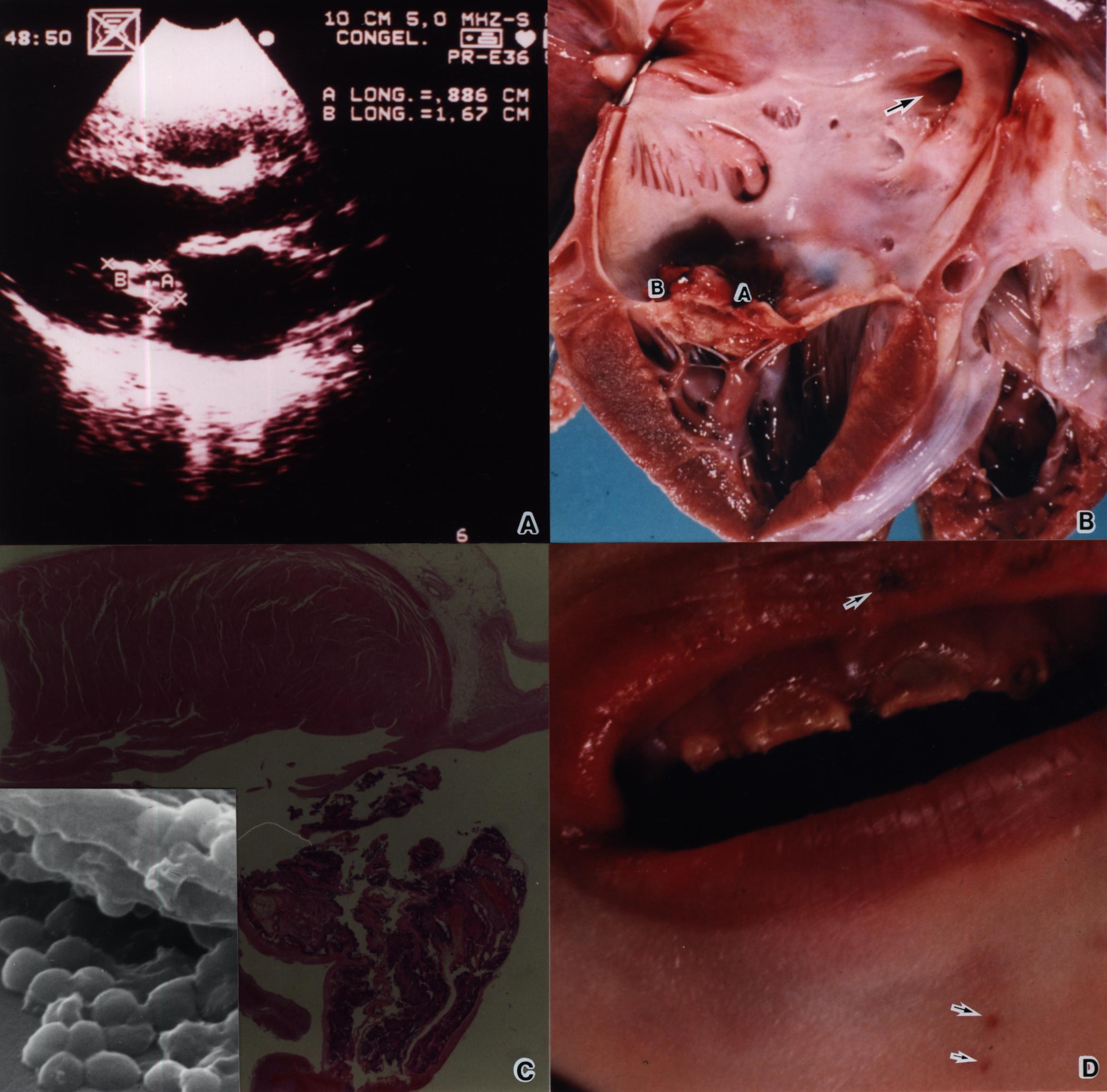
<body><![CDATA[<P><FONT FACE="Arial,Helvetica"><FONT SIZE=-1>El ecocardiograma mostr&oacute; un coraz&oacute;n en "Situs solitus" (normal), levocardia, sin dilataci&oacute;n de las cavidades y sin malformaciones estructurales. En la v&aacute;lvula mitral se observ&oacute; una imagen hiper-ecog&eacute;nica pedinculada, con bordes irregulares semejando una vegetaci&oacute;n, cuyas dimensiones eran de 1 x 2 cm, y estaba adosada a la valva anterior de la mitral; esta lesi&oacute;n prolaps&oacute; tanto hacia atrio como al ventr&iacute;culo izquierdo (<A HREF="#figA,B,C,D">Fig. A</A>). El estudio Doppler demostr&oacute; una insuficiencia mitral moderada. No se observ&oacute; derrame peric&aacute;rdico y la funci&oacute;n sist&oacute;lica del ventr&iacute;culo izquierdo era adecuada.</FONT></FONT>      <P><FONT FACE="Arial,Helvetica"><FONT SIZE=-1>La paciente recibi&oacute; Cefotaxime y de Oxacilina (540mg de cada uno cada 6 horas, v&iacute;a intravenosa). En pocas horas, desarroll&oacute; fallo respiratorio y choque cardiog&eacute;nico, con fibrilaci&oacute;n ventricular y asistolia que no respondi&oacute; a los f&aacute;rmacos y falleci&oacute;.</FONT></FONT>      <P><FONT FACE="Arial,Helvetica"><FONT SIZE=-1>Los hallazgos m&aacute;s relevantes en la autopsia fueron las petequias difusas en ambos pies con lesiones purp&uacute;ricas en plantas, en p&aacute;rpado superior derecho y antebrazo izquierdo; adem&aacute;s, presentaba abundantes caries con compromiso de todas las piezas dentales y gingivitis sangrante (<A HREF="#figA,B,C,D">Fig.D</A>). En la v&aacute;lvula mitral se encontr&oacute; un "trombo mural" o "vegetaci&oacute;n" de 2,5 x 2 x1 cm (<A HREF="#figA,B,C,D">Fig. B</A>), histol&oacute;gicamente mostr&oacute; una gran infiltraci&oacute;n de bacterias cocoides grampositivas (<A HREF="#figA,B,C,D">Fig. C</A>); cuya morfolog&iacute;a se corrobor&oacute; al analizar un corte histol&oacute;gico al microscopio electr&oacute;nico de rastreo (Incerto de la <A HREF="#figA,B,C,D">Fig. C</A>). Como consecuencia de la patolog&iacute;a anterior se encontraron &eacute;mbolos s&eacute;pticos en vasos subcut&aacute;neos de la regi&oacute;n medial del tal&oacute;n derecho, par&eacute;nquima cerebral, ri&ntilde;ones, miocardio, bazo, h&iacute;gado y suprarrenales. Las meninges mostraron opacidad debido a infiltrados de neutr&oacute;filos y macr&oacute;fagos. Se cultiv&oacute; una cepa de <I>Staphylococcus aureus</I> a partir del LCR, sangre y de una punci&oacute;n pulmonar tomadas durante la autopsia. Las causas directas de la muerte fueron los &eacute;mbolos s&eacute;pticos multiorg&aacute;nicos y un choque mixto cardiog&eacute;nico y s&eacute;ptico.</FONT></FONT>     <BR>&nbsp;      <P><B><FONT FACE="Arial,Helvetica"><FONT SIZE=-1>Discusi&oacute;n</FONT></FONT></B>      <P><FONT FACE="Arial,Helvetica"><FONT SIZE=-1>La relaci&oacute;n de endocarditis bacteriana debida a <I>Staphylococcus aureus</I> con la sepsis oral como factor de riesgo es poco frecuente, pues usualmente los agentes involucrados son <I>Streptococcus</I> del grupo <I>viridans</I>, por lo que el tratamiento preventivo en procesos odontol&oacute;gicos usualmente se orienta a esta bacteria; por lo que uno de los antibi&oacute;ticos de elecci&oacute;n es la amoxicilina, el cual no ser&iacute;a el m&aacute;s indicado contra <I>Staphylococcus</I>, como ocurri&oacute; en un caso de endocarditis secundario a un proceso odontol&oacute;gico, en el cual la bacteria incriminada fue <I>Staphylococcus epidermidis</I>, que result&oacute; resistente a la amoxicilina (<A HREF="#6">6</A>). En nuestro caso, la gingivitis pudo representar la puerta de entrada al organismo para la cepa de <I>S. aureus</I>, que se estableci&oacute; en las v&aacute;lvulas cardiacas y se disemin&oacute; a diversos &oacute;rganos. El elemento diagn&oacute;stico m&aacute;s importante fue el ecocardiograma que permiti&oacute; identificar la vegetaci&oacute;n cardiaca, lo cual fue confirmado durante la autopsia. Es importante se&ntilde;alar la importancia de tal herramienta diagn&oacute;stica, lo cual aunado a los an&aacute;lisis de laboratorio, especialmente los hemocultivos confirman el diagn&oacute;stico.</FONT></FONT>      <P><FONT FACE="Arial,Helvetica"><FONT SIZE=-1>Los hallazgos histopatol&oacute;gicos mostraron &eacute;mbolos s&eacute;pticos en diversos &oacute;rganos que condujeron a un choque s&eacute;ptico; una condici&oacute;n que com&uacute;nmente se asocia al lipopolisac&aacute;rido de bacilos gramnegativos. En esta paciente tales hallazgos hacen sospechar la presencia de toxinas estafilococcicas tipo s&uacute;per ant&iacute;geno, capaces de desencadenar un s&iacute;ndrome de choque t&oacute;xico (<A HREF="#25">25</A> -<A HREF="#27">27</A>).</FONT></FONT>      <P><FONT FACE="Arial,Helvetica"><FONT SIZE=-1>La importancia de conocer los factores que predisponen a la endocarditis infecciosa permiten identificar a la poblaci&oacute;n de alto riesgo, promoviendo el diagn&oacute;stico temprano en estos pacientes, lo que a su vez permite el establecimiento de un tratamiento temprano reduciendo las secuelas. En adultos mayores esta patolog&iacute;a se asocia con otros factores de riesgo como la estenosis valvular adquirida, la regurgitaci&oacute;n y las enfermedades degenerativas valvulares, en los cuales la mayor prevalencia se da en el sexo masculino (<A HREF="#5">5</A>, <A HREF="#28">28</A>-<A HREF="#30">30</A>). En todo caso, independientemente de la edad del paciente, el pron&oacute;stico depende de un diagn&oacute;stico oportuno, una selecci&oacute;n apropiada de la terapia antimicrobiana y de la vigilancia adecuada para reconocer cuando el paciente requiere de una intervenci&oacute;n quir&uacute;rgica. Este caso pone de manifiesto como una sepsis oral, asociada a una mala salud e higiene oral puede desencadenar un cuadro fatal, lo que llama imperativamente la atenci&oacute;n hacia este tipo de cuadros, relativamente comunes en nuestro medio.</FONT></FONT>     <CENTER>&nbsp;</CENTER>      <CENTER><A NAME="figA,B,C,D"></A><IMG SRC="/img/fbpe/rccm/v21n3-4/1088i1.JPG" HEIGHT=547 WIDTH=455></CENTER>       
]]></body>
<body><![CDATA[<P><FONT FACE="Arial,Helvetica"><FONT SIZE=-1><B>Figura A</B>: Imagen del ecocardiograma bidimensional en corte longitudinal. Se muestra una estructura con aspecto de "vegetaci&oacute;n" (delimitada por las letras A,B y X), localizada sobre la v&aacute;lvula mitral.</FONT></FONT>      <P><FONT FACE="Arial,Helvetica"><FONT SIZE=-1><B>Figura B</B>. V&aacute;lvula anterior de la Mitral, donde se aprecia la "vegetaci&oacute;n" (letras A y B), proyectada hacia el atrio izquierdo. Se observa el anillo valvular, m&uacute;sculos papilares y las cuerdas tendinosas cuyo aspecto es normal.</FONT></FONT>      <P><FONT FACE="Arial,Helvetica"><FONT SIZE=-1><B>Figura C</B>. Corte histol&oacute;gico te&ntilde;ido con Hematoxilina-Eosina (HE) mostrando acumulaciones (nidos) de bacterias cocoides. El recuadro corresponde a una mayor ampliaci&oacute;n de una acumulaci&oacute;n de bacterias, fotografiada al microscopio electr&oacute;nico de rastreo, a partir de la l&aacute;mina te&ntilde;ida con HE.</FONT></FONT>      <P><FONT FACE="Arial,Helvetica"><FONT SIZE=-1><B>Figura D</B>. Macrofotograf&iacute;a de la enc&iacute;a superior de la paciente, mostrando la franca gingivitis con surcos alveolares llenos de restos alimentarios y 4 piezas dentales con caries evidentes. En ambos labios, barbilla y en la base derecha de la nariz se observan petequias (flechas).</FONT></FONT>     <BR><FONT SIZE=-1>&nbsp;</FONT>      <P><B><FONT FACE="Arial,Helvetica"><FONT SIZE=-1>Referencias</FONT></FONT></B>      <!-- ref --><P><A NAME="1"></A><FONT FACE="Arial,Helvetica"><FONT SIZE=-1>1. Chambers HF Short-course combination and oral therapies of Staphylococcus aureus endocarditis. <I>Infec Dis Clin North Amer</I> 1993; 7: 69.</FONT></FONT>    &nbsp;&nbsp;&nbsp;&nbsp;&nbsp;&nbsp;&nbsp;&nbsp;[&#160;<a href="javascript:void(0);" onclick="javascript: window.open('/scielo.php?script=sci_nlinks&ref=798112&pid=S0253-2948200000020000600001&lng=','','width=640,height=500,resizable=yes,scrollbars=1,menubar=yes,');">Links</a>&#160;]<!-- end-ref --><!-- ref --><P><A NAME="2"></A><FONT FACE="Arial,Helvetica"><FONT SIZE=-1>2. Leung MJ, Nuttull N, Mazur M, Taddeitl, McComish M, Pearman JW Case of Staphylococcus schleiferi Endocarditis and a simple schem to identify clumping factor positive Staphylococci. <I>J Clin Microbiol</I> 1999; 37:3353-3356.</FONT></FONT>    &nbsp;&nbsp;&nbsp;&nbsp;&nbsp;&nbsp;&nbsp;&nbsp;[&#160;<a href="javascript:void(0);" onclick="javascript: window.open('/scielo.php?script=sci_nlinks&ref=798113&pid=S0253-2948200000020000600002&lng=','','width=640,height=500,resizable=yes,scrollbars=1,menubar=yes,');">Links</a>&#160;]<!-- end-ref --><!-- ref --><P><A NAME="3"></A><FONT FACE="Arial,Helvetica"><FONT SIZE=-1>3. Lukes AL, Bright DK, Durak DT Diagnosis of infective endocarditis. <I>Infec Clin North Amer </I>1993; 7:1.</FONT></FONT>    &nbsp;&nbsp;&nbsp;&nbsp;&nbsp;&nbsp;&nbsp;&nbsp;[&#160;<a href="javascript:void(0);" onclick="javascript: window.open('/scielo.php?script=sci_nlinks&ref=798114&pid=S0253-2948200000020000600003&lng=','','width=640,height=500,resizable=yes,scrollbars=1,menubar=yes,');">Links</a>&#160;]<!-- end-ref --><!-- ref --><P><A NAME="4"></A><FONT FACE="Arial,Helvetica"><FONT SIZE=-1>4. Johnson CM. Adherence eventes in the Pathogenesis of infective endocarditis. <I>Infec Dis Clin North Amer.</I> 1993; 7: 21.</FONT></FONT>    &nbsp;&nbsp;&nbsp;&nbsp;&nbsp;&nbsp;&nbsp;&nbsp;[&#160;<a href="javascript:void(0);" onclick="javascript: window.open('/scielo.php?script=sci_nlinks&ref=798115&pid=S0253-2948200000020000600004&lng=','','width=640,height=500,resizable=yes,scrollbars=1,menubar=yes,');">Links</a>&#160;]<!-- end-ref --><!-- ref --><P><A NAME="5"></A><FONT FACE="Arial,Helvetica"><FONT SIZE=-1>5. Steckelberg J.M. and Wilson W.R. Risk factor for infective endocarditis. <I>Infec Clin North Amer </I>1993; 7:9.</FONT></FONT>    &nbsp;&nbsp;&nbsp;&nbsp;&nbsp;&nbsp;&nbsp;&nbsp;[&#160;<a href="javascript:void(0);" onclick="javascript: window.open('/scielo.php?script=sci_nlinks&ref=798116&pid=S0253-2948200000020000600005&lng=','','width=640,height=500,resizable=yes,scrollbars=1,menubar=yes,');">Links</a>&#160;]<!-- end-ref --><!-- ref --><P><A NAME="6"></A><FONT FACE="Arial,Helvetica"><FONT SIZE=-1>6. Pike MG, Warner JO Atopic Dermatitis Complicated by Acute Bacterial Endocarditis. <I>Acta Paediatr Scand</I> 1989; 78: 463-464.</FONT></FONT>    &nbsp;&nbsp;&nbsp;&nbsp;&nbsp;&nbsp;&nbsp;&nbsp;[&#160;<a href="javascript:void(0);" onclick="javascript: window.open('/scielo.php?script=sci_nlinks&ref=798117&pid=S0253-2948200000020000600006&lng=','','width=640,height=500,resizable=yes,scrollbars=1,menubar=yes,');">Links</a>&#160;]<!-- end-ref --><!-- ref --><P><A NAME="7"></A><FONT FACE="Arial,Helvetica"><FONT SIZE=-1>7. Mendelsohn G, Hutchins GM Infective endocarditis during the first decade of life. <I>Am J Dis Child </I>1979; 133: 619.</FONT></FONT>    &nbsp;&nbsp;&nbsp;&nbsp;&nbsp;&nbsp;&nbsp;&nbsp;[&#160;<a href="javascript:void(0);" onclick="javascript: window.open('/scielo.php?script=sci_nlinks&ref=798118&pid=S0253-2948200000020000600007&lng=','','width=640,height=500,resizable=yes,scrollbars=1,menubar=yes,');">Links</a>&#160;]<!-- end-ref --><!-- ref --><P><A NAME="8"></A><FONT FACE="Arial,Helvetica"><FONT SIZE=-1>8. Belik J. Flinn G. Rivera G. Sanyal S. Successeful managemente of bacterial endocarditis of the mitral valve due to Staphylococcus epidemidis en an immunosupressed host. <I>Acta Paediatr Scand</I> 1980; 69:731-734.</FONT></FONT>    &nbsp;&nbsp;&nbsp;&nbsp;&nbsp;&nbsp;&nbsp;&nbsp;[&#160;<a href="javascript:void(0);" onclick="javascript: window.open('/scielo.php?script=sci_nlinks&ref=798119&pid=S0253-2948200000020000600008&lng=','','width=640,height=500,resizable=yes,scrollbars=1,menubar=yes,');">Links</a>&#160;]<!-- end-ref --><!-- ref --><P><A NAME="9"></A><FONT FACE="Arial,Helvetica"><FONT SIZE=-1>9. Etienne J, Fleurette J, Ninet JF, Favet P. Staphylococcal endocarditis after dental extraction<I>. Lancet</I> 1986; ii:511.</FONT></FONT>    &nbsp;&nbsp;&nbsp;&nbsp;&nbsp;&nbsp;&nbsp;&nbsp;[&#160;<a href="javascript:void(0);" onclick="javascript: window.open('/scielo.php?script=sci_nlinks&ref=798120&pid=S0253-2948200000020000600009&lng=','','width=640,height=500,resizable=yes,scrollbars=1,menubar=yes,');">Links</a>&#160;]<!-- end-ref --><!-- ref --><P><A NAME="10"></A><FONT FACE="Arial,Helvetica"><FONT SIZE=-1>10. Abrutyn E. Prevention of bacterial endocarditis. <I>Ann Inter Med</I> 1991; 114:803-804.</FONT></FONT>    &nbsp;&nbsp;&nbsp;&nbsp;&nbsp;&nbsp;&nbsp;&nbsp;[&#160;<a href="javascript:void(0);" onclick="javascript: window.open('/scielo.php?script=sci_nlinks&ref=798121&pid=S0253-2948200000020000600010&lng=','','width=640,height=500,resizable=yes,scrollbars=1,menubar=yes,');">Links</a>&#160;]<!-- end-ref --><!-- ref --><P><A NAME="11"></A><FONT FACE="Arial,Helvetica"><FONT SIZE=-1>11. Blain SM, Trask PA. Dental caries in Hudson TW, Reinhart MA, Rose SD, Stewart GK. Clinical Preventive Medicine: Health promotion and disease prevention. Little Brow and Company, Boston Toronto, USA 1988; pp 326-332.</FONT></FONT>    &nbsp;&nbsp;&nbsp;&nbsp;&nbsp;&nbsp;&nbsp;&nbsp;[&#160;<a href="javascript:void(0);" onclick="javascript: window.open('/scielo.php?script=sci_nlinks&ref=798122&pid=S0253-2948200000020000600011&lng=','','width=640,height=500,resizable=yes,scrollbars=1,menubar=yes,');">Links</a>&#160;]<!-- end-ref --><!-- ref --><P><A NAME="12"></A><FONT FACE="Arial,Helvetica"><FONT SIZE=-1>12. Chaves E, Rojas JA, Rivera P, Hern&aacute;ndez F. Prevalencia de cepas de Staphylococcus productoras de biopel&iacute;cula y con receptores Fc aisladas de muestras cl&iacute;nicas y de individuos sanos. <I>Rev Cost Cienc Med</I> 2000; 21: 51-56.</FONT></FONT>    &nbsp;&nbsp;&nbsp;&nbsp;&nbsp;&nbsp;&nbsp;&nbsp;[&#160;<a href="javascript:void(0);" onclick="javascript: window.open('/scielo.php?script=sci_nlinks&ref=798123&pid=S0253-2948200000020000600012&lng=','','width=640,height=500,resizable=yes,scrollbars=1,menubar=yes,');">Links</a>&#160;]<!-- end-ref --><!-- ref --><P><A NAME="13"></A><FONT FACE="Arial,Helvetica"><FONT SIZE=-1>13. Lockhart PB, Durack RT. Oral microflora as a cause of endocarditis and other distant site Infecctions. <I>Infect Dis Clin North Amer</I> 1999;13:8-33.</FONT></FONT>    &nbsp;&nbsp;&nbsp;&nbsp;&nbsp;&nbsp;&nbsp;&nbsp;[&#160;<a href="javascript:void(0);" onclick="javascript: window.open('/scielo.php?script=sci_nlinks&ref=798124&pid=S0253-2948200000020000600013&lng=','','width=640,height=500,resizable=yes,scrollbars=1,menubar=yes,');">Links</a>&#160;]<!-- end-ref --><!-- ref --><P><A NAME="14"></A><FONT FACE="Arial,Helvetica"><FONT SIZE=-1>14. Baltimore RS. Endocarditis and Intravascular Infections pp 289-298 In: Principles and Practice of pediatric infections diseasses, Long S. S., Pickereing L. K. and Prober C. G. New York. Hurchil Livingstone, 1997.</FONT></FONT>    &nbsp;&nbsp;&nbsp;&nbsp;&nbsp;&nbsp;&nbsp;&nbsp;[&#160;<a href="javascript:void(0);" onclick="javascript: window.open('/scielo.php?script=sci_nlinks&ref=798125&pid=S0253-2948200000020000600014&lng=','','width=640,height=500,resizable=yes,scrollbars=1,menubar=yes,');">Links</a>&#160;]<!-- end-ref --><!-- ref --><P><A NAME="15"></A><FONT FACE="Arial,Helvetica"><FONT SIZE=-1>15. Parras F, Bouza E, Romero J, Quero M, Brito J, Vellibre D. Infectious endocarditis in children. <I>Pediatr Cardiol</I> 1990; 11: 71</FONT></FONT>    &nbsp;&nbsp;&nbsp;&nbsp;&nbsp;&nbsp;&nbsp;&nbsp;[&#160;<a href="javascript:void(0);" onclick="javascript: window.open('/scielo.php?script=sci_nlinks&ref=798126&pid=S0253-2948200000020000600015&lng=','','width=640,height=500,resizable=yes,scrollbars=1,menubar=yes,');">Links</a>&#160;]<!-- end-ref --><!-- ref --><P><A NAME="16"></A><FONT FACE="Arial,Helvetica"><FONT SIZE=-1>16. Saiman L, Prince A, Gersony WM. Pediatric infective endocarditis in the modernera. <I>J Paediatr</I> 1993; 122: 847.</FONT></FONT>    &nbsp;&nbsp;&nbsp;&nbsp;&nbsp;&nbsp;&nbsp;&nbsp;[&#160;<a href="javascript:void(0);" onclick="javascript: window.open('/scielo.php?script=sci_nlinks&ref=798127&pid=S0253-2948200000020000600016&lng=','','width=640,height=500,resizable=yes,scrollbars=1,menubar=yes,');">Links</a>&#160;]<!-- end-ref --><!-- ref --><P><A NAME="17"></A><FONT FACE="Arial,Helvetica"><FONT SIZE=-1>17. Adnan SD, Katharyn AT, Walter Wilson, et all. Prevention of bacterial endocarditis: Recommendation by the American Health Association. <I>Clin Infect Dis</I> 1997;25:1448-58</FONT></FONT>    &nbsp;&nbsp;&nbsp;&nbsp;&nbsp;&nbsp;&nbsp;&nbsp;[&#160;<a href="javascript:void(0);" onclick="javascript: window.open('/scielo.php?script=sci_nlinks&ref=798128&pid=S0253-2948200000020000600017&lng=','','width=640,height=500,resizable=yes,scrollbars=1,menubar=yes,');">Links</a>&#160;]<!-- end-ref --><!-- ref --><P><A NAME="18"></A><FONT FACE="Arial,Helvetica"><FONT SIZE=-1>18. Mortara LA, Bayer AS Staphylococcus aureus bacteremia and endocarditis New diagnostic and therapeutic concepts. <I>Infec Dis Clin North Amer</I> 1993; 7:53.</FONT></FONT>    &nbsp;&nbsp;&nbsp;&nbsp;&nbsp;&nbsp;&nbsp;&nbsp;[&#160;<a href="javascript:void(0);" onclick="javascript: window.open('/scielo.php?script=sci_nlinks&ref=798129&pid=S0253-2948200000020000600018&lng=','','width=640,height=500,resizable=yes,scrollbars=1,menubar=yes,');">Links</a>&#160;]<!-- end-ref --><!-- ref --><P><A NAME="19"></A><FONT FACE="Arial,Helvetica"><FONT SIZE=-1>19. Wilson WA, Infective endocarditis- A changing spectrum? <I>Mayo Clin Proc</I> 1977; 52: 254.</FONT></FONT>    &nbsp;&nbsp;&nbsp;&nbsp;&nbsp;&nbsp;&nbsp;&nbsp;[&#160;<a href="javascript:void(0);" onclick="javascript: window.open('/scielo.php?script=sci_nlinks&ref=798130&pid=S0253-2948200000020000600019&lng=','','width=640,height=500,resizable=yes,scrollbars=1,menubar=yes,');">Links</a>&#160;]<!-- end-ref --><!-- ref --><P><A NAME="20"></A><FONT FACE="Arial,Helvetica"><FONT SIZE=-1>20. Tunkel AR and Kaye D. Endocarditis with negative blood cultures. <I>N Engl J Med</I> 1992; 326:1215.</FONT></FONT>    &nbsp;&nbsp;&nbsp;&nbsp;&nbsp;&nbsp;&nbsp;&nbsp;[&#160;<a href="javascript:void(0);" onclick="javascript: window.open('/scielo.php?script=sci_nlinks&ref=798131&pid=S0253-2948200000020000600020&lng=','','width=640,height=500,resizable=yes,scrollbars=1,menubar=yes,');">Links</a>&#160;]<!-- end-ref --><!-- ref --><P><A NAME="21"></A><FONT FACE="Arial,Helvetica"><FONT SIZE=-1>21. Von Reyn CF, Arbeit RD. Case definitions for Infective Endocarditis. <I>Amer J Med</I> 1994; 96: 220.</FONT></FONT>    &nbsp;&nbsp;&nbsp;&nbsp;&nbsp;&nbsp;&nbsp;&nbsp;[&#160;<a href="javascript:void(0);" onclick="javascript: window.open('/scielo.php?script=sci_nlinks&ref=798132&pid=S0253-2948200000020000600021&lng=','','width=640,height=500,resizable=yes,scrollbars=1,menubar=yes,');">Links</a>&#160;]<!-- end-ref --><!-- ref --><P><A NAME="22"></A><FONT FACE="Arial,Helvetica"><FONT SIZE=-1>22. Dreyfus G, Serraf A, Jebara VA, Delache A, Chauvaud S, Couetil JP, Carpentier A Valve repair in acute endocarditis. <I>Ann Thorac Surg</I> 1990; 49: 706-713.</FONT></FONT>    &nbsp;&nbsp;&nbsp;&nbsp;&nbsp;&nbsp;&nbsp;&nbsp;[&#160;<a href="javascript:void(0);" onclick="javascript: window.open('/scielo.php?script=sci_nlinks&ref=798133&pid=S0253-2948200000020000600022&lng=','','width=640,height=500,resizable=yes,scrollbars=1,menubar=yes,');">Links</a>&#160;]<!-- end-ref --><!-- ref --><P><A NAME="23"></A><FONT FACE="Arial,Helvetica"><FONT SIZE=-1>23. Murphy JP, Foster-Smith K. Management of complications of infective endocarditis with emphasis on echocaardiographic finding. <I>Infec Dis Clin North Amer </I>1993: 7:153-165.</FONT></FONT>    &nbsp;&nbsp;&nbsp;&nbsp;&nbsp;&nbsp;&nbsp;&nbsp;[&#160;<a href="javascript:void(0);" onclick="javascript: window.open('/scielo.php?script=sci_nlinks&ref=798134&pid=S0253-2948200000020000600023&lng=','','width=640,height=500,resizable=yes,scrollbars=1,menubar=yes,');">Links</a>&#160;]<!-- end-ref --><!-- ref --><P><A NAME="24"></A><FONT FACE="Arial,Helvetica"><FONT SIZE=-1>24. Durak DT, Lukes AS, Bright DK. New criteria for diagnosis endocarditis: Utilization of spedific Echocardiographic findings. <I>Amer J Med</I> 1994; 96: 200.</FONT></FONT>    &nbsp;&nbsp;&nbsp;&nbsp;&nbsp;&nbsp;&nbsp;&nbsp;[&#160;<a href="javascript:void(0);" onclick="javascript: window.open('/scielo.php?script=sci_nlinks&ref=798135&pid=S0253-2948200000020000600024&lng=','','width=640,height=500,resizable=yes,scrollbars=1,menubar=yes,');">Links</a>&#160;]<!-- end-ref --><!-- ref --><P><A NAME="25"></A><FONT FACE="Arial,Helvetica"><FONT SIZE=-1>25. Papageorgiou AC, Acharya KR. Microbial superantigens: from structure to function. <I>Trends Microbiol</I> 2000;8:369-375.</FONT></FONT>    &nbsp;&nbsp;&nbsp;&nbsp;&nbsp;&nbsp;&nbsp;&nbsp;[&#160;<a href="javascript:void(0);" onclick="javascript: window.open('/scielo.php?script=sci_nlinks&ref=798136&pid=S0253-2948200000020000600025&lng=','','width=640,height=500,resizable=yes,scrollbars=1,menubar=yes,');">Links</a>&#160;]<!-- end-ref --><!-- ref --><P><A NAME="26"></A><FONT FACE="Arial,Helvetica"><FONT SIZE=-1>26. Krakauer T. Immune response to staphylococcal superantigens. <I>Immunol Res</I> 1999;20:136-173.</FONT></FONT>    &nbsp;&nbsp;&nbsp;&nbsp;&nbsp;&nbsp;&nbsp;&nbsp;[&#160;<a href="javascript:void(0);" onclick="javascript: window.open('/scielo.php?script=sci_nlinks&ref=798137&pid=S0253-2948200000020000600026&lng=','','width=640,height=500,resizable=yes,scrollbars=1,menubar=yes,');">Links</a>&#160;]<!-- end-ref --><!-- ref --><P><A NAME="27"></A><FONT FACE="Arial,Helvetica"><FONT SIZE=-1>27. Schlievert PM, Gahr PJ, Assimacopoulos AP, Dinges MM, Stoehr JA, Harmala JW, Hirt H, Dunny GM. Aggregation and binding substances enhance pathogenicity in rabbit models of Enterococcus faecalis endocarditis. <I>Infect Immun</I> 1998;66:218-223.</FONT></FONT>    &nbsp;&nbsp;&nbsp;&nbsp;&nbsp;&nbsp;&nbsp;&nbsp;[&#160;<a href="javascript:void(0);" onclick="javascript: window.open('/scielo.php?script=sci_nlinks&ref=798138&pid=S0253-2948200000020000600027&lng=','','width=640,height=500,resizable=yes,scrollbars=1,menubar=yes,');">Links</a>&#160;]<!-- end-ref --><!-- ref --><P><A NAME="28"></A><FONT FACE="Arial,Helvetica"><FONT SIZE=-1>28. Vandenesch F, Etienne J, Reverdy ME, Eykyn SJ Endocarditis due to Staphylococcus lugdunesis: Report of 11 cases and review. <I>Clin Infec Dis</I> 1993; 17: 871.</FONT></FONT>    &nbsp;&nbsp;&nbsp;&nbsp;&nbsp;&nbsp;&nbsp;&nbsp;[&#160;<a href="javascript:void(0);" onclick="javascript: window.open('/scielo.php?script=sci_nlinks&ref=798139&pid=S0253-2948200000020000600028&lng=','','width=640,height=500,resizable=yes,scrollbars=1,menubar=yes,');">Links</a>&#160;]<!-- end-ref --><!-- ref --><P><A NAME="29"></A><FONT FACE="Arial,Helvetica"><FONT SIZE=-1>29. Reyes RB, Reyes LP La etiolog&iacute;a de la endocarditis infecciosa en el Instituto Nacionale de Cardiolog&iacute;a "Ignacio Chavez" 1976-1986. <I>Arch Inst Cardio Mex</I> 1989; 59: 287-292.</FONT></FONT>    &nbsp;&nbsp;&nbsp;&nbsp;&nbsp;&nbsp;&nbsp;&nbsp;[&#160;<a href="javascript:void(0);" onclick="javascript: window.open('/scielo.php?script=sci_nlinks&ref=798140&pid=S0253-2948200000020000600029&lng=','','width=640,height=500,resizable=yes,scrollbars=1,menubar=yes,');">Links</a>&#160;]<!-- end-ref --><!-- ref --><P><A NAME="30"></A><FONT FACE="Arial,Helvetica"><FONT SIZE=-1>30. Brook MM. Pediatric Bacterial Endocarditis. <I>Pediat Clin North Amer</I> 1999; 46:275-287.</FONT></FONT>    &nbsp;&nbsp;&nbsp;&nbsp;&nbsp;&nbsp;&nbsp;&nbsp;[&#160;<a href="javascript:void(0);" onclick="javascript: window.open('/scielo.php?script=sci_nlinks&ref=798141&pid=S0253-2948200000020000600030&lng=','','width=640,height=500,resizable=yes,scrollbars=1,menubar=yes,');">Links</a>&#160;]<!-- end-ref --><P><A NAME="1a"></A><FONT FACE="Arial,Helvetica"><SUP><FONT SIZE=-2><A HREF="#1aut">1</A></FONT></SUP><FONT SIZE=-1> Hospital Nacional de Ni&ntilde;os y Universidad de Costa Rica, San Jos&eacute;, Costa Rica. E mail: <A HREF="mailto:secpatologia@hnn.sa.cr">secpatologia@hnn.sa.cr</A></FONT></FONT>      <P><A NAME="2a"></A><FONT FACE="Arial,Helvetica"><SUP><FONT SIZE=-2><A HREF="#2aut">2</A></FONT></SUP><FONT SIZE=-1> Hospital Nacional de Ni&ntilde;os, San Jos&eacute;, Costa Rica.</FONT></FONT>      <P><A NAME="3a"></A><FONT FACE="Arial,Helvetica"><SUP><FONT SIZE=-2><A HREF="#3*au">3</A></FONT></SUP><FONT SIZE=-1> Unidad de Microscopia Electr&oacute;nica y Facultad de Microbiolog&iacute;a,&nbsp; Universidad de Costa Rica, San Jos&eacute;, Costa Rica.</FONT></FONT>     <BR>&nbsp;     ]]></body>
<body><![CDATA[ ]]></body><back>
<ref-list>
<ref id="B1">
<nlm-citation citation-type="journal">
<person-group person-group-type="author">
<name>
<surname><![CDATA[Chambers]]></surname>
<given-names><![CDATA[HF]]></given-names>
</name>
</person-group>
<article-title xml:lang="en"><![CDATA[Short-course combination and oral therapies of Staphylococcus aureus endocarditis]]></article-title>
<source><![CDATA[Infec Dis Clin North Amer]]></source>
<year>1993</year>
<volume>7</volume>
<page-range>69</page-range></nlm-citation>
</ref>
<ref id="B2">
<nlm-citation citation-type="journal">
<person-group person-group-type="author">
<name>
<surname><![CDATA[Leung]]></surname>
<given-names><![CDATA[MJ]]></given-names>
</name>
<name>
<surname><![CDATA[Nuttull]]></surname>
<given-names><![CDATA[N]]></given-names>
</name>
<name>
<surname><![CDATA[Mazur]]></surname>
<given-names><![CDATA[M]]></given-names>
</name>
<name>
<surname><![CDATA[Taddeit]]></surname>
<given-names><![CDATA[l]]></given-names>
</name>
<name>
<surname><![CDATA[McComish]]></surname>
<given-names><![CDATA[M]]></given-names>
</name>
<name>
<surname><![CDATA[Pearman]]></surname>
<given-names><![CDATA[JW]]></given-names>
</name>
</person-group>
<article-title xml:lang="en"><![CDATA[Case of Staphylococcus schleiferi Endocarditis and a simple schem to identify clumping factor positive Staphylococci]]></article-title>
<source><![CDATA[J Clin Microbiol]]></source>
<year>1999</year>
<volume>37</volume>
<page-range>3353-3356</page-range></nlm-citation>
</ref>
<ref id="B3">
<nlm-citation citation-type="journal">
<person-group person-group-type="author">
<name>
<surname><![CDATA[Lukes]]></surname>
<given-names><![CDATA[AL]]></given-names>
</name>
<name>
<surname><![CDATA[Bright]]></surname>
<given-names><![CDATA[DK]]></given-names>
</name>
<name>
<surname><![CDATA[Durak]]></surname>
<given-names><![CDATA[DT]]></given-names>
</name>
</person-group>
<article-title xml:lang="en"><![CDATA[Diagnosis of infective endocarditis]]></article-title>
<source><![CDATA[Infec Clin North Amer]]></source>
<year>1993</year>
<volume>7</volume>
<page-range>1</page-range></nlm-citation>
</ref>
<ref id="B4">
<nlm-citation citation-type="journal">
<person-group person-group-type="author">
<name>
<surname><![CDATA[Johnson]]></surname>
<given-names><![CDATA[CM.]]></given-names>
</name>
</person-group>
<article-title xml:lang="en"><![CDATA[Adherence eventes in the Pathogenesis of infective endocarditis]]></article-title>
<source><![CDATA[Infec Dis Clin North Amer.]]></source>
<year>1993</year>
<volume>7</volume>
<page-range>21</page-range></nlm-citation>
</ref>
<ref id="B5">
<nlm-citation citation-type="journal">
<person-group person-group-type="author">
<name>
<surname><![CDATA[Steckelberg]]></surname>
<given-names><![CDATA[J.M.]]></given-names>
</name>
<name>
<surname><![CDATA[Wilson]]></surname>
<given-names><![CDATA[W.R.]]></given-names>
</name>
</person-group>
<article-title xml:lang="en"><![CDATA[Risk factor for infective endocarditis]]></article-title>
<source><![CDATA[Infec Clin North Amer]]></source>
<year>1993</year>
<volume>7</volume>
<page-range>9</page-range></nlm-citation>
</ref>
<ref id="B6">
<nlm-citation citation-type="journal">
<person-group person-group-type="author">
<name>
<surname><![CDATA[Pike]]></surname>
<given-names><![CDATA[MG]]></given-names>
</name>
<name>
<surname><![CDATA[Warner]]></surname>
<given-names><![CDATA[JO]]></given-names>
</name>
</person-group>
<article-title xml:lang="en"><![CDATA[Atopic Dermatitis Complicated by Acute Bacterial Endocarditis]]></article-title>
<source><![CDATA[Acta Paediatr Scand]]></source>
<year>1989</year>
<volume>78</volume>
<page-range>463-464</page-range></nlm-citation>
</ref>
<ref id="B7">
<nlm-citation citation-type="journal">
<person-group person-group-type="author">
<name>
<surname><![CDATA[Mendelsohn]]></surname>
<given-names><![CDATA[G]]></given-names>
</name>
<name>
<surname><![CDATA[Hutchins]]></surname>
<given-names><![CDATA[GM]]></given-names>
</name>
</person-group>
<article-title xml:lang="en"><![CDATA[Infective endocarditis during the first decade of life]]></article-title>
<source><![CDATA[Am J Dis Child]]></source>
<year>1979</year>
<volume>133</volume>
<page-range>619</page-range></nlm-citation>
</ref>
<ref id="B8">
<nlm-citation citation-type="journal">
<person-group person-group-type="author">
<name>
<surname><![CDATA[Belik]]></surname>
<given-names><![CDATA[J.]]></given-names>
</name>
<name>
<surname><![CDATA[Flinn]]></surname>
<given-names><![CDATA[G]]></given-names>
</name>
<name>
<surname><![CDATA[Rivera]]></surname>
<given-names><![CDATA[G.]]></given-names>
</name>
<name>
<surname><![CDATA[Sanyal]]></surname>
<given-names><![CDATA[S.]]></given-names>
</name>
</person-group>
<article-title xml:lang="en"><![CDATA[Successeful managemente of bacterial endocarditis of the mitral valve due to Staphylococcus epidemidis en an immunosupressed host]]></article-title>
<source><![CDATA[Acta Paediatr Scand]]></source>
<year>1980</year>
<volume>69</volume>
<page-range>731-734</page-range></nlm-citation>
</ref>
<ref id="B9">
<nlm-citation citation-type="journal">
<person-group person-group-type="author">
<name>
<surname><![CDATA[Etienne]]></surname>
<given-names><![CDATA[J]]></given-names>
</name>
<name>
<surname><![CDATA[Fleurette]]></surname>
<given-names><![CDATA[J]]></given-names>
</name>
<name>
<surname><![CDATA[Ninet]]></surname>
<given-names><![CDATA[JF]]></given-names>
</name>
<name>
<surname><![CDATA[Favet]]></surname>
<given-names><![CDATA[P.]]></given-names>
</name>
</person-group>
<article-title xml:lang="en"><![CDATA[Staphylococcal endocarditis after dental extraction]]></article-title>
<source><![CDATA[Lancet]]></source>
<year>1986</year>
<volume>ii</volume>
<page-range>511</page-range></nlm-citation>
</ref>
<ref id="B10">
<nlm-citation citation-type="journal">
<person-group person-group-type="author">
<name>
<surname><![CDATA[Abrutyn]]></surname>
<given-names><![CDATA[E.]]></given-names>
</name>
</person-group>
<article-title xml:lang="en"><![CDATA[Prevention of bacterial endocarditis]]></article-title>
<source><![CDATA[Ann Inter Med]]></source>
<year>1991</year>
<volume>114</volume>
<page-range>803-804</page-range></nlm-citation>
</ref>
<ref id="B11">
<nlm-citation citation-type="book">
<person-group person-group-type="author">
<name>
<surname><![CDATA[Blain]]></surname>
<given-names><![CDATA[SM]]></given-names>
</name>
<name>
<surname><![CDATA[Trask]]></surname>
<given-names><![CDATA[PA]]></given-names>
</name>
</person-group>
<article-title xml:lang="en"><![CDATA[Dental caries]]></article-title>
<person-group person-group-type="editor">
<name>
<surname><![CDATA[Hudson]]></surname>
<given-names><![CDATA[TW]]></given-names>
</name>
<name>
<surname><![CDATA[Reinhart]]></surname>
<given-names><![CDATA[MA]]></given-names>
</name>
<name>
<surname><![CDATA[Rose]]></surname>
<given-names><![CDATA[SD]]></given-names>
</name>
<name>
<surname><![CDATA[Stewart]]></surname>
<given-names><![CDATA[GK]]></given-names>
</name>
</person-group>
<source><![CDATA[Clinical Preventive Medicine: Health promotion and disease prevention]]></source>
<year>1988</year>
<page-range>326-332</page-range><publisher-loc><![CDATA[Boston^eToronto Toronto]]></publisher-loc>
<publisher-name><![CDATA[Little Brow and Company]]></publisher-name>
</nlm-citation>
</ref>
<ref id="B12">
<nlm-citation citation-type="journal">
<person-group person-group-type="author">
<name>
<surname><![CDATA[Chaves]]></surname>
<given-names><![CDATA[E]]></given-names>
</name>
<name>
<surname><![CDATA[Rojas]]></surname>
<given-names><![CDATA[JA]]></given-names>
</name>
<name>
<surname><![CDATA[Rivera]]></surname>
<given-names><![CDATA[P]]></given-names>
</name>
<name>
<surname><![CDATA[Hernández]]></surname>
<given-names><![CDATA[F.]]></given-names>
</name>
</person-group>
<article-title xml:lang="es"><![CDATA[Prevalencia de cepas de Staphylococcus productoras de biopelícula y con receptores Fc aisladas de muestras clínicas y de individuos sanos]]></article-title>
<source><![CDATA[Rev Cost Cienc Med]]></source>
<year>2000</year>
<volume>21</volume>
<page-range>51-56</page-range></nlm-citation>
</ref>
<ref id="B13">
<nlm-citation citation-type="journal">
<person-group person-group-type="author">
<name>
<surname><![CDATA[Lockhart]]></surname>
<given-names><![CDATA[PB]]></given-names>
</name>
<name>
<surname><![CDATA[Durack]]></surname>
<given-names><![CDATA[RT]]></given-names>
</name>
</person-group>
<article-title xml:lang="en"><![CDATA[Oral microflora as a cause of endocarditis and other distant site Infecctions]]></article-title>
<source><![CDATA[Infect Dis Clin North Amer]]></source>
<year>1999</year>
<volume>13</volume>
<page-range>8-33</page-range></nlm-citation>
</ref>
<ref id="B14">
<nlm-citation citation-type="book">
<person-group person-group-type="author">
<name>
<surname><![CDATA[Baltimore]]></surname>
<given-names><![CDATA[RS]]></given-names>
</name>
</person-group>
<article-title xml:lang="en"><![CDATA[Endocarditis and Intravascular Infections]]></article-title>
<person-group person-group-type="editor">
<name>
<surname><![CDATA[Long]]></surname>
<given-names><![CDATA[S. S.]]></given-names>
</name>
<name>
<surname><![CDATA[Pickereing]]></surname>
<given-names><![CDATA[L. K.]]></given-names>
</name>
<name>
<surname><![CDATA[Prober]]></surname>
<given-names><![CDATA[C. G.]]></given-names>
</name>
</person-group>
<source><![CDATA[Principles and Practice of pediatric infections diseasses]]></source>
<year>1997</year>
<page-range>289-298</page-range><publisher-loc><![CDATA[New York ]]></publisher-loc>
<publisher-name><![CDATA[Hurchil Livingstone]]></publisher-name>
</nlm-citation>
</ref>
<ref id="B15">
<nlm-citation citation-type="journal">
<person-group person-group-type="author">
<name>
<surname><![CDATA[Parras]]></surname>
<given-names><![CDATA[F]]></given-names>
</name>
<name>
<surname><![CDATA[Bouza]]></surname>
<given-names><![CDATA[E]]></given-names>
</name>
<name>
<surname><![CDATA[Romero]]></surname>
<given-names><![CDATA[J]]></given-names>
</name>
<name>
<surname><![CDATA[Quero]]></surname>
<given-names><![CDATA[M]]></given-names>
</name>
<name>
<surname><![CDATA[Brito]]></surname>
<given-names><![CDATA[J]]></given-names>
</name>
<name>
<surname><![CDATA[Vellibre]]></surname>
<given-names><![CDATA[D]]></given-names>
</name>
</person-group>
<article-title xml:lang="en"><![CDATA[Infectious endocarditis in children]]></article-title>
<source><![CDATA[Pediatr Cardiol]]></source>
<year>1990</year>
<volume>11</volume><volume>71</volume>
</nlm-citation>
</ref>
<ref id="B16">
<nlm-citation citation-type="journal">
<person-group person-group-type="author">
<name>
<surname><![CDATA[Saiman]]></surname>
<given-names><![CDATA[L]]></given-names>
</name>
<name>
<surname><![CDATA[Prince]]></surname>
<given-names><![CDATA[A]]></given-names>
</name>
<name>
<surname><![CDATA[Gersony]]></surname>
<given-names><![CDATA[WM]]></given-names>
</name>
</person-group>
<article-title xml:lang="en"><![CDATA[Pediatric infective endocarditis in the modernera]]></article-title>
<source><![CDATA[J Paediatr]]></source>
<year>1993</year>
<volume>122</volume>
<page-range>847</page-range></nlm-citation>
</ref>
<ref id="B17">
<nlm-citation citation-type="journal">
<person-group person-group-type="author">
<name>
<surname><![CDATA[Adnan]]></surname>
<given-names><![CDATA[SD]]></given-names>
</name>
<name>
<surname><![CDATA[Katharyn]]></surname>
<given-names><![CDATA[AT]]></given-names>
</name>
<name>
<surname><![CDATA[Wilson]]></surname>
<given-names><![CDATA[Walter]]></given-names>
</name>
</person-group>
<article-title xml:lang="en"><![CDATA[Prevention of bacterial endocarditis: Recommendation by the American Health Association]]></article-title>
<source><![CDATA[Clin Infect Dis]]></source>
<year>1997</year>
<volume>25</volume>
<page-range>1448-58</page-range></nlm-citation>
</ref>
<ref id="B18">
<nlm-citation citation-type="journal">
<person-group person-group-type="author">
<name>
<surname><![CDATA[Mortara]]></surname>
<given-names><![CDATA[LA]]></given-names>
</name>
<name>
<surname><![CDATA[Bayer]]></surname>
<given-names><![CDATA[AS]]></given-names>
</name>
</person-group>
<article-title xml:lang="en"><![CDATA[Staphylococcus aureus bacteremia and endocarditis New diagnostic and therapeutic concepts]]></article-title>
<source><![CDATA[Infec Dis Clin North Amer]]></source>
<year>1993</year>
<volume>7</volume>
<page-range>53</page-range></nlm-citation>
</ref>
<ref id="B19">
<nlm-citation citation-type="journal">
<person-group person-group-type="author">
<name>
<surname><![CDATA[Wilson]]></surname>
<given-names><![CDATA[WA]]></given-names>
</name>
</person-group>
<article-title xml:lang="en"><![CDATA[Infective endocarditis- A changing spectrum?]]></article-title>
<source><![CDATA[Mayo Clin Proc]]></source>
<year>1977</year>
<volume>52</volume>
<page-range>254</page-range></nlm-citation>
</ref>
<ref id="B20">
<nlm-citation citation-type="journal">
<person-group person-group-type="author">
<name>
<surname><![CDATA[Tunkel]]></surname>
<given-names><![CDATA[AR]]></given-names>
</name>
<name>
<surname><![CDATA[Kaye]]></surname>
<given-names><![CDATA[D.]]></given-names>
</name>
</person-group>
<article-title xml:lang="en"><![CDATA[Endocarditis with negative blood cultures]]></article-title>
<source><![CDATA[N Engl J Med]]></source>
<year>1992</year>
<volume>326</volume>
<page-range>1215</page-range></nlm-citation>
</ref>
<ref id="B21">
<nlm-citation citation-type="journal">
<person-group person-group-type="author">
<name>
<surname><![CDATA[Von Reyn]]></surname>
<given-names><![CDATA[CF]]></given-names>
</name>
<name>
<surname><![CDATA[Arbeit]]></surname>
<given-names><![CDATA[RD]]></given-names>
</name>
</person-group>
<article-title xml:lang="en"><![CDATA[Case definitions for Infective Endocarditis]]></article-title>
<source><![CDATA[Amer J Med]]></source>
<year>1994</year>
<volume>96</volume>
<page-range>220</page-range></nlm-citation>
</ref>
<ref id="B22">
<nlm-citation citation-type="journal">
<person-group person-group-type="author">
<name>
<surname><![CDATA[Dreyfus]]></surname>
<given-names><![CDATA[G]]></given-names>
</name>
<name>
<surname><![CDATA[Serraf]]></surname>
<given-names><![CDATA[A]]></given-names>
</name>
<name>
<surname><![CDATA[Jebara]]></surname>
<given-names><![CDATA[VA]]></given-names>
</name>
<name>
<surname><![CDATA[Delache]]></surname>
<given-names><![CDATA[A]]></given-names>
</name>
<name>
<surname><![CDATA[Chauvaud]]></surname>
<given-names><![CDATA[S]]></given-names>
</name>
<name>
<surname><![CDATA[Couetil]]></surname>
<given-names><![CDATA[JP]]></given-names>
</name>
<name>
<surname><![CDATA[Carpentier]]></surname>
<given-names><![CDATA[A]]></given-names>
</name>
</person-group>
<article-title xml:lang="en"><![CDATA[Valve repair in acute endocarditis]]></article-title>
<source><![CDATA[Ann Thorac Surg]]></source>
<year>1990</year>
<volume>49</volume>
<page-range>706-713</page-range></nlm-citation>
</ref>
<ref id="B23">
<nlm-citation citation-type="journal">
<person-group person-group-type="author">
<name>
<surname><![CDATA[Murphy]]></surname>
<given-names><![CDATA[JP]]></given-names>
</name>
<name>
<surname><![CDATA[Foster-Smith]]></surname>
<given-names><![CDATA[K.]]></given-names>
</name>
</person-group>
<article-title xml:lang="en"><![CDATA[Management of complications of infective endocarditis with emphasis on echocaardiographic finding]]></article-title>
<source><![CDATA[Infec Dis Clin North Amer]]></source>
<year>1993</year>
<volume>7</volume>
<page-range>153-165</page-range></nlm-citation>
</ref>
<ref id="B24">
<nlm-citation citation-type="journal">
<person-group person-group-type="author">
<name>
<surname><![CDATA[Durak]]></surname>
<given-names><![CDATA[DT]]></given-names>
</name>
<name>
<surname><![CDATA[Lukes]]></surname>
<given-names><![CDATA[AS]]></given-names>
</name>
<name>
<surname><![CDATA[Bright]]></surname>
<given-names><![CDATA[DK]]></given-names>
</name>
</person-group>
<article-title xml:lang="en"><![CDATA[New criteria for diagnosis endocarditis: Utilization of spedific Echocardiographic findings]]></article-title>
<source><![CDATA[Amer J Med]]></source>
<year>1994</year>
<volume>96</volume>
<page-range>200</page-range></nlm-citation>
</ref>
<ref id="B25">
<nlm-citation citation-type="journal">
<person-group person-group-type="author">
<name>
<surname><![CDATA[Papageorgiou]]></surname>
<given-names><![CDATA[AC]]></given-names>
</name>
<name>
<surname><![CDATA[Acharya]]></surname>
<given-names><![CDATA[KR]]></given-names>
</name>
</person-group>
<article-title xml:lang="en"><![CDATA[Microbial superantigens: from structure to function]]></article-title>
<source><![CDATA[Trends Microbiol]]></source>
<year>2000</year>
<volume>8</volume>
<page-range>369-375</page-range></nlm-citation>
</ref>
<ref id="B26">
<nlm-citation citation-type="journal">
<person-group person-group-type="author">
<name>
<surname><![CDATA[Krakauer]]></surname>
<given-names><![CDATA[T.]]></given-names>
</name>
</person-group>
<article-title xml:lang="en"><![CDATA[Immune response to staphylococcal superantigens]]></article-title>
<source><![CDATA[Immunol Res]]></source>
<year>1999</year>
<volume>20</volume>
<page-range>136-173</page-range></nlm-citation>
</ref>
<ref id="B27">
<nlm-citation citation-type="journal">
<person-group person-group-type="author">
<name>
<surname><![CDATA[Schlievert]]></surname>
<given-names><![CDATA[PM]]></given-names>
</name>
<name>
<surname><![CDATA[Gahr]]></surname>
<given-names><![CDATA[PJ]]></given-names>
</name>
<name>
<surname><![CDATA[Assimacopoulos]]></surname>
<given-names><![CDATA[AP]]></given-names>
</name>
<name>
<surname><![CDATA[Dinges]]></surname>
<given-names><![CDATA[MM]]></given-names>
</name>
<name>
<surname><![CDATA[Stoehr]]></surname>
<given-names><![CDATA[JA]]></given-names>
</name>
<name>
<surname><![CDATA[Harmala]]></surname>
<given-names><![CDATA[JW]]></given-names>
</name>
<name>
<surname><![CDATA[Hirt]]></surname>
<given-names><![CDATA[H]]></given-names>
</name>
<name>
<surname><![CDATA[Dunny]]></surname>
<given-names><![CDATA[GM]]></given-names>
</name>
</person-group>
<article-title xml:lang="en"><![CDATA[Aggregation and binding substances enhance pathogenicity in rabbit models of Enterococcus faecalis endocarditis]]></article-title>
<source><![CDATA[Infect Immun]]></source>
<year>1998</year>
<volume>66</volume>
<page-range>218-223</page-range></nlm-citation>
</ref>
<ref id="B28">
<nlm-citation citation-type="journal">
<person-group person-group-type="author">
<name>
<surname><![CDATA[Vandenesch]]></surname>
<given-names><![CDATA[F]]></given-names>
</name>
<name>
<surname><![CDATA[Etienne]]></surname>
<given-names><![CDATA[J]]></given-names>
</name>
<name>
<surname><![CDATA[Reverdy]]></surname>
<given-names><![CDATA[ME]]></given-names>
</name>
<name>
<surname><![CDATA[Eykyn]]></surname>
<given-names><![CDATA[SJ]]></given-names>
</name>
</person-group>
<article-title xml:lang="en"><![CDATA[Endocarditis due to Staphylococcus lugdunesis: Report of 11 cases and review]]></article-title>
<source><![CDATA[Clin Infec Dis]]></source>
<year>1993</year>
<volume>17</volume>
<page-range>871</page-range></nlm-citation>
</ref>
<ref id="B29">
<nlm-citation citation-type="journal">
<person-group person-group-type="author">
<name>
<surname><![CDATA[Reyes]]></surname>
<given-names><![CDATA[RB]]></given-names>
</name>
<name>
<surname><![CDATA[Reyes]]></surname>
<given-names><![CDATA[LP]]></given-names>
</name>
</person-group>
<article-title xml:lang="es"><![CDATA[La etiología de la endocarditis infecciosa en el Instituto Nacionale de Cardiología "Ignacio Chavez" 1976-1986]]></article-title>
<source><![CDATA[Arch Inst Cardio Mex]]></source>
<year>1989</year>
<volume>59</volume>
<page-range>287-292</page-range></nlm-citation>
</ref>
<ref id="B30">
<nlm-citation citation-type="journal">
<person-group person-group-type="author">
<name>
<surname><![CDATA[Brook]]></surname>
<given-names><![CDATA[MM.]]></given-names>
</name>
</person-group>
<article-title xml:lang="en"><![CDATA[Pediatric Bacterial Endocarditis]]></article-title>
<source><![CDATA[Pediat Clin North Amer]]></source>
<year>1999</year>
<volume>46</volume>
<page-range>275-287</page-range></nlm-citation>
</ref>
</ref-list>
</back>
</article>
