<?xml version="1.0" encoding="ISO-8859-1"?><article xmlns:mml="http://www.w3.org/1998/Math/MathML" xmlns:xlink="http://www.w3.org/1999/xlink" xmlns:xsi="http://www.w3.org/2001/XMLSchema-instance">
<front>
<journal-meta>
<journal-id>0253-2948</journal-id>
<journal-title><![CDATA[Revista Costarricense de Ciencias Médicas]]></journal-title>
<abbrev-journal-title><![CDATA[Rev. costarric. cienc. méd]]></abbrev-journal-title>
<issn>0253-2948</issn>
<publisher>
<publisher-name><![CDATA[Editorial Nacional de Salud y Seguridad Social]]></publisher-name>
</publisher>
</journal-meta>
<article-meta>
<article-id>S0253-29481999000200002</article-id>
<title-group>
<article-title xml:lang="es"><![CDATA[Medición del aumento en volumen celular inducido por alfa hemolisina de E. Coli (HL y A) en leucocitos polimorfonucleares neutrófilos: efecto del calcio extracelular]]></article-title>
</title-group>
<contrib-group>
<contrib contrib-type="author">
<name>
<surname><![CDATA[García]]></surname>
<given-names><![CDATA[Jorge]]></given-names>
</name>
<xref ref-type="aff" rid="A01"/>
</contrib>
</contrib-group>
<aff id="A01">
<institution><![CDATA[,Universidad de Costa Rica Departamento de Bioquímica ]]></institution>
<addr-line><![CDATA[ ]]></addr-line>
</aff>
<pub-date pub-type="pub">
<day>00</day>
<month>12</month>
<year>1999</year>
</pub-date>
<pub-date pub-type="epub">
<day>00</day>
<month>12</month>
<year>1999</year>
</pub-date>
<volume>20</volume>
<numero>3-4</numero>
<fpage>141</fpage>
<lpage>152</lpage>
<copyright-statement/>
<copyright-year/>
<self-uri xlink:href="http://www.scielo.sa.cr/scielo.php?script=sci_arttext&amp;pid=S0253-29481999000200002&amp;lng=en&amp;nrm=iso"></self-uri><self-uri xlink:href="http://www.scielo.sa.cr/scielo.php?script=sci_abstract&amp;pid=S0253-29481999000200002&amp;lng=en&amp;nrm=iso"></self-uri><self-uri xlink:href="http://www.scielo.sa.cr/scielo.php?script=sci_pdf&amp;pid=S0253-29481999000200002&amp;lng=en&amp;nrm=iso"></self-uri><abstract abstract-type="short" xml:lang="es"><p><![CDATA[Leucocitos polimorfonucleares neutrófilos (PMN) sufrieron aumento en su volumen celular medio (VCM) cuando se expusieron a sobrenadantes hemolíticos como fuente de alfa hemolisina (HlyA) de Escherichia coli, una citotoxina que se inserta en la membrana celular y forma poros hidrofílicos, que permiten la redistribución de cationes a través de la membrana y ulterior movimiento de agua hacia el interior de la célula. En este estudio se cuantificó el incremento en VCM causado por Hly A, el cual fue proporcional a la dosis hemolítica, y se estableció que al no adicionar calcio a la suspensión de PMN, el incremento en VCM fue más pronunciado, lo que indica un efecto protector del calcio extracelular ante el efecto de Hly A. Tanto el ionóforo de calcio A23187 como Hly A disminuyeron la granularidad citoplásmica de PMN, lo que sugiere que el flujo de calcio hacia el citosol a través de los poros formados por Hly A puede alterar de manera importante la fisiología de PMN, probablemente interferir con la función fagocitaria y amplificar la respuesta inflamatoria a la bacteria hemolítica.]]></p></abstract>
<abstract abstract-type="short" xml:lang="en"><p><![CDATA[Mean cell volume (MCV ) of polymorphonuclear neutrophils exposed to E. coli hemolytic supernatants increased in a dose dependent way. E. coli alpha hemolysin (Hly A) is a membrane active cytotoxin which forms hydrophilic, cation selective pores upon insertion. Cation movement through hemolisin pores result in net water movement into the target cell. lncreases in MCV were more pronounced when no calcium was added to the buffer on the PMN suspension, indicating calcium exerts a protective effect against Hly A induced damage. Both the calcium ionophore A23187 and Hly A decreased cytoplasmic granularity of PMN, suggesting calcium influx through Hly A pores may interfere with PMN function in vivo and could amplify the inflammatory response to hemolytic E. coli.]]></p></abstract>
<kwd-group>
<kwd lng="la"><![CDATA[Escherichia coli]]></kwd>
<kwd lng="es"><![CDATA[alfa hemolisina]]></kwd>
<kwd lng="es"><![CDATA[leucocitos polimortonucleares neutrófilos]]></kwd>
<kwd lng="la"><![CDATA[Escherichia coli]]></kwd>
<kwd lng="en"><![CDATA[alpha hemolysin]]></kwd>
<kwd lng="en"><![CDATA[Polymorphonuclear neutrophils]]></kwd>
</kwd-group>
</article-meta>
</front><body><![CDATA[ <CENTER><B><FONT FACE="Arial,Helvetica">Medici&oacute;n del aumento en volumen celular inducido por alfa hemolisina</FONT></B></CENTER>      <CENTER><B><FONT FACE="Arial,Helvetica">de <I>e. coli</I> (HLY A) en leucocitos polimorfonucleares neutr&oacute;filos:</FONT></B></CENTER>      <CENTER><B><FONT FACE="Arial,Helvetica">efecto del calcio extracelular</FONT></B></CENTER> &nbsp;     <BR>&nbsp;     <CENTER><B><FONT FACE="Arial,Helvetica"><FONT SIZE=-1>Jorge Garc&iacute;a&nbsp;<A NAME="*a"></A><A HREF="#*">*</A></FONT></FONT></B></CENTER>      <CENTER>&nbsp;</CENTER>       <P><B><FONT FACE="Arial,Helvetica"><FONT SIZE=-1>Resumen</FONT></FONT></B><B><FONT FACE="Arial,Helvetica"><FONT SIZE=-1></FONT></FONT></B>      <P><FONT FACE="Arial,Helvetica"><FONT SIZE=-1>Leucocitos polimorfonucleares neutr&oacute;filos (PMN) sufrieron aumento en su volumen celular medio (VCM) cuando se expusieron a sobrenadantes hemol&iacute;ticos como fuente de alfa hemolisina (HlyA) de <I>Escherichia coli</I>, una citotoxina que se inserta en la membrana celular y forma poros hidrof&iacute;licos, que permiten la redistribuci&oacute;n de cationes a trav&eacute;s de la membrana y ulterior movimiento de agua hacia el interior de la c&eacute;lula.&nbsp; En este estudio se cuantific&oacute; el incremento en VCM causado por Hly A, el cual fue proporcional a la dosis hemol&iacute;tica, y se estableci&oacute; que al no adicionar calcio a la suspensi&oacute;n de PMN, el incremento en VCM fue m&aacute;s pronunciado, lo que indica un efecto protector del calcio extracelular ante el efecto de Hly A. Tanto el ion&oacute;foro de calcio A23187 como Hly A disminuyeron la granularidad citopl&aacute;smica de PMN, lo que sugiere que el flujo de calcio hacia el citosol a trav&eacute;s de los poros formados por Hly A puede alterar de manera importante la fisiolog&iacute;a de PMN, probablemente interferir con la funci&oacute;n fagocitaria y amplificar la respuesta inflamatoria a la bacteria hemol&iacute;tica.</FONT></FONT><FONT SIZE=-1></FONT>      <P><B><FONT FACE="Arial,Helvetica"><FONT SIZE=-1>Palabras clave</FONT></FONT></B><I><FONT FACE="Arial,Helvetica"><FONT SIZE=-1></FONT></FONT></I>      <P><FONT SIZE=-1><FONT FACE="Arial,Helvetica"><I>Escherichia coli</I>, alfa hemolisina, leucocitos polimortonucleares neutr&oacute;filos.</FONT></FONT>     ]]></body>
<body><![CDATA[<BR><FONT SIZE=-1>&nbsp;</FONT>     <BR><B><FONT FACE="Arial,Helvetica"><FONT SIZE=-1>Abstract</FONT></FONT></B><FONT SIZE=-1></FONT>      <P><FONT SIZE=-1><FONT FACE="Arial,Helvetica">Mean cell volume (MCV ) of polymorphonuclear neutrophils exposed to <I>E. coli</I> hemolytic supernatants increased in a dose dependent way.&nbsp;<I> E. coli</I> alpha hemolysin (Hly A) is a membrane active cytotoxin which forms hydrophilic, cation selective pores upon insertion.&nbsp; Cation movement through hemolisin pores result in net water movement into the target cell. lncreases in MCV were more pronounced when no calcium was added to the buffer on the PMN suspension, indicating calcium exerts a protective effect against Hly A induced damage.&nbsp; Both the calcium ionophore A23187 and Hly A decreased cytoplasmic granularity of PMN, suggesting calcium influx through Hly A pores may interfere with PMN function in vivo and could amplify the inflammatory response to hemolytic <I>E. coli.</I></FONT></FONT><FONT SIZE=-1></FONT>      <P><B><FONT FACE="Arial,Helvetica"><FONT SIZE=-1>Key Words</FONT></FONT></B><I><FONT FACE="Arial,Helvetica"><FONT SIZE=-1></FONT></FONT></I>      <P><FONT SIZE=-1><FONT FACE="Arial,Helvetica"><I>Escherichia coli</I>, alpha hemolysin, Polymorphonuclear neutrophils.</FONT></FONT>     <BR><FONT SIZE=-1>&nbsp;</FONT>     <BR><FONT SIZE=-1>&nbsp;</FONT>     <BR><B><FONT FACE="Arial,Helvetica"><FONT SIZE=-1>Introducci&oacute;n</FONT></FONT></B><FONT SIZE=-1></FONT>      <P><FONT SIZE=-1><FONT FACE="Arial,Helvetica"><I>Escherichia coli</I> es una bacteria oportunista que causa infecciones tanto en tracto intestinal como en otros tejidos (<A HREF="#1"> 1 </A>). Entre los factores de virulencia que expresa se encuentra la hemolisina alfa, o Hly A (<A HREF="#2">2</A>), prote&iacute;na que la bacteria sintetiza como pro-toxina y que activa por acilaci&oacute;n antes de secretaria al medio externo como lipoprote&iacute;na, y cuya actividad depende de la presencia de iones calcio en soluci&oacute;n (<A HREF="#3">3</A>-<A HREF="#5">5</A>).&nbsp; El papel de Hly A como factor de virulencia se ha demostrado mediante estudios que indican que las cepas de <I>Escherichia coli</I> a las cuales se transforma con los genes codificantes de HlyA se tornan virulentas en modelos animales (<A HREF="#6">6</A>), y que la eliminaci&oacute;n de dichos genes reduce dr&aacute;sticamente su virulencia (<A HREF="#4">4</A>).</FONT></FONT><FONT SIZE=-1></FONT>      <P><FONT SIZE=-1><FONT FACE="Arial,Helvetica">HlyA es una citotoxina que ejerce su acci&oacute;n en varios tipos de c&eacute;lulas, incluyendo eritrocitos (<A HREF="#7">7</A>), fibroblastos (<A HREF="#8">8</A>), leucocitos polimorfonucleares neutr&oacute;filos (<A HREF="#9">9</A>, <A HREF="#10">10</A>), macr&oacute;fagos (<A HREF="#11">11</A>), monocitos ( <A HREF="#19">19</A>,<A HREF="#12">12</A> ) y linfocitos (<A HREF="#11">11</A>, <A HREF="#12">12</A> ). La asociaci&oacute;n y posterior inserci&oacute;n de Hly A en membranas de c&eacute;lulas blanco (<A HREF="#13">13</A>) causa la formaci&oacute;n de poros (<A HREF="#14">14</A>, <A HREF="#15">15</A>), lo cual explica la actividad citot&oacute;xica.&nbsp; Los poros formados en la membrana se comportan como canales hidrof&iacute;licos con selectividad can&oacute;nica (<A HREF="#11">11</A>).&nbsp; Aunque no son visibles al microscopio electr&oacute;nico (16), permiten el paso de cationes (<A HREF="#17">17</A>), ATP (<A HREF="#18">18</A>) y probablemente otras mol&eacute;culas.&nbsp; El da&ntilde;o causado a la membrana eventualmente conduce a muerte celular.&nbsp; La evidencia obtenida por Bhakdi et al (<A HREF="#18">18</A>) muestra que Hly A es una leucocidina muy potente, de acci&oacute;n irreversible, que causa la degranulaci&oacute;n de Polimorfonucleares neutr&oacute;filos, y seg&uacute;n datos obtenidos por medio de citometr&iacute;a de flujo, hace que las c&eacute;lulas aumenten su volumen.</FONT></FONT><FONT SIZE=-1></FONT>      ]]></body>
<body><![CDATA[<P><FONT SIZE=-1><FONT FACE="Arial,Helvetica">Este estudio pretende cuantificar el aumento de volumen celular promedio en leucocitos polimorfonucleares neutr&oacute;filos expuestos a Hly A y estudiar la posible influencia de los iones calcio del medio extracelular en dicho efecto.&nbsp; Adem&aacute;s, se complement&oacute; con mediciones utilizando citometr&iacute;a de flujo para corroborar el aumento en volumen celular, y tratar de definir una posible participaci&oacute;n del calcio externo en la degranulaci&oacute;n causada por HlyA.</FONT></FONT>     <BR><FONT SIZE=-1>&nbsp;</FONT>     <BR><FONT SIZE=-1>&nbsp;</FONT>     <BR><B><FONT FACE="Arial,Helvetica"><FONT SIZE=-1>Materiales y m&eacute;todos</FONT></FONT></B>     <CENTER><FONT SIZE=-1>&nbsp;</FONT></CENTER>      <CENTER><FONT SIZE=-1>&nbsp;</FONT></CENTER> <FONT SIZE=-1><B><FONT FACE="Arial,Helvetica">Cepas de <I>Escherichia coli</I></FONT></B></FONT>     <BR><FONT SIZE=-1>&nbsp;</FONT>     <BR><FONT SIZE=-1><FONT FACE="Arial,Helvetica">Las cepas usadas en esta investigaci&oacute;n se obtuvieron del laboratorio del Dr. R.A. Welch (U. of Wisconsin, Madison, Wl) y son todas derivadas de la <I>Escherichia coli</I> DH 1.&nbsp; La cepa hemol&iacute;tica WAM 589 se transform&oacute; con el pl&aacute;smido pWAM04, (pl&aacute;smido pUC 19 en el cual se subclonaron los genes de hemolisina, contenidos en el operon hly CABD proveniente del pl&aacute;smido pSF4000).&nbsp; La cepa control WAM 675 no es hemol&iacute;tica y fue transformada con el pl&aacute;smido pUC 19 desprovisto de genes de hemolisina.&nbsp; La cepa control WAM 783 es la DH1 transformada con un derivado del pl&aacute;smido pWAMO4, el cual porta una deleci&oacute;n en el gen hlyC.&nbsp; Este gen es responsable de la acilaci&oacute;n de la pro HlyA (<A HREF="#20">20</A>).&nbsp; En consecuencia, WAM 783 secreta una hemolisina no acilada, incapaz de insertarse en membranas de la c&eacute;lula blanco.</FONT></FONT>     <BR><FONT SIZE=-1>&nbsp;&nbsp;</FONT>     <BR><FONT SIZE=-1>&nbsp;</FONT>     ]]></body>
<body><![CDATA[<BR><FONT SIZE=-1><B><FONT FACE="Arial,Helvetica">Preparaci&oacute;n de sobrenadantes de cultivos bacterianos</FONT></B></FONT><FONT SIZE=-1></FONT>      <P><FONT SIZE=-1><FONT FACE="Arial,Helvetica">Se usaron sobrenadantes crudos de cultivos de la cepa WAM 589 como fuente de hemolisina y sobrenadantes crudos de las cepas WAM 675 y 783 como controles, a diluciones elevadas con respecto a los vol&uacute;menes de reacci&oacute;n (m&aacute;s de 1: 500).</FONT></FONT><FONT SIZE=-1></FONT>      <P><FONT SIZE=-1><FONT FACE="Arial,Helvetica">Las cepas de <I>Escherichia coli</I> se cultivaron en caldo Luria-Bertani (LB), con ampicilina (100 ug/mi) para seleccionar en cultivo las cepas transformadas WAM 589 y 675, y cloramfenicol (20 &micro;g/mi) para seleccionar la cepa WAM 783.&nbsp; Vol&uacute;menes de 100 ml de caldo LB se inocularon con 1-3 ml de un cultivo estacionario de la cepa correspondiente (incubado a 37&deg; C durante 12 horas), luego de lo cual se incubaron a 37&deg; C con agitaci&oacute;n (200 rpm) en un incubador rotatorio G25 ( New Brunswick, Edison, NJ).&nbsp; Se tomaron muestras de 10-15 ml de los cultivos cuando &eacute;stos alcanzaron una densidad &oacute;ptica (DO ) de 0,3-0,4&nbsp;<IMG SRC="/img/fbpe/rccm/v20n3-4/symbol1.GIF" HEIGHT=25 WIDTH=13 ALIGN=ABSCENTER>(600 nm) y las bacterias se sedimentaron por centrifugaci&oacute;n (7 min, 800xg) en una centr&iacute;fuga Sorvall SS1 (Dupont lnstruments, Wilmington, DE).&nbsp; El sobrenadante se esteriliz&oacute; por filtraci&oacute;n a trav&eacute;s de una membrana est&eacute;ril con porosidad de 0,4 um (Millipore), y se mantuvo en hielo previo a su utilizaci&oacute;n.</FONT></FONT>     
<BR><FONT SIZE=-1>&nbsp;</FONT>     <BR><FONT SIZE=-1>&nbsp;</FONT>     <BR><FONT SIZE=-1><B><FONT FACE="Arial,Helvetica">Determinaci&oacute;n de actividad hemol&iacute;tica</FONT></B></FONT><FONT SIZE=-1></FONT>      <P><FONT SIZE=-1><FONT FACE="Arial,Helvetica">A cada ml de diluciones dobles seriadas de sobrenadante bacteriano crudo y est&eacute;ril en soluci&oacute;n salina 0,85% con calcio (10 mM CaCl<SUB>2</SUB>) se a&ntilde;adi&oacute; 1 ml de suspensi&oacute;n al 1% de eritrocitos de carnero suspendidos soluci&oacute;n salina. Se incubaron a 37&deg; C durante 1 h. Al finalizar la incubaci&oacute;n, se a&ntilde;adi&oacute; a cada diluci&oacute;n 2 ml de soluci&oacute;n salina fr&iacute;a para detener la reacci&oacute;n; se centrifug&oacute; a 700xg por 5 min en una centr&iacute;fuga Sorvali GLC-2B (Dupont lnstruments, Wilmington, DE) y se ley&oacute; la DO del fluido sobrenadante a&nbsp;<IMG SRC="/img/fbpe/rccm/v20n3-4/symbol1.GIF" HEIGHT=25 WIDTH=13 ALIGN=ABSCENTER> 545 nm para estimar la liberaci&oacute;n de hemoglobina.&nbsp; Se determin&oacute; la diluci&oacute;n de sobrenadante que caus&oacute; 50% de lisis eritrocitaria mediante interpolaci&oacute;n en papel semilogar&iacute;tmico en el cual se hab&iacute;a trazado una curva patr&oacute;n de hem&oacute;lisis (hipot&oacute;nica, en agua destilada) obtenida independientemente con el mismo lote de eritrocitos.&nbsp; El t&iacute;tulo hemol&iacute;tico (HU50/ml) se defini&oacute; como el inverso de la diluci&oacute;n de sobrenadante de cultivo bacteriano que caus&oacute; 50% de hem&oacute;lisis.&nbsp; Dado que los ensayos con c&eacute;lulas se efectuaron en buffer HC (1 mM Ca Cl<SUB>2</SUB> y 0,1% gelatina, pH 7,4), se hicieron titulaciones de sobrenadantes hemol&iacute;ticos en el mismo buffer, para descartar variaciones en el t&iacute;tulo hemol&iacute;tico debidas al cambio de soluci&oacute;n.&nbsp; El t&iacute;tulo hemol&iacute;tico no vari&oacute; con respecto al obtenido en soluci&oacute;n salina con calcio.</FONT></FONT>     
<BR><FONT SIZE=-1>&nbsp;</FONT>     <BR><FONT SIZE=-1>&nbsp;</FONT>     <BR><FONT SIZE=-1><B><FONT FACE="Arial,Helvetica">Obtenci&oacute;n de leucocitos polimorfonucleares neutr&oacute;filos (PMN)</FONT></B></FONT>     ]]></body>
<body><![CDATA[<BR><FONT SIZE=-1>&nbsp;</FONT>     <BR><FONT SIZE=-1><FONT FACE="Arial,Helvetica">Se us&oacute; sangre venosa heparinizada obtenida de voluntarios sanos, la cual se coloc&oacute; en tubos de ensayo sobre medio Mono Poly (ICN-Flow) y se centrifug&oacute; de acuerdo con instrucciones del fabricante.&nbsp; Se recogi&oacute; la banda de PMN y &eacute;stos se lavaron usando 20 ml de buffer HEPES sin calcio (HNC: HEPES 10 mM, NaCl 144 mM, KCI 5 mM, Glucosa 5,5 mM, gelatina para cultivo de tejidos 0,5% p/v, pH 7,4).&nbsp; Los PMN se resuspendieron luego en NH<SUB>4</SUB>CI (0,83% en HEPES 10 mM, pH 7,4) durante 7 minutos, para lisar eritrocitos contaminantes.&nbsp; Los PMN se lavaron una vez m&aacute;s con HNC y se resuspendieron luego en 1 mi de buffer (0,8 mi de HNC + 0,2 ml de buffer HEPES con calcio, o HC.&nbsp; El conteo de c&eacute;lulas en la suspensi&oacute;n se efectu&oacute; usando una c&aacute;mara cuenta gl&oacute;bulos (Hemacytometer).</FONT></FONT>     <BR><FONT SIZE=-1>&nbsp;</FONT>     <BR><FONT SIZE=-1>&nbsp;</FONT>     <BR><FONT SIZE=-1><B><FONT FACE="Arial,Helvetica">Medici&oacute;n de volumen celular medio (VCM) y citometr&iacute;a de flujo</FONT></B></FONT><FONT SIZE=-1></FONT>      <P><FONT SIZE=-1><FONT FACE="Arial,Helvetica">Se agregaron vol&uacute;menes de 2-4 ml de buffer HC est&eacute;ril a viales de vidrio siliconizado para uso en contadores de centelleo.&nbsp; Se a&ntilde;adi&oacute; luego el volumen de suspensi&oacute;n celular necesario para obtener 1 x 10<SUP>6</SUP> c&eacute;lulas/ml.&nbsp; El experimento se inici&oacute; adicionando el volumen de sobrenadante crudo est&eacute;ril, sus controles, o reactivos seg&uacute;n se indica, a las suspensiones celulares.&nbsp; Los viales se mantuvieron a 370 C, con rotaci&oacute;n a 100-150 rpm (G76 Gyrotory Water Bath Shaker, New Brunswick, Edison, NJ).&nbsp; A intervalos definidos, se recogieron al&iacute;quotas de 200 ul , las cuales se diluyeron en soluci&oacute;n lsoton (Beckton-Dickinson), de acuerdo con instrucciones del fabricante, para conteo total y medici&oacute;n de VCM en un contador Coulter, modelo ZBI, con un canalizador 256 (Coulter lnstruments, Hialeah, FL), conectado a una computadora IBM con programa Accu 256.
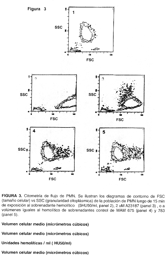
</FONT></FONT><FONT SIZE=-1></FONT>      <P><FONT SIZE=-1><FONT FACE="Arial,Helvetica">Para citometr&iacute;a de flujo, se tomaron al&iacute;quotas de 500 ul de los viales y se analizaron en un cit&oacute;metro FACSCAN (Beckton-Dickinson), con excitaci&oacute;n laser de X 488 nm, graficando el contorno de la poblaci&oacute;n de PMN con respecto a "Forward Scatter'y "Side Scatter".</FONT></FONT>     <BR><FONT SIZE=-1>&nbsp;</FONT>     <BR><FONT SIZE=-1>&nbsp;</FONT>     <BR><B><FONT FACE="Arial,Helvetica"><FONT SIZE=-1>Resultados</FONT></FONT></B><FONT SIZE=-1></FONT>      ]]></body>
<body><![CDATA[<P><FONT SIZE=-1><FONT FACE="Arial,Helvetica">El VCM de PMN expuestos a sobrenadantes hemol&iacute;ticos aument&oacute; en relaci&oacute;n directa a la dosis empleada, alcanzando un m&aacute;ximo de m&aacute;s de 200% por sobre el valor basal en aquellos expuestos a 5 HU50/ml o valores superiores (<A HREF="#fig1">Fig. 1</A>). El efecto de la hemolisina se detect&oacute; a diluciones extremadamente altas de sobrenadante hemol&iacute;tico.&nbsp; Por ejemplo, &eacute;ste debi&oacute; diluirse en razones del orden de 1: 20 000 para dar 0,1 HU50/ml, dosis</FONT> <FONT FACE="Arial,Helvetica">ante la cual los PMN aumentaron su VCM de manera leve pero significativa.&nbsp; En presencia de sobrenadantes de las cepas control WAM 675 y 783 los PMN no aumentaron su VCM con respecto al valor basal.&nbsp; Para estudiar el efecto del calcio extracelular en el VCM (<A HREF="#fig2">Fig. 2</A>), al&iacute;cuotas de PMN suspendidos en HNC (sin calcio agregado) se expusieron a 2 HU50/ml; en este caso, se adicion&oacute; previamente CACI<SUB>2</SUB> (5 mM) al sobrenadante bacteriano para asegurar la activaci&oacute;n de la hemolisina.&nbsp; Dada la diluci&oacute;n tan elevada del sobrenadante en la suspensi&oacute;n de PMN, la contribuci&oacute;n del calcio agregado al sobrenadante al volumen de reacci&oacute;n fue despreciable.&nbsp; Para estudiar el efecto de la transiocaci&oacute;n selectiva de calcio hacia el interior de la c&eacute;lula en el VCM, los PMN se expusieron tambi&eacute;n a 1,2 uM del ion&oacute;foro de calcio A23187 estando suspendidos en HC, o en HNC, para controlar con &eacute;ste &uacute;ltimo el efecto de A23187 sin calcio externo.&nbsp; Los PMN resuspendidos en HNC y expuestos a 2 HU50/mi mostraron un aumento muy pronunciado en VCM, mucho mayor que el de los expuestos a la misma dosis hemol&iacute;tica en presencia de HC. Los PMN expuestos al ion&oacute;foro A23187 aumentaron leve pero significativamente su VCM con respecto al valor basal s&oacute;lo en presencia de calcio en soluci&oacute;n (suspendidos en HC).&nbsp; Los resultados de la citometr&iacute;a de flujo se presentan en la Fig. 3. El panel 1 ilustra la posici&oacute;n del contorno de los PMN usados como control.</FONT></FONT><FONT SIZE=-1></FONT>      <P><FONT SIZE=-1><FONT FACE="Arial,Helvetica">Aquellos expuestos a 5 HU 50/ml (panel 2) mostraron aumento en tama&ntilde;o celular, evidenciado por el aumento en FSC (Forward Scatter), as&iacute; como disminuci&oacute;n en granularidad citopl&aacute;smica, seg&uacute;n se desprende de la disminuci&oacute;n en SSC (Side Scatter); ambos efectos se notaron tambi&eacute;n en PMN tratados con 2 uM de ion&oacute;foro A23187 (panel 3).&nbsp; Sin embargo, y en concordancia con los hallazgos en las determinaciones de MCV, el tama&ntilde;o de los PMN expuestos a 2uM A23187 no aument&oacute; tanto como en el caso de los expuestos a 5 HU/mi.&nbsp; Los PMN expuestos a sobrenadantes de la cepa WAM 675 (panel 4) o WAM 783 (panel 5) no mostraron cambios importantes en FSC o SSC, excepto por un porcentaje peque&ntilde;o de c&eacute;lulas (5-10%) que aumentan aparentemente en tama&ntilde;o, debido a cambios en forma celular inducidos por el LPS presente en el sobrenadante de cultivo bacteriano.</FONT></FONT>     <BR><FONT SIZE=-1>&nbsp;</FONT>     <BR><FONT SIZE=-1>&nbsp;</FONT>     <BR><B><FONT FACE="Arial,Helvetica"><FONT SIZE=-1>Discusi&oacute;n</FONT></FONT></B><FONT SIZE=-1></FONT>      <P><FONT SIZE=-1><FONT FACE="Arial,Helvetica">Los sobrenadantes de cultivos bacterianos son una mezcla compleja de componentes del medio de cultivo y de productos bacterianos.&nbsp; Su uso como fuente de hemolisina se justifica en esta investigaci&oacute;n en vista de la complejidad estructural de la alfa hemolisina, la cual no ha podido ser totalmente purificada, ya que forma complejos con el lipopolisac&aacute;rido ( LPS ) presente en los mismos medios de cultivo (<A HREF="#21">21</A>).&nbsp; Sobrenadantes de las cepas isog&eacute;nicas WAM 675 y 783 se usaron como controles para asegurar que los efectos observados son atribuibles &uacute;nicamente a Hly A. Se efectuaron tambi&eacute;n experimentos de inactivaci&oacute;n t&eacute;rmica y enzim&aacute;tica, sometiendo los sobrenadantes hemol&iacute;ticos en &eacute;ste &uacute;ltimo caso a la acci&oacute;n de lipasa, proteinasa K y tripsina.&nbsp; Ambos tratamientos, t&eacute;rmico y enzim&aacute;tico, eliminaron por completo la actividad hemol&iacute;tica de los sobrenadantes.&nbsp; Adem&aacute;s, la actividad hemol&iacute;tica se bloque&oacute; por completo cuando se les incub&oacute; previamente con D12, un anticuerpo monocional anti HlyA, pero no disminuy&oacute; cuando se les incub&oacute; con el anticuerpo monocional D1, que posee especificidad anti LPS.&nbsp; La obtenci&oacute;n de los anticuerpos D1 y D12 ha sido reportada previamente (<A HREF="#21">21</A>).&nbsp; Todo lo anterior demuestra de manera concluyente que el principio activo en el sobrenadante de la cepa WAM 589 es Hly A, y valida su uso en este estudio.</FONT></FONT><FONT SIZE=-1></FONT>      <P><FONT SIZE=-1><FONT FACE="Arial,Helvetica">
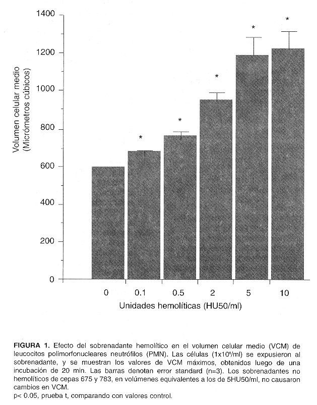
Los PMN aumentaron su MCV en respuesta al sobrenadante hemol&iacute;tico, a&uacute;n a diluciones altas, sin verse afectados por los sobrenadantes control.&nbsp; Ello es congruente con el mecanismo de acci&oacute;n propuesto para la alfa hemolisina, a saber, la formaci&oacute;n de poros en membrana (<A HREF="#11">11</A>,<A HREF="#15">15</A>).&nbsp; La aparici&oacute;n de tales lesiones permite la redistribuci&oacute;n de cationes a trav&eacute;s de la membrana de acuerdo a su gradiente de concentraci&oacute;n, en particular de sodio, potasio y calcio (<A HREF="#22">22</A>, <A HREF="#23">23</A>), y hace que la c&eacute;lula afectada entre en equilibrio de Donnan.&nbsp; La presencia de prote&iacute;nas intracelulares como aniones no difusibles causa entonces un incremento en la concentraci&oacute;n de cationes intracelulares, que conduce a movimiento de agua hacia el interior de la c&eacute;lula, y explica el incremento en VCM.&nbsp; Observaciones al microscopio de luz, no documentadas en el presente trabajo, mostraron que las c&eacute;lulas afectadas aumentan su volumen, pierden su forma, su adherencia al sustrato y eventualmente pierden citoplasma, el cual se proyecta como abultamientos por sobre la membrana celular y se expulsa al exterior.
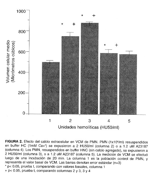
</FONT></FONT><FONT SIZE=-1></FONT>      <P><FONT SIZE=-1><FONT FACE="Arial,Helvetica">Los PMN se expusieron tambi&eacute;n a la hemolisina suspendidos en buffer sin calcio con el fin de explorar el posible efecto del mismo en el VCM.&nbsp; La disminuci&oacute;n en calcio externo acent&uacute;a el aumento en VCM inducido por Hly A, en concordancia con los resultados de Menestrina et al: concentraciones fisiol&oacute;gicas de calcio aumentan la probabilidad de que el poro formado por la hemolisina se encuentre moment&aacute;neamente cerrado, mientras que concentraciones muy bajas lo mantienen abierto con m&aacute;s frecuencia, y la redistribuci&oacute;n i&oacute;nica a trav&eacute;s de la membrana se acelera como resultado (<A HREF="#14">14</A>).&nbsp; Otros autores (<A HREF="#24">24</A>) apuntan que tanto el calcio como el zinc contribuyen a la protecci&oacute;n de membranas celulares sometidas a la acci&oacute;n de agentes hemol&iacute;ticos.&nbsp; Tal efecto podr&iacute;a jugar incluso un efecto protector en infecciones.&nbsp; PMN expuestos a ion&oacute;foro de calcio aumentan levemente su volumen, ya que el aumento en calcio intracelular induce abultamientos en la membrana de las c&eacute;lulas, relacionados probablemente con alteraciones en el citoesqueleto, pero es patente que el efecto del incremento en calcio no influencia el VCM como lo hace la hemolisina (<A HREF="#fig2">Fig. 2</A>).</FONT></FONT><FONT SIZE=-1></FONT>      <P><FONT SIZE=-1><FONT FACE="Arial,Helvetica">Los resultados de la citometr&iacute;a de flujo concuerdan con las mediciones de VCM, ya que el incremento de FSC en PMN expuestos a hemolisina indica aumento en tama&ntilde;o celular.&nbsp; El ion&oacute;foro de calcio A23187 produjo una transiocaci&oacute;n de calcio al interior de la c&eacute;lula, y caus&oacute; en consecuencia una disminuci&oacute;n en la granularidad citopl&aacute;smica (degranulaci&oacute;n) de los PMN que es igual a la inducida por la Hly A. Esta degranulaci&oacute;n, por lo tanto, es debida al aumento en el calcio citopi&aacute;smico inducido por ambos agentes ( <A HREF="#fig3">Fig. 3</A>).&nbsp; De hecho, experimentos no reportados en este trabajo comprobaron que Hly A causa incrementos en calcio intracelular, y que, en efecto, Hly A induce liberaci&oacute;n al medio externo de enzimas contenidas en los gr&aacute;nulos de PMN, de acuerdo con los resultados de Bhakdi et al (<A HREF="#18">18</A>).</FONT></FONT><FONT SIZE=-1></FONT>      <P><FONT SIZE=-1><FONT FACE="Arial,Helvetica">Estos hallazgos indican que Hly A puede alterar la fisiolog&iacute;a de la c&eacute;lula blanco debido, entre otras causas, al incremento en el calcio intracelular posterior la aparici&oacute;n de poros en la membrana.&nbsp; Como la concentraci&oacute;n fisiol&oacute;gica de calcio extracelular oscila en rango milimolar, y la concentraci&oacute;n intracelular en rango micromolar o nanomolar, la apertura de poros no fisiol&oacute;gicos permite el paso de calcio al citoplasma.&nbsp; El aumento no fisiol&oacute;gico en el calcio citopl&aacute;smico puede activar fosfolipasas y quinasas, trastornando seriamente el metabolismo y funci&oacute;n celular.&nbsp; Las observaciones al microscopio de luz que mostraron p&eacute;rdida de forma y adhesi&oacute;n de PMN al sustrato son consistentes con una posible desorganizaci&oacute;n del citoesqueleto, cuya funci&oacute;n depende de la concentraci&oacute;n intracelular de calcio.</FONT></FONT><FONT SIZE=-1></FONT>      ]]></body>
<body><![CDATA[<P><FONT SIZE=-1><FONT FACE="Arial,Helvetica">Esta investigaci&oacute;n ampl&iacute;a reportes previos a&ntilde;adiendo la cuantificaci&oacute;n del efecto de Hly A en el VCM de PMN; la evidencia obtenida incluye a HlyA dentro del grupo de agentes cuyo efecto es modulado por el calcio extracelular, y postula que el incremento causado por HlyA en el calcio intracelular contribuye a las alteraciones en la fisiolog&iacute;a celular inducidas por la hemolisina.&nbsp; En PMN, tales alteraciones, podr&iacute;an explicar los defectos en quimiotaxis y fagocitosis reportados por Cavalieri y Snyder (<A HREF="#25">25</A>) y amplificar&iacute;an la respuesta inflamatoria a las bacterias hemol&iacute;ticas y el da&ntilde;o tisular al causar liberaci&oacute;n de contenidos granulares de PMN.</FONT></FONT>     <BR><FONT SIZE=-1>&nbsp;</FONT>     <BR><FONT SIZE=-1>&nbsp;</FONT>     <BR><B><FONT FACE="Arial,Helvetica"><FONT SIZE=-1>Referencias</FONT></FONT></B>     <BR><FONT SIZE=-1>&nbsp;</FONT>     <!-- ref --><BR><A NAME="1"></A><FONT SIZE=-1><FONT FACE="Arial,Helvetica">1) Eisenstein, B.I., Jones, G.W. The spectrum of infectious and pathogenic mechanisms of<I> Escher&iacute;chia coli Adv lnt Med </I>1988;33:231-252.</FONT></FONT><FONT SIZE=-1></FONT>    &nbsp;&nbsp;&nbsp;&nbsp;&nbsp;&nbsp;&nbsp;&nbsp;[&#160;<a href="javascript:void(0);" onclick="javascript: window.open('/scielo.php?script=sci_nlinks&ref=771436&pid=S0253-2948199900020000200001&lng=','','width=640,height=500,resizable=yes,scrollbars=1,menubar=yes,');">Links</a>&#160;]<!-- end-ref --><!-- ref --><P><A NAME="2"></A><FONT SIZE=-1><FONT FACE="Arial,Helvetica">2) Cavalieri, S.J., Bohach, G.A., Snyder, l.S. <I>Escherichia coli</I> alpha hemolysin: characteristics and probable role in pathogenicity.&nbsp;<I> Microbiol Rev </I>1984; 48: 326-343.</FONT></FONT><FONT SIZE=-1></FONT>    &nbsp;&nbsp;&nbsp;&nbsp;&nbsp;&nbsp;&nbsp;&nbsp;[&#160;<a href="javascript:void(0);" onclick="javascript: window.open('/scielo.php?script=sci_nlinks&ref=771437&pid=S0253-2948199900020000200002&lng=','','width=640,height=500,resizable=yes,scrollbars=1,menubar=yes,');">Links</a>&#160;]<!-- end-ref --><!-- ref --><P><A NAME="3"></A><FONT SIZE=-1><FONT FACE="Arial,Helvetica">3) Snyder, l.S., Zwadyk, P. Some factors affecting production and assay of<I> Escherichia coli</I> hemolysins.&nbsp; J Gen Microbiol 1969; 55: 139-143.</FONT></FONT><FONT SIZE=-1></FONT>    &nbsp;&nbsp;&nbsp;&nbsp;&nbsp;&nbsp;&nbsp;&nbsp;[&#160;<a href="javascript:void(0);" onclick="javascript: window.open('/scielo.php?script=sci_nlinks&ref=771438&pid=S0253-2948199900020000200003&lng=','','width=640,height=500,resizable=yes,scrollbars=1,menubar=yes,');">Links</a>&#160;]<!-- end-ref --><!-- ref --><P><A NAME="4"></A><FONT SIZE=-1><FONT FACE="Arial,Helvetica">4) Chakraborty, T, Kathariou, S. Hacker, J., Hof, H., Huhle, B., Wagner, W., Kuhn, M., Goebel, W.&nbsp; Molecular analysis of bacterial cytolisins.&nbsp; <I>Rev Infec Dis</I> 1987; 9 (S5):S456-S466.</FONT></FONT>    &nbsp;&nbsp;&nbsp;&nbsp;&nbsp;&nbsp;&nbsp;&nbsp;[&#160;<a href="javascript:void(0);" onclick="javascript: window.open('/scielo.php?script=sci_nlinks&ref=771439&pid=S0253-2948199900020000200004&lng=','','width=640,height=500,resizable=yes,scrollbars=1,menubar=yes,');">Links</a>&#160;]<!-- end-ref --><BR><FONT SIZE=-1>&nbsp;</FONT>     ]]></body>
<body><![CDATA[<BR><A NAME="5"></A><FONT SIZE=-1><FONT FACE="Arial,Helvetica">5) lssartel, J.P., Koronakis, V., Hughes, C. Activation of <I>Escherichia coli prohaemolysin</I> to the mature toxin by acyl carrier protein-dependent fatty acid acyiation.&nbsp; Nature 1991;</FONT></FONT>     <BR><FONT SIZE=-1><FONT FACE="Arial,Helvetica">351: 759-761.</FONT></FONT>     <BR><FONT SIZE=-1>&nbsp;</FONT>     <!-- ref --><BR><A NAME="6"></A><FONT SIZE=-1><FONT FACE="Arial,Helvetica">6) Welch, R.A., Dellinger, E.P., Minshew, B., Falkow, S. Haemolysin contributes to viruience of extra intestinal<I> E. coli</I> infections.&nbsp; Nature 1981; 294: 665- 667.</FONT></FONT>    &nbsp;&nbsp;&nbsp;&nbsp;&nbsp;&nbsp;&nbsp;&nbsp;[&#160;<a href="javascript:void(0);" onclick="javascript: window.open('/scielo.php?script=sci_nlinks&ref=771444&pid=S0253-2948199900020000200006&lng=','','width=640,height=500,resizable=yes,scrollbars=1,menubar=yes,');">Links</a>&#160;]<!-- end-ref --><BR><FONT SIZE=-1>&nbsp;</FONT>     <!-- ref --><BR><A NAME="7"></A><FONT SIZE=-1><FONT FACE="Arial,Helvetica">7) Snyder, l.S., Koch, N.A. Production and characteristics of</FONT> <FONT FACE="Arial,Helvetica">hemolysins of<I> Escherichia coli</I>.</FONT> <FONT FACE="Arial,Helvetica"><I>J Bacteriol</I> 1966; 91: 763-767.</FONT></FONT>    &nbsp;&nbsp;&nbsp;&nbsp;&nbsp;&nbsp;&nbsp;&nbsp;[&#160;<a href="javascript:void(0);" onclick="javascript: window.open('/scielo.php?script=sci_nlinks&ref=771446&pid=S0253-2948199900020000200007&lng=','','width=640,height=500,resizable=yes,scrollbars=1,menubar=yes,');">Links</a>&#160;]<!-- end-ref --><BR><FONT SIZE=-1>&nbsp;</FONT>     <!-- ref --><BR><A NAME="8"></A><FONT SIZE=-1><FONT FACE="Arial,Helvetica">8) Cavalieri, S.J., Snyder, l.S. Cytotoxic activity of partially purified <I>Escherichia coli</I> alpha <I>hemolysin J Med Microbiol</I> 1982; 15: 11-21.</FONT></FONT>    &nbsp;&nbsp;&nbsp;&nbsp;&nbsp;&nbsp;&nbsp;&nbsp;[&#160;<a href="javascript:void(0);" onclick="javascript: window.open('/scielo.php?script=sci_nlinks&ref=771448&pid=S0253-2948199900020000200008&lng=','','width=640,height=500,resizable=yes,scrollbars=1,menubar=yes,');">Links</a>&#160;]<!-- end-ref --><BR><FONT SIZE=-1>&nbsp;</FONT>     <BR><A NAME="9"></A><FONT SIZE=-1><FONT FACE="Arial,Helvetica">9) Cavalieri, S.J., Snyder, l.S. Effect of Escherichia coll aipha hemolysin on. human peripheral leukocyte viability in vitro.&nbsp; <I>Infect Immun</I>. 1982; 36:455-461.</FONT></FONT>     ]]></body>
<body><![CDATA[<BR><FONT SIZE=-1>&nbsp;</FONT>     <BR><A NAME="10"></A><FONT SIZE=-1><FONT FACE="Arial,Helvetica">10) Gadeberg, O., Orskov, I. In vitro cytotoxic effect of aipha hemolytic Escherichia coll on human blood granulocytes. <I>Infect Immun</I> 1984; 45: 255-</FONT> <FONT FACE="Arial,Helvetica">260.</FONT></FONT>     <BR><FONT SIZE=-1>&nbsp;</FONT>     <BR><A NAME="11"></A><FONT SIZE=-1><FONT FACE="Arial,Helvetica">11) Menestrina, G., Dalla Serra, M., Pederzoili, C., Bregante, M., Gambale, F. Bacterial hemolysins and leukotoxins affect target cells by forming large exogenous pores into their plasma membrane.&nbsp;<I> Escherichia coli</I> hemolysin A as a case example.&nbsp; Bioscience Reports 1995; 15: 543-551.</FONT></FONT>     <BR><FONT SIZE=-1>&nbsp;</FONT>     <!-- ref --><BR><A NAME="12"></A><FONT SIZE=-1><FONT FACE="Arial,Helvetica">12) Garc&iacute;a, J.D. Efecto citot&oacute;xico de la hemolisina de<I> Escherichia coli</I> (parcialmente purificada) en linfocitos y monocitos in vitro.&nbsp; Rev Cost Cienc Med 1984; 5: 170-174.</FONT></FONT>    &nbsp;&nbsp;&nbsp;&nbsp;&nbsp;&nbsp;&nbsp;&nbsp;[&#160;<a href="javascript:void(0);" onclick="javascript: window.open('/scielo.php?script=sci_nlinks&ref=771456&pid=S0253-2948199900020000200012&lng=','','width=640,height=500,resizable=yes,scrollbars=1,menubar=yes,');">Links</a>&#160;]<!-- end-ref --><BR><FONT SIZE=-1>&nbsp;</FONT>     <!-- ref --><BR><A NAME="13"></A><FONT SIZE=-1><FONT FACE="Arial,Helvetica">13) Bauer, M.E., Weich, R.A. Association of RTX toxins with erythrocytes.<I> lnfect lmmun </I>1996; 64: 4665-4672.</FONT></FONT><FONT SIZE=-1></FONT>    &nbsp;&nbsp;&nbsp;&nbsp;&nbsp;&nbsp;&nbsp;&nbsp;[&#160;<a href="javascript:void(0);" onclick="javascript: window.open('/scielo.php?script=sci_nlinks&ref=771458&pid=S0253-2948199900020000200013&lng=','','width=640,height=500,resizable=yes,scrollbars=1,menubar=yes,');">Links</a>&#160;]<!-- end-ref --><!-- ref --><P><A NAME="14"></A><FONT SIZE=-1><FONT FACE="Arial,Helvetica">14) Menestrina, G., Bashford, C.L., Pasternak, C.A. Pore-forming</FONT> <FONT FACE="Arial,Helvetica">toxins: experiments with S. aureus alpha-toxin, <I>C. perfringens</I> theta-toxin and <I>E. coli</I> hemolysin in lipid bilayers, liposomes and intact cells. <I>Toxicon</I> 1990; 28: 477-491.</FONT></FONT>    &nbsp;&nbsp;&nbsp;&nbsp;&nbsp;&nbsp;&nbsp;&nbsp;[&#160;<a href="javascript:void(0);" onclick="javascript: window.open('/scielo.php?script=sci_nlinks&ref=771459&pid=S0253-2948199900020000200014&lng=','','width=640,height=500,resizable=yes,scrollbars=1,menubar=yes,');">Links</a>&#160;]<!-- end-ref --><BR><FONT SIZE=-1>&nbsp;</FONT>     ]]></body>
<body><![CDATA[<!-- ref --><BR><A NAME="15"></A><FONT SIZE=-1><FONT FACE="Arial,Helvetica">15) Bhakdi, S., Mackman, N., Nicaud, J.M., Holland, l.B<I>. Escherichia coli</I> hemolysin may damage target cell membranas by generating transmembrane pores. lnfect Immun 1986; 52: 63-69.</FONT></FONT>    &nbsp;&nbsp;&nbsp;&nbsp;&nbsp;&nbsp;&nbsp;&nbsp;[&#160;<a href="javascript:void(0);" onclick="javascript: window.open('/scielo.php?script=sci_nlinks&ref=771461&pid=S0253-2948199900020000200015&lng=','','width=640,height=500,resizable=yes,scrollbars=1,menubar=yes,');">Links</a>&#160;]<!-- end-ref --><BR><FONT SIZE=-1>&nbsp;</FONT>     <BR><A NAME="16"></A><FONT SIZE=-1><FONT FACE="Arial,Helvetica">16) Bhakdi, S., Tranum-Jensen, J. Damage to mammalian celis by proteins that form transmembrane pores.&nbsp; <I>Rev Physiol Biochem Pharmacol </I>1987; 107:147-223.</FONT></FONT>     <BR><FONT SIZE=-1>&nbsp;</FONT>     <BR><A NAME="17"></A><FONT SIZE=-1><FONT FACE="Arial,Helvetica">17) Jorgensen, S.E., Hammer, R.F, Wu, G.F. Effects of a single hit from the aipha hemolysin produced by Escherich&iacute;a col&iacute; on the morphology of sheep erythrocytes. <I>lnfect lmmun</I> 1980;</FONT> <FONT FACE="Arial,Helvetica">27: 988-994.</FONT></FONT>     <BR><FONT SIZE=-1>&nbsp;</FONT>     <BR><A NAME="18"></A><FONT SIZE=-1><FONT FACE="Arial,Helvetica">18) Bhakdi, S., Greulich, S., MuHly, M., Eberspacher, B., Becker, H., Thiele, A., Hugo, F. Potent lescocidal action of Escherich&iacute;a coll hemolysin mediated by permeabilization of target cell membranas<I>.&nbsp; J Exp Med </I>1989;</FONT></FONT>     <BR><FONT SIZE=-1><FONT FACE="Arial,Helvetica">169: 737-754.</FONT></FONT>     <BR><FONT SIZE=-1>&nbsp;</FONT>     <!-- ref --><BR><A NAME="19"></A><FONT SIZE=-1><FONT FACE="Arial,Helvetica">19) Bhakdi, S., Muhly, M., Korom, S., Schmidt, G. Effects of <I>Escherichia coli </I>hemolysin on human monocytes.&nbsp; <I>J Clin lnvest</I> 1990; 85: 1746-1753.</FONT></FONT>    &nbsp;&nbsp;&nbsp;&nbsp;&nbsp;&nbsp;&nbsp;&nbsp;[&#160;<a href="javascript:void(0);" onclick="javascript: window.open('/scielo.php?script=sci_nlinks&ref=771470&pid=S0253-2948199900020000200019&lng=','','width=640,height=500,resizable=yes,scrollbars=1,menubar=yes,');">Links</a>&#160;]<!-- end-ref --><BR><FONT SIZE=-1>&nbsp;</FONT>     <!-- ref --><BR><A NAME="20"></A><FONT SIZE=-1><FONT FACE="Arial,Helvetica">20) Hardie, K.R., lssartel, J.P., Koronakis, E., Hughes, C., Koronakis, V. In vitro activation of <I>Escherichia coli </I>prohaemolysin to the mature membrane-targeted toxin requires HlyC and a low-molecularweight cytosolic poiypeptide. <I>Molecular M&iacute;crobiology</I> 1991;</FONT> <FONT FACE="Arial,Helvetica">5: 1669-1679.</FONT></FONT>    &nbsp;&nbsp;&nbsp;&nbsp;&nbsp;&nbsp;&nbsp;&nbsp;[&#160;<a href="javascript:void(0);" onclick="javascript: window.open('/scielo.php?script=sci_nlinks&ref=771472&pid=S0253-2948199900020000200020&lng=','','width=640,height=500,resizable=yes,scrollbars=1,menubar=yes,');">Links</a>&#160;]<!-- end-ref --><BR><FONT SIZE=-1>&nbsp;</FONT>     <!-- ref --><BR><A NAME="21"></A><FONT SIZE=-1><FONT FACE="Arial,Helvetica">21) Bohach, G.A., Snyder, l.S. Chemical and immunologicalanalysis of the complex structure of <I>Escherichia coli </I>alpha hemolysin.&nbsp; J Bacteriol 1985;</FONT> <FONT FACE="Arial,Helvetica">164: 1071-1080.</FONT></FONT>    &nbsp;&nbsp;&nbsp;&nbsp;&nbsp;&nbsp;&nbsp;&nbsp;[&#160;<a href="javascript:void(0);" onclick="javascript: window.open('/scielo.php?script=sci_nlinks&ref=771474&pid=S0253-2948199900020000200021&lng=','','width=640,height=500,resizable=yes,scrollbars=1,menubar=yes,');">Links</a>&#160;]<!-- end-ref --><BR><FONT SIZE=-1>&nbsp;</FONT>     <BR><A NAME="22"></A><FONT SIZE=-1><FONT FACE="Arial,Helvetica">22) Jorgensen, S.E., Mulcahy, P.F., Louis, C.F. Effect of Escherich&iacute;a col&iacute; hemolysin on permeability of erythrocyte membranes to calcium.&nbsp;<I> Toxicon</I> 1986;</FONT> <FONT FACE="Arial,Helvetica">24: 559-566.</FONT></FONT>     <BR><FONT SIZE=-1>&nbsp;</FONT>     <BR><A NAME="23"></A><FONT SIZE=-1><FONT FACE="Arial,Helvetica">23) Jorgensen, S.E., Mulcahy, P.F., Wu, G.K., Louis, C.F. Calcium accumulation in human and sheep erythrocytes that is induced by <I>Escherichia coli</I> hemolysin.&nbsp; Tox&iacute;con 1983; 21:717-727.</FONT></FONT>     <BR><FONT SIZE=-1>&nbsp;</FONT>     <!-- ref --><BR><A NAME="24"></A><FONT SIZE=-1><FONT FACE="Arial,Helvetica">24) Pasternak, C.A. A novel role of Ca+<SUP>2</SUP> and Zn +<SUP>2</SUP>: protection of cells against membrane damage.&nbsp; <I>Bioscience Reports</I> 1988; 6: 579-583.</FONT></FONT>    &nbsp;&nbsp;&nbsp;&nbsp;&nbsp;&nbsp;&nbsp;&nbsp;[&#160;<a href="javascript:void(0);" onclick="javascript: window.open('/scielo.php?script=sci_nlinks&ref=771480&pid=S0253-2948199900020000200024&lng=','','width=640,height=500,resizable=yes,scrollbars=1,menubar=yes,');">Links</a>&#160;]<!-- end-ref --><BR><FONT SIZE=-1>&nbsp;</FONT>     <!-- ref --><BR><A NAME="25"></A><FONT SIZE=-1><FONT FACE="Arial,Helvetica">25) Cavalieri, S.J., Snyder, l.S. Effect of<I> Escherichia coli </I>alpha hemolysin on human peripheral leukocyte function in vitro. Infect lmmun 1982; 37: 966974.</FONT></FONT>    &nbsp;&nbsp;&nbsp;&nbsp;&nbsp;&nbsp;&nbsp;&nbsp;[&#160;<a href="javascript:void(0);" onclick="javascript: window.open('/scielo.php?script=sci_nlinks&ref=771482&pid=S0253-2948199900020000200025&lng=','','width=640,height=500,resizable=yes,scrollbars=1,menubar=yes,');">Links</a>&#160;]<!-- end-ref --><BR><FONT SIZE=-1>&nbsp;</FONT>     <BR><FONT SIZE=-1>&nbsp;</FONT>     <BR><FONT SIZE=-1>&nbsp;</FONT>     <BR>&nbsp;<A NAME="*"></A><A HREF="#*a">*</A> <FONT FACE="Arial,Helvetica"><FONT SIZE=-1>Departamento de Bioqu&iacute;mica, Facultad de Medicina, Universidad de Costa Rica.</FONT></FONT>     <BR><FONT SIZE=-1>&nbsp;</FONT>     <BR><FONT SIZE=-1>&nbsp;</FONT>     <BR><FONT SIZE=-1>&nbsp;</FONT>     <CENTER><A NAME="fig1"></A><IMG SRC="/img/fbpe/rccm/v20n3-4/0665i1.GIF" HEIGHT=802 WIDTH=629></CENTER>      
]]></body>
<body><![CDATA[<CENTER><FONT SIZE=-1>&nbsp;</FONT></CENTER>      <CENTER><FONT SIZE=-1>&nbsp;</FONT></CENTER>      <CENTER><FONT SIZE=-1>&nbsp;</FONT></CENTER>      <CENTER><FONT SIZE=-1>&nbsp;</FONT></CENTER>      <CENTER><FONT SIZE=-1>&nbsp;</FONT></CENTER>      <CENTER><FONT SIZE=-1>&nbsp;</FONT></CENTER>      <CENTER><A NAME="fig2"></A><IMG SRC="/img/fbpe/rccm/v20n3-4/0665i2.GIF" HEIGHT=701 WIDTH=597></CENTER>      
<CENTER><FONT SIZE=-1>&nbsp;</FONT></CENTER>      <CENTER><FONT SIZE=-1>&nbsp;</FONT></CENTER>      <CENTER><FONT SIZE=-1>&nbsp;</FONT></CENTER>      ]]></body>
<body><![CDATA[<CENTER><FONT SIZE=-1>&nbsp;</FONT></CENTER>      <CENTER><FONT SIZE=-1>&nbsp;</FONT></CENTER>      <CENTER><FONT SIZE=-1>&nbsp;</FONT></CENTER>      <CENTER><A NAME="fig3"></A><IMG SRC="/img/fbpe/rccm/v20n3-4/0665i3.GIF" HEIGHT=890 WIDTH=582></CENTER>      
<CENTER><FONT SIZE=-1>&nbsp;</FONT></CENTER>       ]]></body><back>
<ref-list>
<ref id="B1">
<nlm-citation citation-type="journal">
<person-group person-group-type="author">
<name>
<surname><![CDATA[Eisenstein]]></surname>
<given-names><![CDATA[B.I.]]></given-names>
</name>
<name>
<surname><![CDATA[Jones]]></surname>
<given-names><![CDATA[G.W.]]></given-names>
</name>
</person-group>
<article-title xml:lang="en"><![CDATA[The spectrum of infectious and pathogenic mechanisms of Escheríchia coli]]></article-title>
<source><![CDATA[Adv lnt Med]]></source>
<year>1988</year>
<volume>33</volume>
<page-range>231-252</page-range></nlm-citation>
</ref>
<ref id="B2">
<nlm-citation citation-type="journal">
<person-group person-group-type="author">
<name>
<surname><![CDATA[Cavalieri]]></surname>
<given-names><![CDATA[S.J.]]></given-names>
</name>
<name>
<surname><![CDATA[Bohach]]></surname>
<given-names><![CDATA[G.A.]]></given-names>
</name>
<name>
<surname><![CDATA[Snyder]]></surname>
<given-names><![CDATA[l.S]]></given-names>
</name>
</person-group>
<article-title xml:lang="en"><![CDATA[Escherichia coli alpha hemolysin: characteristics and probable role in pathogenicity]]></article-title>
<source><![CDATA[Microbiol Rev]]></source>
<year>1984</year>
<volume>48</volume>
<page-range>326-343</page-range></nlm-citation>
</ref>
<ref id="B3">
<nlm-citation citation-type="journal">
<person-group person-group-type="author">
<name>
<surname><![CDATA[Snyder]]></surname>
<given-names><![CDATA[l.S.]]></given-names>
</name>
<name>
<surname><![CDATA[Zwadyk]]></surname>
<given-names><![CDATA[P.]]></given-names>
</name>
</person-group>
<article-title xml:lang="en"><![CDATA[Some factors affecting production and assay of Escherichia coli hemolysins]]></article-title>
<source><![CDATA[J Gen Microbiol]]></source>
<year>1969</year>
<volume>55</volume>
<page-range>139-143</page-range></nlm-citation>
</ref>
<ref id="B4">
<nlm-citation citation-type="journal">
<person-group person-group-type="author">
<name>
<surname><![CDATA[Chakraborty]]></surname>
<given-names><![CDATA[T]]></given-names>
</name>
<name>
<surname><![CDATA[Kathariou]]></surname>
<given-names><![CDATA[S]]></given-names>
</name>
<name>
<surname><![CDATA[Hacker]]></surname>
<given-names><![CDATA[J.]]></given-names>
</name>
<name>
<surname><![CDATA[Hof]]></surname>
<given-names><![CDATA[H.]]></given-names>
</name>
<name>
<surname><![CDATA[Huhle]]></surname>
<given-names><![CDATA[B.]]></given-names>
</name>
<name>
<surname><![CDATA[Wagner]]></surname>
<given-names><![CDATA[W.]]></given-names>
</name>
<name>
<surname><![CDATA[Kuhn]]></surname>
<given-names><![CDATA[M.]]></given-names>
</name>
<name>
<surname><![CDATA[Goebel]]></surname>
<given-names><![CDATA[W.]]></given-names>
</name>
</person-group>
<article-title xml:lang="en"><![CDATA[Molecular analysis of bacterial cytolisins]]></article-title>
<source><![CDATA[Rev Infec Dis]]></source>
<year>1987</year>
<volume>9</volume>
<numero>^sS5</numero>
<issue>^sS5</issue>
<supplement>S5</supplement>
<page-range>S456-S466</page-range></nlm-citation>
</ref>
<ref id="B5">
<nlm-citation citation-type="journal">
<person-group person-group-type="author">
<name>
<surname><![CDATA[lssartel]]></surname>
<given-names><![CDATA[J.P.]]></given-names>
</name>
<name>
<surname><![CDATA[Koronakis]]></surname>
<given-names><![CDATA[V.]]></given-names>
</name>
<name>
<surname><![CDATA[Hughes]]></surname>
<given-names><![CDATA[C.]]></given-names>
</name>
</person-group>
<article-title xml:lang="en"><![CDATA[Activation of Escherichia coli prohaemolysin to the mature toxin by acyl carrier protein-dependent fatty acid acyiation]]></article-title>
<source><![CDATA[Nature]]></source>
<year>1991</year>
<volume>351</volume>
<page-range>759-761</page-range></nlm-citation>
</ref>
<ref id="B6">
<nlm-citation citation-type="journal">
<person-group person-group-type="author">
<name>
<surname><![CDATA[Welch]]></surname>
<given-names><![CDATA[R.A.]]></given-names>
</name>
<name>
<surname><![CDATA[Dellinger]]></surname>
<given-names><![CDATA[E.P.]]></given-names>
</name>
<name>
<surname><![CDATA[Minshew]]></surname>
<given-names><![CDATA[B.]]></given-names>
</name>
<name>
<surname><![CDATA[Falkow]]></surname>
<given-names><![CDATA[S.]]></given-names>
</name>
</person-group>
<article-title xml:lang="en"><![CDATA[Haemolysin contributes to viruience of extra intestinal E. coli infections]]></article-title>
<source><![CDATA[Nature]]></source>
<year>1981</year>
<volume>294</volume>
<page-range>665- 667</page-range></nlm-citation>
</ref>
<ref id="B7">
<nlm-citation citation-type="journal">
<person-group person-group-type="author">
<name>
<surname><![CDATA[Snyder]]></surname>
<given-names><![CDATA[l.S.]]></given-names>
</name>
<name>
<surname><![CDATA[Koch]]></surname>
<given-names><![CDATA[N.A.]]></given-names>
</name>
</person-group>
<article-title xml:lang="en"><![CDATA[Production and characteristics of hemolysins of Escherichia coli]]></article-title>
<source><![CDATA[J Bacteriol]]></source>
<year>1966</year>
<volume>91</volume>
<page-range>763-767</page-range></nlm-citation>
</ref>
<ref id="B8">
<nlm-citation citation-type="journal">
<person-group person-group-type="author">
<name>
<surname><![CDATA[Cavalieri]]></surname>
<given-names><![CDATA[S.J.]]></given-names>
</name>
<name>
<surname><![CDATA[Snyder]]></surname>
<given-names><![CDATA[l.S.]]></given-names>
</name>
</person-group>
<article-title xml:lang="en"><![CDATA[Cytotoxic activity of partially purified Escherichia coli alpha hemolysin]]></article-title>
<source><![CDATA[J Med Microbiol]]></source>
<year>1982</year>
<volume>15</volume>
<page-range>11-21</page-range></nlm-citation>
</ref>
<ref id="B9">
<nlm-citation citation-type="journal">
<person-group person-group-type="author">
<name>
<surname><![CDATA[Cavalieri]]></surname>
<given-names><![CDATA[S.J.]]></given-names>
</name>
<name>
<surname><![CDATA[Snyder]]></surname>
<given-names><![CDATA[l.S.]]></given-names>
</name>
</person-group>
<article-title xml:lang="en"><![CDATA[Effect of Escherichia coli alpha hemolysin on. human peripheral leukocyte viability in vitro]]></article-title>
<source><![CDATA[Infect Immun]]></source>
<year>1982</year>
<volume>36</volume>
<page-range>455-461</page-range></nlm-citation>
</ref>
<ref id="B10">
<nlm-citation citation-type="journal">
<person-group person-group-type="author">
<name>
<surname><![CDATA[Gadeberg]]></surname>
<given-names><![CDATA[O.]]></given-names>
</name>
<name>
<surname><![CDATA[Orskov]]></surname>
<given-names><![CDATA[I.]]></given-names>
</name>
</person-group>
<article-title xml:lang="en"><![CDATA[In vitro cytotoxic effect of aipha hemolytic Escherichia coli on human blood granulocytes]]></article-title>
<source><![CDATA[Infect Immun]]></source>
<year>1984</year>
<volume>45</volume>
<page-range>255- 260</page-range></nlm-citation>
</ref>
<ref id="B11">
<nlm-citation citation-type="journal">
<person-group person-group-type="author">
<name>
<surname><![CDATA[Menestrina]]></surname>
<given-names><![CDATA[G.]]></given-names>
</name>
<name>
<surname><![CDATA[Dalla Serra]]></surname>
<given-names><![CDATA[M.]]></given-names>
</name>
<name>
<surname><![CDATA[Pederzolli]]></surname>
<given-names><![CDATA[C.]]></given-names>
</name>
<name>
<surname><![CDATA[Bregante]]></surname>
<given-names><![CDATA[M.]]></given-names>
</name>
<name>
<surname><![CDATA[Gambale]]></surname>
<given-names><![CDATA[F.]]></given-names>
</name>
</person-group>
<article-title xml:lang="en"><![CDATA[Bacterial hemolysins and leukotoxins affect target cells by forming large exogenous pores into their plasma membrane. Escherichia coli hemolysin A as a case example]]></article-title>
<source><![CDATA[Bioscience Reports]]></source>
<year>1995</year>
<volume>15</volume>
<page-range>543-551</page-range></nlm-citation>
</ref>
<ref id="B12">
<nlm-citation citation-type="journal">
<person-group person-group-type="author">
<name>
<surname><![CDATA[García]]></surname>
<given-names><![CDATA[J.D]]></given-names>
</name>
</person-group>
<article-title xml:lang="es"><![CDATA[Efecto citotóxico de la hemolisina de Escherichia coli (parcialmente purificada) en linfocitos y monocitos in vitro]]></article-title>
<source><![CDATA[Rev Cost Cienc Med]]></source>
<year>1984</year>
<volume>5</volume>
<page-range>170-174</page-range></nlm-citation>
</ref>
<ref id="B13">
<nlm-citation citation-type="journal">
<person-group person-group-type="author">
<name>
<surname><![CDATA[Bauer]]></surname>
<given-names><![CDATA[M.E]]></given-names>
</name>
<name>
<surname><![CDATA[Weich]]></surname>
<given-names><![CDATA[R.A]]></given-names>
</name>
</person-group>
<article-title xml:lang="en"><![CDATA[Association of RTX toxins with erythrocytes]]></article-title>
<source><![CDATA[lnfect lmmun]]></source>
<year>1996</year>
<volume>64</volume>
<page-range>4665-4672</page-range></nlm-citation>
</ref>
<ref id="B14">
<nlm-citation citation-type="journal">
<person-group person-group-type="author">
<name>
<surname><![CDATA[Menestrina]]></surname>
<given-names><![CDATA[G]]></given-names>
</name>
<name>
<surname><![CDATA[Bashford]]></surname>
<given-names><![CDATA[C.L.]]></given-names>
</name>
<name>
<surname><![CDATA[Pasternak]]></surname>
<given-names><![CDATA[C.A.]]></given-names>
</name>
</person-group>
<article-title xml:lang="en"><![CDATA[Pore-forming toxins: experiments with S. aureus alpha-toxin, C. perfringens theta-toxin and E. coli hemolysin in lipid bilayers, liposomes and intact cells]]></article-title>
<source><![CDATA[Toxicon]]></source>
<year>1990</year>
<volume>28</volume>
<page-range>477-491</page-range></nlm-citation>
</ref>
<ref id="B15">
<nlm-citation citation-type="journal">
<person-group person-group-type="author">
<name>
<surname><![CDATA[Bhakdi]]></surname>
<given-names><![CDATA[S.]]></given-names>
</name>
<name>
<surname><![CDATA[Mackman]]></surname>
<given-names><![CDATA[N.]]></given-names>
</name>
<name>
<surname><![CDATA[Nicaud]]></surname>
<given-names><![CDATA[J.M.]]></given-names>
</name>
<name>
<surname><![CDATA[Holland]]></surname>
<given-names><![CDATA[l.B]]></given-names>
</name>
</person-group>
<article-title xml:lang="en"><![CDATA[Escherichia coli hemolysin may damage target cell membranas by generating transmembrane pores]]></article-title>
<source><![CDATA[lnfect Immun]]></source>
<year>1986</year>
<volume>52</volume>
<page-range>63-69</page-range></nlm-citation>
</ref>
<ref id="B16">
<nlm-citation citation-type="journal">
<person-group person-group-type="author">
<name>
<surname><![CDATA[Bhakdi]]></surname>
<given-names><![CDATA[S.]]></given-names>
</name>
<name>
<surname><![CDATA[Tranum-Jensen]]></surname>
<given-names><![CDATA[J.]]></given-names>
</name>
</person-group>
<article-title xml:lang="en"><![CDATA[Damage to mammalian cells by proteins that form transmembrane pores]]></article-title>
<source><![CDATA[Rev Physiol Biochem Pharmacol]]></source>
<year>1987</year>
<volume>107</volume>
<page-range>147-223</page-range></nlm-citation>
</ref>
<ref id="B17">
<nlm-citation citation-type="journal">
<person-group person-group-type="author">
<name>
<surname><![CDATA[Jorgensen]]></surname>
<given-names><![CDATA[S.E.]]></given-names>
</name>
<name>
<surname><![CDATA[Hammer]]></surname>
<given-names><![CDATA[R.F]]></given-names>
</name>
<name>
<surname><![CDATA[Wu]]></surname>
<given-names><![CDATA[G.F]]></given-names>
</name>
</person-group>
<article-title xml:lang="en"><![CDATA[Effects of a single hit from the alpha hemolysin produced by Escherichia coli on the morphology of sheep erythrocytes]]></article-title>
<source><![CDATA[lnfect lmmun]]></source>
<year>1980</year>
<volume>27</volume>
<page-range>988-994</page-range></nlm-citation>
</ref>
<ref id="B18">
<nlm-citation citation-type="journal">
<person-group person-group-type="author">
<name>
<surname><![CDATA[Bhakdi]]></surname>
<given-names><![CDATA[S.]]></given-names>
</name>
<name>
<surname><![CDATA[Greulich]]></surname>
<given-names><![CDATA[S.]]></given-names>
</name>
<name>
<surname><![CDATA[MuHly]]></surname>
<given-names><![CDATA[M.]]></given-names>
</name>
<name>
<surname><![CDATA[Eberspacher]]></surname>
<given-names><![CDATA[B.]]></given-names>
</name>
<name>
<surname><![CDATA[Becker]]></surname>
<given-names><![CDATA[H.]]></given-names>
</name>
<name>
<surname><![CDATA[Thiele]]></surname>
<given-names><![CDATA[A.]]></given-names>
</name>
<name>
<surname><![CDATA[Hugo]]></surname>
<given-names><![CDATA[F.]]></given-names>
</name>
</person-group>
<article-title xml:lang="en"><![CDATA[Potent lescocidal action of Escherichia coll hemolysin mediated by permeabilization of target cell membranas]]></article-title>
<source><![CDATA[J Exp Med]]></source>
<year>1989</year>
<volume>169</volume>
<page-range>737-754</page-range></nlm-citation>
</ref>
<ref id="B19">
<nlm-citation citation-type="journal">
<person-group person-group-type="author">
<name>
<surname><![CDATA[Bhakdi]]></surname>
<given-names><![CDATA[S.]]></given-names>
</name>
<name>
<surname><![CDATA[Muhly]]></surname>
<given-names><![CDATA[M.]]></given-names>
</name>
<name>
<surname><![CDATA[Korom]]></surname>
<given-names><![CDATA[S.]]></given-names>
</name>
<name>
<surname><![CDATA[Schmidt]]></surname>
<given-names><![CDATA[G.]]></given-names>
</name>
</person-group>
<article-title xml:lang="en"><![CDATA[Effects of Escherichia coli hemolysin on human monocytes]]></article-title>
<source><![CDATA[J Clin lnvest]]></source>
<year>1990</year>
<volume>85</volume>
<page-range>1746-1753</page-range></nlm-citation>
</ref>
<ref id="B20">
<nlm-citation citation-type="journal">
<person-group person-group-type="author">
<name>
<surname><![CDATA[Hardie]]></surname>
<given-names><![CDATA[K.R.]]></given-names>
</name>
<name>
<surname><![CDATA[lssartel]]></surname>
<given-names><![CDATA[J.P.]]></given-names>
</name>
<name>
<surname><![CDATA[Koronakis]]></surname>
<given-names><![CDATA[E.]]></given-names>
</name>
<name>
<surname><![CDATA[Hughes]]></surname>
<given-names><![CDATA[C.]]></given-names>
</name>
<name>
<surname><![CDATA[Koronakis]]></surname>
<given-names><![CDATA[V.]]></given-names>
</name>
</person-group>
<article-title xml:lang="en"><![CDATA[In vitro activation of Escherichia coli prohaemolysin to the mature membrane-targeted toxin requires HlyC and a low-molecularweight cytosolic poiypeptide]]></article-title>
<source><![CDATA[Molecular Mícrobiology]]></source>
<year>1991</year>
<volume>5</volume>
<page-range>1669-1679</page-range></nlm-citation>
</ref>
<ref id="B21">
<nlm-citation citation-type="journal">
<person-group person-group-type="author">
<name>
<surname><![CDATA[Bohach]]></surname>
<given-names><![CDATA[G.A.]]></given-names>
</name>
<name>
<surname><![CDATA[Snyder]]></surname>
<given-names><![CDATA[l.S.]]></given-names>
</name>
</person-group>
<article-title xml:lang="en"><![CDATA[Chemical and immunologicalanalysis of the complex structure of Escherichia coli alpha hemolysin]]></article-title>
<source><![CDATA[J Bacteriol]]></source>
<year>1985</year>
<volume>164</volume>
<page-range>1071-1080</page-range></nlm-citation>
</ref>
<ref id="B22">
<nlm-citation citation-type="journal">
<person-group person-group-type="author">
<name>
<surname><![CDATA[Jorgensen]]></surname>
<given-names><![CDATA[S.E.]]></given-names>
</name>
<name>
<surname><![CDATA[Mulcahy]]></surname>
<given-names><![CDATA[P.F.]]></given-names>
</name>
<name>
<surname><![CDATA[Louis]]></surname>
<given-names><![CDATA[C.F.]]></given-names>
</name>
</person-group>
<article-title xml:lang="en"><![CDATA[Effect of Escherichia coli hemolysin on permeability of erythrocyte membranes to calcium]]></article-title>
<source><![CDATA[Toxicon]]></source>
<year>1986</year>
<volume>24</volume>
<page-range>559-566</page-range></nlm-citation>
</ref>
<ref id="B23">
<nlm-citation citation-type="journal">
<person-group person-group-type="author">
<name>
<surname><![CDATA[Jorgensen]]></surname>
<given-names><![CDATA[S.E.]]></given-names>
</name>
<name>
<surname><![CDATA[Mulcahy]]></surname>
<given-names><![CDATA[P.F.]]></given-names>
</name>
<name>
<surname><![CDATA[Wu]]></surname>
<given-names><![CDATA[G.K.]]></given-names>
</name>
<name>
<surname><![CDATA[Louis]]></surname>
<given-names><![CDATA[C.F.]]></given-names>
</name>
</person-group>
<article-title xml:lang="en"><![CDATA[Calcium accumulation in human and sheep erythrocytes that is induced by Escherichia coli hemolysin]]></article-title>
<source><![CDATA[Toxicon]]></source>
<year>1983</year>
<volume>21</volume>
<page-range>717-727</page-range></nlm-citation>
</ref>
<ref id="B24">
<nlm-citation citation-type="journal">
<person-group person-group-type="author">
<name>
<surname><![CDATA[Pasternak]]></surname>
<given-names><![CDATA[C.A]]></given-names>
</name>
</person-group>
<article-title xml:lang="en"><![CDATA[A novel role of Ca+² and Zn +²: protection of cells against membrane damage]]></article-title>
<source><![CDATA[Bioscience Reports]]></source>
<year>1988</year>
<volume>6</volume>
<page-range>579-583</page-range></nlm-citation>
</ref>
<ref id="B25">
<nlm-citation citation-type="journal">
<person-group person-group-type="author">
<name>
<surname><![CDATA[Cavalieri]]></surname>
<given-names><![CDATA[S.J.]]></given-names>
</name>
<name>
<surname><![CDATA[Snyder]]></surname>
<given-names><![CDATA[l.S.]]></given-names>
</name>
</person-group>
<article-title xml:lang="en"><![CDATA[Effect of Escherichia coli alpha hemolysin on human peripheral leukocyte function in vitro]]></article-title>
<source><![CDATA[Infect lmmun]]></source>
<year>1982</year>
<volume>37</volume>
<page-range>966-974</page-range></nlm-citation>
</ref>
</ref-list>
</back>
</article>
