Peripheral blood samples from five adult specimens of rainbow trout (Oncorhynchus mykiis) from an introduced population cultured in the Neusas Experimental Fish Hatchery (73°3828" W; 5°830" N) were studied with the electron microscope. The blood was fixed in 3% buffered glutaraldehyde, followed by 1% buffered osmium tetraoxide, dehydrated in ascending alcohols, and embedded in resines. Cells observed were erythrocytes, lymphocytes, monocytes, polimorphonuclear leucocytes (PMN) and plateletes. Erythrocytes had picnotic nucleus, poliribosomes and mitocondria; lymphocytes were of two types acording to nuclear morphology, one with rounded nucleus and other with indented, notched nucleus. Both had dense cytoplasm without membranes, ribosomes, and mitocondria gathered in clumps of 5 or 6 located in a paranuclear position. Monocytes presented an iregular nucleus and a cytoplasm with ribosomes and granules of variable density, grouped with mitocondria along the concave face of the nucleus. PMN have lobated nucleous, numerous dense granules rounded and elipsoid granules of diferent sizes concentrated towards the notched nucleous, were abundant mitocondria are also present. Platelets were occasionally seen as vacuolated non nucleated cells with small, oval dense granules. The results are similar to those of temperate populations.
Key words
Oncorhynchus mykiss, citohematology, ultracitology.
La trucha arco iris Oncorhynchus mykiss es la especie más utilizada con fines piscícolas en las aguas frías continentales de Colombia. La tecnología de su cultivo es conocida ampliamente. Aún cuando se han presentado enfermedades mortales, éstas no han generado pérdidas graves, sin embargo, es necesario conocer los parámetros normales para tener una base comparativa en el momento en que se desencadene un proceso patológico. Hasta ahora nada se conocía sobre la hematología de la trucha en Colombia, por lo cual se estudió su cuadro hemático, la histología de los órganos hematopoyéticos (Rodríguez 1995) y su ultraestructura, objeto de este estudio, que pretende hacer una descripción detallada de los componentes sanguíneos celulares. ]]>
Se tomaron 3 ml de sangre heparinizada de 5 truchas arco iris adultas con longitud y peso promedio de 32.6 cm y 430.5 g, respectivamente. El plasma fue separado y las células se fijaron en glutaraldehído al 3 % en amortiguador de fosfatos (48h, 4 ºC). Se retiró la capa de leucocitos y se cortó en bloques de 1mm3 que se fijaron con tetraóxido de osmio al 1% durante 2h. Se deshidrataron en gradientes ascendentes de etanol, seguidos por óxido de propileno y se incluyeron en resinas epóxicas. Se obtuvieron cortes de 0.5*m (CAMBIE MICRAS POR *m) para microscopia de luz de alta resolución (MOAR) y de las áreas seleccionadas se realizaron cortes ultrafinos para microscopia electrónica (Anderson 1965, Watanabe et al. 1967 Lentz 1971, Mokotoff 1973, Campbell 1985, Heming 1989, Rodríguez 1991).Se identificaron cinco tipos de células sanguíneas en la trucha: eritrocitos, linfocitos, monocitos, polimorfonucleares y plaquetas:
| Parámetro | ]]> Media | |
| Glóbulos rojos | | |
| Glóbulos blancos | | |
| Linfocitos ( 8.5 *m) (CAMBIE MICRAS POR *m) | | |
| Monocitos ( 8 *m) (CAMBIE MICRAS POR *m) | | |
| Polimorfonucleares (9*m) (CAMBIE MICRAS POR *m) | ]]> 24 % | |
| Hemoglobina | | |
| Hematocrito | | |
| Volumen corpuscular medio | | |
| Hemoglobina corpuscular media | | |
| Concentración de hemoglobina ]]> Corpuscular media | | |
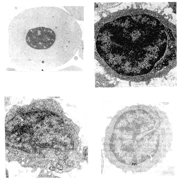
Los eritrocitos elipsoidales, alargados, tienen de esferoide hasta rectangular, con heterocromatina densa y picnótica, con citoplasma denso, homogéneo y mitocondrias dispersas escasas (Fig. 1).
Fig. 1 Ultramicrofotografías de células sanguíneas de trucha arco iris (Oncorynchus mykiis), fijadas en glutaraldehído y tetraóxido de osmio y teñidas con acetato de uranilo y citrato de plomo. Se observan: eritrocito (e), linfocitos (l), monocito (m) (a y b). X 32.000.
Los linfocitos, redondeados, presentan dos tipos según su morfología nuclear: unos con núcleos redondeados y otros con núcleos indentados; ambos densos, grandes, ocupando la mayor parte de la célula; la heterocromatina se distribuye centralmente y sobre la membrana nuclear dejando visibles pequeñas áreas de cromatina clara o eucromatina. No hay nucleolo. El escaso citoplasma es denso, homogéneo, granular por la presencia de innumerables ribosomas, sin membranas del retículo endoplásmico, con aparato de Golgi, lisosomas y un conglomerado paranuclear de 5 a 7 mitocondrias, en el lado indentado del núcleo en aquellos linfocitos con ésta característica nuclear. Hay vesículas de pinocitosis vecinas a la membrana citoplasmática. Numerosas microvellosidades de diferente tamaño se desprenden de la membrana nuclear.
Los monocitos redondeados, con núcleo circular tienen una zona que tiende a ser convexa. Es heterocromático, con más eucromatina que los linfocitos. Sin nucleolo característico. En el citoplasma se ven cisternas periféricas de retículo endoplásmico granuloso, un aparato de Golgi y varios gránulos de densidad variable, localizados en el lado convexo del núcleo, con algunas mitocondrias. Las vesículas de pinocitosis son abundantes y la membrana celular origina pequeñas microvellosidades menos abundantes y prominentes que las de los linfocitos. En algunos monocitos se observan vacuolas que sugieren origen lipídico y que se correlacionan con su aspecto vacuolado visto con las técnicas de MOAR y en los frotis de sangre periférica teñidos con la tinción de Giemsa (Rodríguez 1995). ]]>
Los polimorfonucleares son redondeados con núcleos bilobulados o trilobulados, con heterocromatina sobre la membrana nuclear y eucromatina central. No tienen nucleolo y su membrana nuclear conforma una cisterna perinuclear entre las células sanguíneas. En un corte se observa el citoplasma con más de dos docenas de gránulos densos, de tamaño variable, redondeados y fusiformes, pequeños y grandes, agrupados hacia el lado citoplásmico de la mayor escotadura nuclear, con vacuolas y mitocondrias. La membrana celular origina numerosas microvellosidades, algunas ramificadas sugiriendo pseudópodos. Los gránulos densos se agrupan de forma radiada alrededor de vacuolas lipídicas (Fig. 2).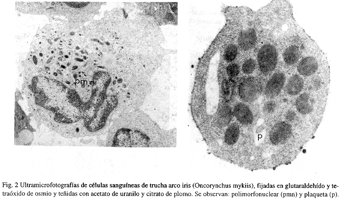
Las plaquetas, anucleadas, están cubiertas por una membrana celular que origina microvellosidades y pseudópodos. Contienen mitocondrias y numerosos gránulos densos redondeados. Formaciones concéntricas circulares de microtúbulos dispuestos en cuatro capas, se observan en el ectoplasma de éstas plaquetas.
La ultraestructura evidencia las características morfológicas de las cinco células sanguíneas de la trucha arco iris (eritrocitos, linfocitos, monocitos, polimorfonucleares y plaquetas). No se observaron eosinófilos.
Los resultados ponen en evidencia la similitud de la morfología de las células sanguíneas de la especie aclimatada en Colombia y la de la misma especie en países donde ésta es nativa; la morfología leucocitaria es semejante a la apreciada en otras especies, incluyendo los mamíferos (Bogner & Ellis 1977, Fawcett 1981, Fawcett 1986), especialmente aquella de las plaquetas. Sin embargo, Jagoe & Welter (1995), encuentran variación significativa entre la forma y tamaño de los núcleos de los eritrocitos de Oncorhynchus mykiss, Micropterus salmoides, Lepomis macrochirus, Esox niger, Perca flavescens, Gambusia holbrooki y Micropterus coosae, aunque esta variación es pequeña dentro de las especies al ser comparada con las diferencias encontradas inter-especies.
La morfología debe sugerir la función o inducir a la explicación de ésta, así, los exuberantes gránulos de los neutrófilos y de las plaquetas constituyen una fuente para estudios sobre su composición y su papel defensivo. ]]>
Resumen
Se estudió por ultraestructura la sangre periférica de 5 adultos de una población introducida de trucha arco iris (Oncorhynchus mykiis) cultivados en el Centro de Piscicultura Experimental del Neusa, (73°38´28"w; 5°8´30"N), para describir la morfología de los elementos que la componen. La sangre fue fijada con glutaraldehído, coloreada con tetraóxido de osmio al 1%, deshidratada en gradientes de alcoholes, centrifugada e incluida en resinas. Se observaron eritrocitos, linfocitos, monocitos, leucocitos polimorfonucleares (PMN) y plaquetas. Los eritrocitos mostraron núcleo picnótico y mitocondrias. Se vieron linfocitos de dos tipos: con núcleo redondeado y con núcleo indentado, ambos con ribosomas y acúmulos de 5 a 6 mitocondrias. Los monocitos poseen ribosomas y gránulos agrupados junto con mitocondrias a lo largo del núcleo irregular. Los PMN tienen mitocondrias y numerosos gránulos densos, redondeados y elipsoidales concentrados a lo largo de la ensenada nuclear lobulada. Las plaquetas se vieron ocasionalmente, como células anucleadas, vacuoladas con pequeños gránulos densos, ovalados.
Referencias
Anderson, D.R. 1965. A method of preparing peripheral leucocytes for electron microscopy. Ultrastruc. Res. 13: 263-268. [ Links ]
Bogner, K. & A. Ellis. 1977. Propiedades y funciones de los linfocitos y tejidos linfoides de los peces teleósteos. Acribia, Zarargoza. p. 71 85. [ Links ]
Campbell, T. W. 1985. Veterinary clinics of North America: Small Animal Practice Rev. 18: 349-363. [ Links ]
Fawcett, 1981. The cell. Saunders, Filadelfia. 757 p. [ Links ]
Fawcett, 1986. A textbook of histology. Saunders, Filadelfia. 879 p. [ Links ]
Heming, T.A. 1989. Clinical studies on fish blood: importance of sample collection and measurement techniques. Am. J. Vet. Res. 50: 93-97. [ Links ]
Jagoe, C.H. & D.A. Welter. 1995. Quantitative comparisons of the morphology and ultraestructure of erythrocyte nuclei from seven freswater fish species. Can. J. Zool. Rev. Can. Zool. 73: 1951-1959. [ Links ]
Lentz, T. 1971. Cell fine structure. An atlas of Drawings of whole-cell structure. Saunders, Filadelfia. 437 p. [ Links ]
Mokotoff, G. 1973. Electron microscopy laboratory techniques. Library Research Associates. Nueva York. 138 p. [ Links ]
Nonaka, M., N. Yamaguchi, S. Natsuume & M. Takahashi. 1981. The complement system of rainbow trout (Salmo gairdnerii). J. Inmunol. 126: 1489 1494. [ Links ]
Rodríguez, A. 1991. Hematología de la trucha arco iris. Valores normales, morfología celular sanguínea y hematopoyética con microscopio de luz y electrónico. Modificaciones en algunas enfermedades. Tesis Universidad de Bogotá Jorge Tadeo Lozano. Bogotá, Colombia. 144 p. [ Links ]
Rodríguez, A. 1995. Determinación de algunos aspectos hematológicos Oncorhynchus mykiis (Salmonidae), en Cundinamarca, Colombia. Rev. Biol. Trop. 43: 283-288. [ Links ]
Watanabe, I., S. Donahue & N. Hoggartt. 1967. Method for electron microscopic studies of circulating human leucocytes and observation of their fine structure. J. Ultrastruct. Res. 20: 366-382. [ Links ]
1 Universidad de Bogotá Jorge Tadeo Lozano, Centro de Investigaciones Científicas.
Santafé de Bogotá, Colombia. Fax 57 1- 281 28 40 ]]>