


 Inglês (pdf)
 Artigo em XML
 Referências do artigo
 Enviar este artigo por email
 Citado por SciELO
  Similares em
SciELO
Similares em
SciELO 
 Permalink
Introduction
Elimination of microorganisms in root canals is one of the primary goals for successful root canal therapy (1). During mechanical preparation, uninstrumented areas may remain in root canals (2). Although the use of irrigants aids to disinfect the untouched surfaces of root canals, bacteria in dentinal tubules cannot be completely removed (3). Sealer penetration into dentinal tubules during canal obturation may prevent growth of the remanent bacteria inside the tubules (4). Therefore, dentinal tubule penetration of irrigant and sealer is considered clinically important (5).
Various methods such as cold lateral condensation and warm obturation techniques are used to hermetically fill root canals in endodontic practice. These techniques have an important role in filling root canal irregularities, improving sealer penetration and preventing microleakage. The main objective of this research was to compare the effect of cold lateral condensation, continuous wave obturation and core-carrier based techniques on sealer penetration. Earlier studies indicated that the removal of smear layer from dentinal tubules positively affects sealer penetration (6,7). Conventional needle irrigation (CNI), a frequently used irrigation method, presents inadequate dentinal tubule penetration (8). Therefore, different irrigant agitation techniques can be used for final irrigation to remove the smear layer from the dentinal tubules and to more clearly determine the effect of obturation techniques. Ultrasonic tips activate irrigation by producing acoustic microstreaming and cavitation, which improves irrigants' cleaning abilities (9). XP-Endo Finisher is made of shape memory alloy and the manufacturer asserts that it cleans the root canal system effectively without damaging dentine (10). There are many studies about the effectiveness of multiple irrigation systems on dentinal tubule penetration of materials in the literature. But, no studies have been published about the efficiency of XP-Endo Finisher on dentinal tubule penetration of irrigant. Hence, the purpose of the first part of this research was to compare the effectiveness of XP-Endo Finisher with passive ultrasonic irrigation (PUI) and CNI on dentinal tubule penetration of irrigation solution using confocal laser scanning microscopy (CLSM). In the second part of this study, the effect of cold lateral condensation, continuous wave obturation and core-carrier based techniques on sealer penetration were compared. Also, the most effective activation method, which emerged as a result of the first part of this study, was used as the final irrigation method to remove the smear layer in the second part of this study.
Materials and methods
A power analysis was carried out utilizing data from a previous study (11). The computation revealed that there should be at least 3 teeth in each group (power=0.95, α=0.05, effect size=2.15). However, this study was designed with 10 teeth in each group. After ethics committee approval was obtained for this research (protocol no: 39-2016), all endodontic procedures were conducted by an experienced single operator. Sixty extracted mandibular premolars with straight, single-rooted and round-shaped canals were used. Each tooth was analysed under a stereomicroscope to extract any root with immature root apices, cracks, or fractures. The length of samples was standardized to 16mm using a diamond disc.
The working length (WL) of teeth was established 1mm from the apex using a size 15 stainless-steel K-file. Each canal was prepared by using Twisted File Adaptive (Kerr Co., Orange, CA) instruments in the sequence ML1 (25/.08), ML2 (35/.06) and ML3 (50/.04). All instruments were performed with the TF-Adaptive pre-set program and the Elements Motor (Kerr Co., Orange, CA). The canals were irrigated with 2mL of 5.25% NaOCl using a syringe with a 27-gauge side-vented needle (Endo-Eze; Ultradent Products, Salt Lake City, UT) between each file. The needle was positioned 2mm from the WL. After instrumentation, the root canals were flushed with 5mL of 17% EDTA for 1 minute for removal of the smear layer from dentin walls. The canals were rinsed subsequently 5mL of distilled water and dried using sterile paper points. The roots were randomly assigned into two main groups (n=30) as the final irrigation technique experiment and obturation technique experiment (Figure 1). The simple randomization method was used for the teeth allocation in the required steps.
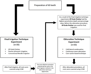
Final irrigation tecnique experiment
Before the final irrigation procedure, 2% chlorhexidine (CHX) was mixed with 0.1% rhodamine-B fluorescent dye (Merck, Darmstadt, Germany) for visualisation of its dentinal tubule penetration under CLSM. The roots were randomly allocated into three groups (n=10) according to the final irrigation methods.
XP-Endo Finisher: The canals were irrigated with CHX+Rhodamine-B using the XP-Endo Finisher file. The XP-Endo Finisher file was inserted 1mm from the WL. The endodontic motor was adjusted at 800rpm/0,8Ncm and operated in 3 cycles of 20 seconds with an up and down motion. In each cycle, the canals were irrigated using 5mL CHX+Rhodamine-B for 1 minute.
Passive ultrasonic irrigation (PUI): The canals were irrigated with CHX+Rhodamine-B using an ultrasonic device (NSKVarios, NSK, Japan). E11 ultrasonic tip and U-file (NSKVarios, NSK, Japan) were set at 2mm from the WL and operated at Endo mode. Ultrasonic irrigation was performed in 3 cycles of 20 seconds. In each cycle, the canals were irrigated using 5mL CHX+Rhodamine-B for 1 minute.
Conventional Needle irrigation (CNI): The canals were irrigated with 5mL CHX+Rhodamine-B for 1 minute using a syringe with a 27-gauge sidevented needle. The needle was positioned 2mm from the WL and used with an up and down motion.
Obturation tecnique experiment
As a result of the final irrigation system experiment, XP-Endo Finisher had the highest penetration depth and percentage value (Table 1). Therefore, in the second part of this research, the final irrigation was performed using XP-Endo Finisher as described above. However, chlorhexidine was not mixed with 0.1% rhodamine-B fluorescent dye. After final irrigation, all teeth were dried with sterile paper points and randomly allocated into three groups (n=10) according to the obturation technique. Before root canal filling, AH Plus sealer (Dentsply Sirona, York, PA, USA) was mixed with 0.1% rhodamine-B fluorescent dye.
Cold lateral condensation technique (CLC): A size 50/0.02 gutta-percha cone with the labeled sealer was placed into the root canal. Then, the canal was filled with a size 20/0.02 accessory gutta-percha cones using a size 25 finger spreader (Mani Inc., Tochigi, Japan) until obturation of the root canal was completed.
Continuous-wave obturation technique (CW): A size 50/0.04 gutta-percha cone (Dentsply Sirona, York, PA, USA) with the labeled sealer was placed into the root canal. The down-pack unit of the Elements Free (Kerr Co., Orange, CA) was placed 3-4mm from the WL and then the remaining material was removed. Then the canal was obturated using the Elements Free Backfill unit until the canal orifice was reached.
Core-carrier-based technique (GC): A size 50 GuttaCore obturator (Dentsply Sirona, York, PA, USA) was heated in the Thermaprep 2 (Dents- ply Sirona, York, PA, USA) oven and inserted into the canal. The part of the obturator at the canal entrance was removed.
After the obturation procedures, the canal entrance was filled with Cavit (3M ESPE, Seefeld, Germany) and the quality of the canal filling was affirmed by radiograph. Then, the roots were stored in an incubator at 37°C and 100% humidity for 7 days.
Table 1 Means and standard deviations of penetration depth (μm) and percentage (%) of irrigant according to the final irrigation techniques.
| - | Group | - | - | - |
|---|---|---|---|---|
| - | XP-Endo Finisher | Passive ultrasonic irrigation (PUI) | Conventional needle irrigation (CNI) | p |
| Penetration depth | 425.82 ± 119.53‡ | 357.04 ± 145.61‡ | 152.49 ± 80.10† | 0.001 |
| Penetration percentage | 86.70 ± 18.66‡ | 77.60 ± 28.75‡ | 51.20 ± 31.96† | 0.018 |
Different superscript footnotes show significant differences in each parameter (p<0.05).
Confocal laser scanning microscopic analysis
All roots were horizontally sectioned in 1-mm-thick slices with a water-cooled Isomet machine (Isomet Buehler, Lake Bluff, IL, USA) at 3mm from the root apex. Then, the slices were polished with silicon carbide abrasive paper and mounted on glass slides. All specimens were scanned using CLSM (Leica, Mannheim, Germany) at x5 magnification under Ar/HeNe laser excitation with a wavelength of 565 to 600nm.
All images were imported to an Adobe Photoshop Software program (Adobe Systems Inc., San Jose, CA, USA) to analyse the depth and percentage of irrigant and sealer penetration into dentinal tubules. For the depth of penetration, the deepest point of penetration was measured from the canal wall to the point of the maximum material penetration. Also, to calculate the penetration percentage, the penetration depth was divided by the circumference of the root canal. All measurements were performed by a single operator who was blinded to the groups.
Statistical analysis
The data were analysed with the SPSS program (Version 24; SPSS Inc., Chicago, IL, USA). The normality of the data distribution was tested using the Shapiro-Wilk test. Because the data were not normally distributed, the Kruskal-Wallis test was used to compare variables between groups. The Bonferroni's post-hoc test was used for all pairwise comparisons. The level of significance was set at p<0.05.
Results
Final irrigation tecnique experiment
The results of irrigant penetration depth and percentage are presented in Table 1 and the representative CLSM images of each group are shown in Figure 2. According to CLSM analysis, XP-Endo Finisher had the highest penetration depth and percentage value, followed by PUI group and CNI. However, there was no significant difference between XP-Endo Finisher and PUI in terms of depth and percentage of irrigant penetration (p>.05). Also, CNI group exhibited significantly the lowest penetration depth and percentage (p<.05).
Obturation tecnique experiment
The results of sealer penetration depth and percentage are presented in Table 2 and the repre- sentative CLSM images of each group are shown in Figure 2. CLSM analysis revealed that there was no significant difference among the obturation techniques with respect to depth and percentage of sealer penetration (p>.05).
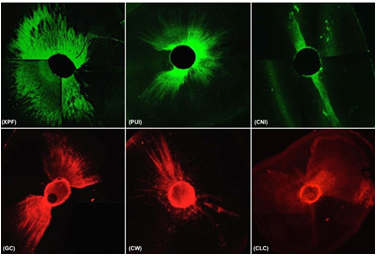
Figure 2 The representative confocal laser scanning microscopic images of each group: (XPF) XP-Endo Finisher, (PUI) passive ultrasonic irrigation, (CNI) conventional needle irrigation, (GC) core-carrier-based technique, (CW) continuous-wave obturation technique, (CLC) cold lateral condensation technique.
Table 2 Means and standard deviations of penetration depth (μm) and percentage (%) of sealer accor- ding to the obturation techniques.
| - | Group | - | - | - |
|---|---|---|---|---|
| - | Core-carrier-based technique (GC) | Continuous-wave obturation technique (CW) | Cold lateral condensation technique (CLC) | p |
| Penetration depth | 283.92 ± 221.35 | 259.29 ± 127.12 | 260.65 ± 130.36 | 0.993 |
| Penetration percentage | 50 ± 39.1 | 49.6 ± 37.77 | 77.5 ± 30.9 | 0.160 |
The level of significance was set at p<0.05.
Discussion
The removal of the smear layer within complex canal anatomy is a clinical challenge for dentists. The deeper the irrigant penetrates the dentinal tubules, the more it removes the smear layer from the dentinal tubules. Therefore, the first objective of this research was to assess the efficiency of XP-Endo Finisher, PUI and CNI on dentinal tubule penetration of irrigant. As a result of this assessment, XP-Endo Finisher had the highest penetration depth and percentage value. Previous studies have shown that removing the smear layer from dentinal tubules improves sealer penetration (6,7). Thus, the main purpose of this research was to investigate the efficiency of various obturation methods on dentinal tubule penetration of sealer after the smear layer was removed with XP-Endo Finisher.
The smear layer that forms on canal walls during instrumentation reduces dentinal permeability and dentinal tubule penetration of materials. Chelating agents and NaOCl combination is recommended for removal of the smear layer from dentin walls (7). As a final irrigation solution, CHX is suggested due to its long-lasting antibacterial activity. However, CHX and NaOCl can react with each other and produce Parachloroaniline (PCA) which obstructs the dentinal tubules. Before final CHX irrigation, root canals should be rinsed with distilled water to avoid PCA formation (12).
Dentinal tubule penetration of materials might be observed with microscopes such as stereomicroscope, scanning electron microscopy (SEM) and CLSM. Earlier studies suggested CLSM as it provides 3D analysis and quantitative evaluation and does not require special sample preparation before scanning, unlike the SEM technique (13,14). In CLSM analysis, irrigation solution or sealer is mixed with rhodamine-B fluorescent dye for visualisation of material penetration into dentinal tubules and the samples are horizontally sectioned. Under the CLSM analysis test conditions, the samples used to examine dentin tubule penetration of irrigant cannot be reused to examine sealer penetration. Thus, this study was divided into two parts as the final irrigation technique experiment and the obturation technique experiment. Furthermore, viscosity and surface tension of irrigants or flow and film thickness of sealers can influence their penetration into dentinal tubules (14,15). But, the small amount of the dye (0,1%) does not affect the physical properties of materials.
Many studies have confirmed that material penetration into dentinal tubules is more difficult in the apical region (5,16,17). This can be attributed to tubular sclerosis, anatomic irregularities, the smaller diameter of dentinal tubules in this area (18). Additionally, the smear layer in the apical region is more difficult to remove because of reduced irrigation delivery (19). Because of these difficulties, dentin slices were obtained at 3mm from the root apex in this study.
We found that dentinal tubule penetration in the buccolingual direction was higher than in the mesiodistal direction similar to previous studies (20,21). This is explained by the'butterfly effect' phenomenon that appears as a result of higher tubular sclerosis on the mesiodistal surfaces of the canal lumen (22). This non-homogenous dentinal tubule penetration might influence the results. Thus, irrigant penetration should not be evaluated only with the'maximum penetration depth' parameter. In our study, penetration percentage also was used as the second parameter and calculated using the method described by Gharib et al. (23).
Previous studies indicated that XP-Endo Finisher and PUI showed same efficacy in removing hard tissue debris (24) and calcium hydroxide from canals (25,26). Also, another study reported that XP-Endo Finisher was more successful than PUI in point of antibiotic paste removal from root canals (27). However, there have been no studies regarding the effectiveness of XP-Endo Finisher on dentin tubule penetration of irrigant in the literature. According to the findings of the current study, XP-Endo Finisher and PUI had similar effects on irrigant penetration.
The XP-Endo Finisher is produced from an exclusive alloy (Martensite-Austenite Electropolish-FleX) which is straight in its martensite phase at room temperature, but its shape changes to be more contact with root canal walls at body temperature because of its austenite phase (27). For this reason, the manufacturer claims that it can clean areas that previous systems could not reach. In accordance with this expectation, we found that the XP-Endo Finisher is more successful than CNI. But, no statistical difference was observed between the XP-Endo Finisher and PUI. The shape of the root canals may have led to this result because in this research, straight, round-shaped mandibular premolars were used to eliminate anatomical differences. In curved canals, XP-Endo Finisher can be in touch with root canal walls more than other irrigation systems due to its alloy feature. To support this argument, further studies are needed.
Dentinal tubule penetration of sealer may be affected by the physical properties of endodontic sealers such as setting time, flow and film thickness (13). Camilleri (28) reported that the properties of sealers were adversely changed by heat. However, little information is available on the comparison of the effect of warm obturation techniques and cold obturation methods on dentinal tubule penetration of sealer and these studies revealed that sealer penetration into dentinal tubules is independent of filling techniques similar to our results (21,29,30). However, the results of our study showed high standard deviations. This can be attributed to tubular sclerosis and anatomic irregularities in the apical area. Increasing the number of samples in future investigations may help to avoid this problem. Besides, De Deus et al. (11) indicated that warm obturation techniques may have a beneficial impact on sealer's penetration ability. The explanation for this discrepancy between the results might be that the root canals were obturated after final irrigation with XP-Endo Finisher to extermi- nate the negatory impact of the smear layer on sealer penetration in our study. Because XP-Endo Finisher removes smear layer and hard tissue debris from dentinal tubules better than CNI (24,31). Also, this study showed that XP-Endo Finisher enables the irrigant to penetrate deeper into the dentinal tubules in comparison to CNI.
Conclusion
Under the limitations of the current study, XP-Endo Finisher was as effective as that of PUI on dentinal tubule penetration of irrigant and can be used for irrigation activation instead of CNI in order to enhance dentinal tubule penetration of irrigants. Also, the canal filling techniques did not play an effective role in sealer penetration into dentinal tubules.
Author contribution statment
Conceptualization and design: E.D. and D.T.
Literature review: E.D. and D.T. and S.G.K.
Methodology and validation: E.D. and D.T. and S.G.K.
Formal analysis: E.D.
Investigation and data collection: E.D. and D.T.
Resources: E.D. and D.T.
Data analysis and interpretation: E.D.
Writing-original draft preparation: S.G.K. and D.T. and E.D.
Writing-review & editing: S.G.K. and D.T.
Supervision: S.G.K. and D.T. and E.D.
Project administration: E.D. and D.T.
Funding acquisition: E.D. and D.T.

