Inglês (pdf)
 Artigo em XML
 Referências do artigo
 Enviar este artigo por email
 Citado por SciELO
  Similares em
SciELO
Similares em
SciELO 
 Permalink
PhD. Jessie Reyes Carmona Editor Jefe
ODOVTOS-International Journal of Dental Sciences Universidad de Costa Rica
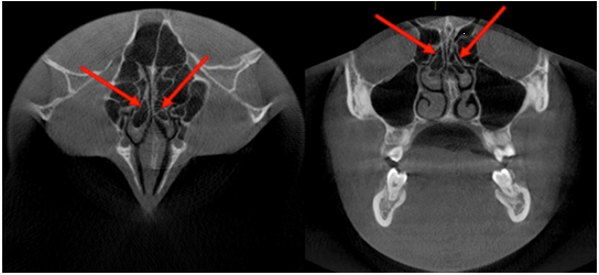
The nasal turbinates, also called nasal shells, are attached to the lateral and/ or superior walls of the nostril and can present several anatomical variations. Thus, we can mention the concha bullosa, the paradoxical curvature of the middle turbinate (both common anatomical variants) and the secondary middle turbinate, the latter in turn, can be pneumatized (very rare) (1).
The term ''concha bullosa'' generally refers to pneumatization of the middle turbinate, however, pneumatization of the superior and inferior turbinates can also occur (2). Some authors mention it as a "variation in the pneumatization of the bone plate caused by an extension of the ethmoid cells" (3). The literature reports 3 types of concha bullosa: lamellar, bulbous and true; the latter refers to the complete pneumatization of the anterior third of the turbinate. The origin of this condition is undetermined (4), but it can be considered as predisposing causes to trauma, deviation of the nasal septum and mouth breathing (3). The frequency of pneumatization of the middle turbinate is in a range from 14% to 53%, while in the inferior turbinate it is 1% (2,4).
The anatomical variants of the nasal turbinates are important in the pathogenesis of the inflammatory processes of the paranasal sinuses, such as for the etiology of sinusitis associated with the middle turbinate (4,5). A significant correlation between the palatal dimensions, the presence of the concha bullosa and the deviation of the nasal septum that can contribute to the establishment of the skeletal malocclusion is also mentioned (6). In addition, the presence of the concha bullosa and deviation of the nasal septum would be related to compensatory hypertrophy of the contralateral turbinate (7). Most patients with concha bullosa of the middle turbinate are asymptomatic, but cases of blockade of the osteomeatal complex that could lead to chronic sinusitis have begun to be reported, in addition to reports of headaches in patients with pneumatization or the presence of concha bullosa in the superior turbinate (3,8).
Knowledge of anatomical structures and their spatial relationships is essential for diagnosis, as well as treatment planning. With the development and widespread use of new imaging technologies such as cone beam computed tomography (CBCT) and the analysis tools that it includes, it is possi- ble to observe anatomical structures outside the dentomaxillary complex; being the nasoethmoidal region (nose and ethmoid bone) represented in detail when the field of vision is wide enough (2). In addition, this technique offers multiple advantages such as costs and low radiation dose compared to other imaging techniques, high resolution and definition of details that could not be observed in two-dimensional radiographs. Likewise, it allows observing findings, from anatomical variants, benign lesions to pathologies of importance for the patient's health (9).
The analysis and interpretation of the images by the specialist in Oral and Maxillofacial Radiology must contemplate the description of the anatomical variants, which depending on the case could be the origin of some non-specific symptomatology of the patient. The identification of these will allow complete diagnostic evaluations and adequate treatment plans, leaving the decision to refer to the corresponding specialty to the clinician (9).
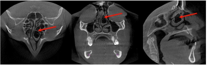
Figure 1 Images taken from the archive of the Oral and Maxillofacial Radiology Service of the Teaching Dental Center of the Universidad Peruana Cayetano Heredia.
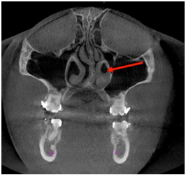
Figure 2 Images taken from the archive of the Oral and Maxillofacial Radiology Service of the Teaching Dental Center of the Universidad Peruana Cayetano Heredia.