


 English (pdf)
 Article in xml format
 Article references
 Send this article by e-mail
 Cited by SciELO
  Similars in
SciELO
Similars in
SciELO 
 Permalink
Introduction
Root canal morphology and morphological anatomical variations significantly affect the success of endodontic treatment (1). Awareness of the variations in the root canal can avoid endodontic treatment failure and determine the best treatment plan (2,3). Canine teeth are strategically important in the dental arch. It has long and stable roots and is the longest remaining teeth in the mouth. They guide occlusion during eccentric movements and chewing so it is very important for prosthetic support (4). The most common root canal pattern in the canine teeth is a single root and a single canal but some may have two roots and two canals (5,6). The inability to find this additional canal leads to failure of endodontic treatment. Race and genetic factors are thought to affect root canal morphology. Therefore, the root canal morphologies of different ethnic populations should also be considered in order to increase the success rate in endodontic treatments (7,8).
In recent years, cone beam computed tomography (CBCT) has gained increasing importance in endodontics in vivo for the three-dimensional evaluation of the root canal system. It has been stated to be as reliable as the modified canal dyeing and transparency technique (9,10). The data acquired by CBCT presents coronal, sagittal, and axial sections, decreasing the superposition of anatomical structures. Additionally, CBCT is preferred due to its low cost, high resolution, a lower dose of radiation, and better image quality (11,12). Conventional radiography, root canal staining methods, clearing methods, tooth sectioning, microscopic observation, and CBCT have been used in previous investigations (10,13,14,15). There are limited studies of different populations in the literature evaluating root and canal morphology of canine teeth using CBCT (2,3,5,6,16,17,18). In addition, there is no study examining the apical foramen localization and bifurcation point in root canal morphology of canine teeth using CBCT in the Turkish population. Therefore, the aim of this study was to investigate the number of roots and root canals in the canine teeth in the Turkish population and the bifurcation point with two canals, and the position of apical foramen using CBCT images.
Material and methods
In this retrospective study, CBCT data of 300 patients between 2016 and 2018 were screened and evaluated. A total of 235 patients, 100 males, and 135 females were included in this study. A total of 426 maxillary and mandibular canine teeth were analyzed. The ethical approval was obtained from the Clinical Research Ethics Committee of Altınbaş University (approval number: 2020/26). The CBCT was performed to evaluate patients requiring diffe- rent indications such as dental implant planning, impacted third molar localization, jaw lesions, trauma, or the maxillary sinus pathologies. Before radiographic examinations, informed consent was received from all patients. Inclusion criteria were patients with the presence of at least one mandibular anterior tooth, absence of root canal fillings, coronal and post restorations, periapical lesions, fixed crown restorations, orthodontic braces, and high quality CBCT images. After excluding images that did not meet the inclusion criteria, 235 CBCT images were included in the study.
CBCT images of all patients were obtained with NewTom VGi evo (CeflaGroup, Verona, Italy) device. After setting the device to 1-32mA and 110 kV, images with a voxel size of 0.3mm were created with a single 360-degree rotation. Radiological images were evaluated on a 22" high image quality and 1920x1080 display resolution Barco medical monitor to provide an effective evaluation by an oral radiologist (OO) and an endodontist (ANK). NNT Viewer (CeflaGroup, Verona, Italy) was used for the reconstruction and evaluation of all projections. The brightness and contrast of the images were adjusted to ensure optimal visualization. The maxillary and mandibular canines of the 20% of the images were assessed separately by two observers to evaluate inter-observer compliance. When disagreements occurred, it was discussed and reached a final consensus.
Maxillary and mandibular canine teeth were evaluated for the number of roots, root canals, and canal morphology based on the Vertucci's method (18). All root canal configurations were recorded as two root canals, except type I (single canal). Also, the beginning and end points of the separation in cases with more than one single canal were analyzed.
The canal configuration was classified based on the following criteria of Vertucci's method (18);
Type I: Only one canal extending from the pulp chamber to the apex.
Type II: Two canals, which leave the pulp chamber separately, are joined in the apical region and terminate as a single canal.
Type III: The single canal that leaves the pulp chamber is divided into two, and then ends up as a single canal in the apical region.
Type IV: Two separate canals that leave the pulp chamber terminate in two separate canals in the apical region.
Type V: The canal that leaves the pulp chamber as a single canal terminates by dividing into two canals.
Type VI: Two distinct canals that leave the pulp chamber first merge into a single canal and separate again in the apical region and end in two separate canals.
Type VII: A single canal leaving the pulp chamber is divided into two, then rejoined to form a single canal, and then re-divided at the apical region and terminates with two separate foramens.
Type VIII: Three distinct canals leave the pulp chamber and terminate separately.
The root canal separation was grouped as the cervical, middle and apical third of the root in cases of type II-type VII.
For the statistical analyses, the IBM SPSS Statistics 22 (IBM SPSS, Turkey) program was used while assessing the findings of the study. Chi-square test, Fisher Freeman Halton test and Yates's continuity correction were used to compare qualitative data as well as descriptive statistical methods (mean, standard deviation (SD), and frequency). Values of <0.05 were considered as significant statistically (P<0.05).
Results
Among 300 patients, 426 maxillary and mandibular canine teeth in 235 patients matched with our inclusion criteria were evaluated, 100 (42,6%) were male, and 135 (57,4%) were female. The age range was 14-76 years (mean age ± SD: 37.27±13.40 years).
A total of 191 (44,8%) maxillary canine teeth and 235 (55,2%) mandibular canine teeth were evaluated (Table 1). All the maxillary canines had a single root and single canal. None of them have bifurcation points. In the majority of maxillary canines, the apical foramen is positioned centrally at 70,2% and laterally at 29,8%. (Table 2).
Table 1 Vertucci classification, number of canals, number of roots, bifurcation point, and apical foramen distributions in all canine teeth.
| - | - | n | % |
|---|---|---|---|
| Vertucci classification (n=426) | Type I | 409 | 96,0 |
| - | Type II | 1 | 0,2 |
| - | Type III | 16 | 3,8 |
| Number of canals (n=426) | Single canal | 409 | 95,9 |
| - | Double canal | 17 | 4,1 |
| Number of roots (n=426) | Single root | 417 | 97,9 |
| - | Double root | 9 | 2,1 |
| Bifurcation point (n=17) | Middle | 9 | 53,8 |
| - | Servical | 8 | 46,2 |
| Apical Foramen (n=426) | Central | 291 | 68,3 |
| - | Lateral | 135 | 31,7 |
Table 2 Vertucci classification, number of canals, number of roots, bifurcation point, and apical foramen distributions in the maxillary canines (Root canal classification of maxillary canines).
| - | - | n | % |
|---|---|---|---|
| Vertucci classification (n=191) | Type I | 191 | 100 |
| - | Type II | 0 | 0 |
| - | Type III | 0 | 0 |
| Number of canals (n=191) | Single canal | 191 | 100 |
| - | Double canal | 0 | 0 |
| Number of roots (n=191) | Single root | 191 | 100 |
| - | Double root | 0 | 0 |
| Bifurcation point (n=0) | Middle | 0 | 0 |
| - | Servical | 0 | 0 |
| Apical Foramen (n=191) | Central | 134 | 70,2 |
| - | Lateral | 57 | 29,8 |
The mandibular canine teeth had a single root of 96,2%, and a single canal of 88,9%. The majority of the teeth had a Type I canal configuration in mandibular canines (92,8%). The prevalence of the second canal in mandibular canine teeth was 7,2% and the other canal patterns found were Type III (6,8%) and Type II (0,4%). In the majority of mandibular canines, the apical foramen is positioned centrally at 66,8% and laterally at 33,2%. The root canal separation in two root canals of mandibular canine was detected in the middle third of the root with a 53,8% ratio and in the cervical third of the root with 46,2% ratio (Table 3).
Table 3 Vertucci classification, number of canals, number of roots, bifurcation point, and apical foramen distributions in the mandibular canines (Root canal classification of mandibular canines).
| - | - | n | % |
|---|---|---|---|
| Vertucci classification (n=235) | Type I | 218 | 92,8 |
| - | Type II | 1 | 0,4 |
| - | Type III | 16 | 6,8 |
| Number of canals (n=235) | Single canal | 218 | 92,8 |
| - | Double canal | 17 | 7,2 |
| Number of roots (n=235) | Single root | 226 | 96,2 |
| - | Double root | 9 | 3,8 |
| Bifurcation point (n=17) | Middle | 9 | 53,8 |
| - | Servical | 8 | 46,2 |
| Apical Foramen (n=235) | Central | 157 | 66,8 |
| - | Lateral | 78 | 33,2 |
There was no statistically significant difference between the Vertucci classifications and root canal numbers by gender (p>0.05). The incidence of Type I morphology was found at 96,2% and 95,9% in males and females, respectively. While 93,4% of females had one canal and 6,6% of them had two canals, 94,5% of males had one canal and 5,5% of them had two canals. There was also no statistically significant difference between genders in root canal separation and the position of the apical foramen (Table 4).
Table 4 Distribution of maxillary and mandibular canines teeth by gender.
| - | - | Maxillary | - | - | Mandibular | - | - | Total | - | - |
| - | - | Female | Male | P | Female | Male | p | Female | Male | P |
| - | - | n (%) | n (%) | - | n (%) | n (%) | - | n (%) | n (%) | - |
| Vertucci classification | Type I | 109 (%100) | 82 (%100) | - | 125 (%92,6) | 93 (%93) | ¹1,000 | 234 (%95,9) | 175 (%96,2) | ¹1,000 |
| - | Type II | - | - | - | 1 (%0,7) | 0 (%0) | - | 1 (%0,4) | 0 (%0) | - |
| - | Type III | - | - | 9 (%6,7) | 7 (%7) | - | 9 (%3,7) | 7 (%3,8) | - | |
| Canal number | Single canal | 109 (%100) | 82 (%100) | - | 125 (%92,6) | 93 (%93) | ²0,813 | 234 (%95,9) | 175 (%96,2) | ²0,804 |
| - | Double canal | - | - | - | 10 (%7,4) | 7 (%7) | - | 10 (%4,1) | 7 (%3,8) | - |
| Root number | Single root | 109 (%100) | 82 (%100) | - | 129 (%95,6) | 97 (%97) | ³0,736 | 238 (%97,5) | 179 (%98,4) | ³0,738 |
| - | Double root | - | - | - | 6 (%4,4) | 3 (%3) | - | 6 (%2,5) | 3 (%1,6) | - |
| Bifurcation point | Middle | - | - | - | 4 (%50) | 3 (%60) | ³0,701 | 8 (%50) | 6 (%60) | ³0,701 |
| Servical | - | - | - | 4 (%50) | 2 (%40) | - | 8 (%50) | 4 (%40) | - | |
| Apical Foramen | Central | 76 (%69,7) | 58 (%70,7) | ⁴0,880 | 95 (%70,4) | 62 (%62) | ⁴0,178 | 171 (%70,1) | 120 (%65,9) | ⁴0,363 |
| - | Lateral | 33 (%30,3) | 24 (%29,3) | - | 40 (%29,6) | 38 (%38) | - | 73 (%29,9) | 62 (%34,1) | - |
¹Fisher Freeman Halton Test. ²Continuity (yates) correction. ³Fisher's Exact Test. ⁴Ki-kare te.
Discussion
In endodontic treatment, failure to identify extra canals, being unaware of the presence an additional canal and insufficient debridement are the most common causes of failure. Therefore, in order to be successful in endodontic treatment, root canal morphology must be well known (6). Analysis of root canal morphologies of teeth belonging to different populations and ethnic origins and preoperative evaluation of root canal systems are essential in this respect (10). Staining and cleaning technique on extracted teeth has been used in previous studies to evaluate root canal anatomy of canine teeth (16,17,18,19,20). CBCT is a low radiation dose technique that has been proven to be accurate in evaluating root canal systems, frequently used today. In addition, there are few studies examining root and canal morphology of canine teeth using CBCT in the Turkish population (21,22,23,24).
In the present study, the most common root canal pattern in the maxillary canine was a Type I in 100% of the samples (Figure 1). Similar findings were reported by Pineda and Kuttler (100%) (15), Vertucci (100%) (18), and Mağat (0,1%) (23) but the prevalence of an additional canal in maxillary canines, in this study, was lower than Amardeep et al. (20,4%) (5), Çalışkan et al. (6,52%) (19), Sert and Bayırlı (6,5%) (20), Altunsoy (female 1,3%, male 3,2%) (21), and Büyükbayram et al. (2,44%) (25), in maxillary canines (Table 5). The difference in the findings of the other authors may be explained by study techniques, number of patients, the number of teeth and different populations.
The most common root canal pattern in the mandibular canines had the same pattern, with a single root and a single canal. Type III was observed most frequently among teeth with two root canals, followed by Types II; Types IV, V, VI, VII, and VIII were not observed (Figure 2). The prevalence of an additional canal in the mandibular canine teeth in this study was 7,2%, which was close to the findings of Han et al. (6,3%) (2), Rahimi et al. (9,4%) (17), Kayaoğlu et al. (6,1%) (22), Altunsoy (female 6%, male 8,4%) (21), and Mağat (9,4%) (23), but lower than those of Amardeep (20,4%) (5), Vertucci (22%) (18), Çalışkan et al. (19,6%) (19) and Sert and Bayırlı (24%) (20), and Büyükbayram et al. (15,84%) (24) (Table 6).
Most of the additional canals did not course along the root length; in most cases, the canals began at one orifice, divided into two, and then continued as a single canal with one foramen. However, in some cases, two canals began from two orifices and fused into one and then continued as a single canal. Dentists in clinical practice should therefore take into account the position of all root canals to debride and remove pulp tissue.
In all other studies, Vertucci Type I has been observed as the most prevalent, as in our study. Of the double root canals, Types II and III were the most observed canal types in previous studies. In studies conducted with CBCT (2,5,18,19,20,21,22,23,24) the most common type of double root canal was Type III, as in our study.
In the present study, gender was not related to the presence of double root canals. There was no statistically significant difference between the Vertucci classifications and root canal numbers by gender (p>0.05). In addition, there was also no statistically significant difference between genders in root canal separation and the position of the apical foramen.
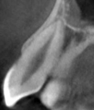
Table 5 Prevalence of a additional canal in maxillary canine teeth.
| Previous Studies | Population | Technique | Year | Number of Teeth | % |
|---|---|---|---|---|---|
| Pineda and Kuttler | Mexico | Radiographs | 1972 | 260 | 0 |
| Vertucci | USA | Staining and Clearing | 1984 | 100 | 0 |
| Caliskan et al. | Turkey | Staining and Clearing | 1995 | 200 | 6,52 |
| Sert and Bayırlı | Turkey | Staining and Clearing | 2004 | 200 | 6,5 |
| Altunsoy et al. (male) | Turkey | CBCT | 2014 | 773 | 3,2 |
| Altunsoy et al. (female) | Turkey | CBCT | 2014 | 750 | 1,3 |
| Amardeep et al. | India | CBCT | 2014 | 250 | 8,4 |
| Büyükbayram et al. | Turkey | CBCT | 2015 | 82 | 2,44 |
| Mağat | Turkey | CBCT | 2019 | 820 | 0,1 |
| Present Study | Turkey | CBCT | 2020 | 191 | 0 |

Figure 2 CBCT cross sectional images of mandibular canine teeth (A) Type I, (B) Type II, (C) Type III.
Table 6 Prevalence of a additional canal in mandibular canine teeth.
| Previous Studies | Population | Technique | Year | Number of Teeth | % |
|---|---|---|---|---|---|
| Pineda and Kuttler | Mexico | Radiographs | 1972 | 187 | 13,5 |
| Vertucci | USA | Staining and Clearing | 1984 | 100 | 22 |
| Pecora et al. | Brazil | Staining and Clearing | 1993 | 149 | 7,8 |
| Caliskan et al. | Turkey | Staining and Clearing | 1995 | 100 | 19,6 |
| Sert and Bayırlı | Turkey | Staining and Clearing | 2004 | 200 | 24 |
| Rahimi et al. | India | Staining and Clearing | 2013 | 131 | 9,4 |
| Han et al. | China | CBCT | 2014 | 1210 | 6,3 |
| Amardeep et al. | India | CBCT | 2014 | 250 | 20,4 |
| Kayaoğlu et al. | Turkey | CBCT | 2015 | 134 | 6,1 |
| Büyükbayram et al. | Turkey | Staining and Clearing | 2015 | 101 | 15,84 |
| Mağat | Turkey | CBCT | 2019 | 820 | 9,4 |
| Present Study | Turkey | CBCT | 2020 | 235 | 7,2 |
Conclusion
The following conclusions may be reached from this retrospective study:
In a Turkish subpopulation, the maxillary canines showed mostly single root and single canal morphology, however, the prevalence of two root canals in mandibular canines was 7,2%.
Apical foramen was centrally positioned in the majority of the teeth, 70,2% and 66,8% in maxillary and mandibular canines, respectively.
In the mandibular canine teeth, the root canal separation was located in the middle third of the root in almost 53,.8% of cases.
Author contribution statement
Conceptualization and design: Ö.O. and A.N.K.
Literature review: Ö.O. and A.N.K.
Methodology and validation: Ö.O. and A.N.K.
Investigation and data collection: Ö.O. and A.N.K.
Data analysis and interpretation: Ö.O. and A.N.K.
Writing-original draft preparation: A.N.K.
Writing-review & editing: Ö.O. and A.N.K.

