


 English (pdf)
 Article in xml format
 Article references
 Send this article by e-mail
 Cited by SciELO
  Similars in
SciELO
Similars in
SciELO 
 Permalink
Introduction
Advancements in technology and its advantages have increased the use of digital systems in dentistry (1,2). Improvements in digital imaging systems and software have expanded the possibility of image enhancement and diagnostic task specific post-processing adjustments (3). Digital imaging systems also enable a long-term online imaging storage and simplify communication with professionals and patients (4). The lower radiation dose required by digital image receptor is another advantage of digital imaging systems compared with conventional radiographic films (1), which minimize the radiation-related risks and optimizing the outcomes for both professionals and patients (5). Moreover, it presents a lower risk of environmetal contamination due to the non-use of chemical solutions used in conventional radiographic processing, as well as a decrease in imaging exams repetition due to errors in this processing (1).
In the United States and in many European countries, digital radiology is a reality (5). In Belgium, for example, 90% of dentists use digital imaging systems (4). Such a reality, however, may be different in developing countries like Brazil, country of huge dimensions and regions with different socioeconomic conditions. It is important to verify the realities of access to digital systems to establish guidelines for digital radiology, communication and radiological protection adequate for each region. The main objective of the present study was, therefore, to analyze the use of digital imaging systems by dentists, assessing the types of image receptors, imaging exams and digital imaging enhancement tools used in Brazilian dental offices. In addition, the methods of sharing the imaging exams with colleagues and patients was assessed to achieve a overview of the current use of dental radiographic in Brazil.
Material and methods
This questionnaire-based cross-sectional study was approved without restrictions by the Institutional Ethics Committee (protocol: #23706819.7.0000.5418), and written informed consent was obtained from volunteers. Participation was confidential, and the questionnaires were evaluated anonymously.
Based on the questionnarie proposed by Snell et al. (2018) (4), multiple-choice questions were divided into: demographic data (gender, age, professional experience), professional status (dental specialty and type of clinic where dentist works), radiographic system used (conventional and/or digital; if digital, photostimulable phosphor plate (PSP) and/or solid sensor; and intra and/or extraoral exams), and dental image communication (printout, email, cloud storage, portable storage, social media, online applications (app) or website.
Two oral radiologists adjusted the questionnaire to address topics not covered in the original, as the use of digital image enhancement tools and the sharing of imaging exams on social media. Ten oral radiology professors with experience in digital and conventional imaging systems evaluated the questionnaire to confirm the appropriateness of each multiple-choice question.
Then, a pre-test was conducted with 10 dentists from different dental specialties to verify the participants' understanding of the questionnaire. Each dentist filled one questionnaire. Anyone have doubts nor made suggestions. The final version of the questionnaire comprised 8 questions. Questions 1 and 2 were common to all dentists, while questions 3 to 8 were answered only by dentists who used digital radiography systems. Fully or almost fully completed questionnaires were considered valid.
This final version was applied to a convenience sample of dentists between September (2019) and June (2020). Manually, during dental meetings in Brazil, and digitally, by emailling and messaging (WhatsApp®) through an access link to the questionnaire on the Google Forms platform (Alphabet Co., Mountain View, California, USA). The questionnaire was completed in approximately five minutes. The final sample consisted of 478 valid questionnaires (372 paper-based and 126 online- based) (Figure 1). Most participants were woman (n=315, 65.9%), with average age of 33.8±9.2 years.
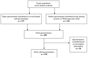
Figure 1 Flowchart showing the inclusion and exclusion criteria and study population (questionnaires considered for analysis) n=numbers.
Data were underwent descriptive and frequency analysis. Statistical significance was calculated by chi-square and Fisher's exact test, using SPSS software version 24.0 (IBM Corp., Armonk, NY) and significance level of 5%.
Results
Table 1 shows the sample distribution accor- ding to sex, age group and workplace. Most dentists are between 20 and 29 years old (43.7%, n=209) and work in shared dental clinics (34.7%, n=166).
Regarding the use of intraoral image receptor, 51% (n=244) of the dentists uses digital receptors (PSP and or solid sensor), while 68.4% (n=327) uses radiographic films. Some dentists use exclusively radiographic films (48.9%, n=234) and some others use exclusively digital receptors (31.6%, n=151). Among digital image receptors, PSP is more used than solid sensors (Table 2). Dentists between 20 and 29 years old use more radiographic films than other age groups (p=0.001). General practitioners, implantodontists and orthodontists use more radiographic films than other dental specialties (p<0.001), whereas oral radiologists use more digital image receptors (mainly PSP) than radiographic films (p<0.001) (Table 2).
As for extraoral imaging exams, dentists use more panoramic radiography (PAN) than skull radiographies (44%, n=245). Considering the age group and type of image receptor, dentists between 20 and 29 years old use more radiographic film than digital receptors for both PAN and skull radiography (p=0.004). This same age group uses less skull radiography and cone-beam computed tomography (CBCT) than the others (p=0.007). PAN is less used by edodontists and more used by oral radiologists (p<0.001). When compared to endodontists, general practitioners, periodon- tists and prosthodontists, oral radiologists perform digital skull radiography more often (p<0.001). CBCT is most often performed by oral radiologists and oral and maxillofacial surgeons (p<0.001) (Table 3).
Of those dentists who use digital receptors, most use digital image enhancement tools (87.8%, n=209 from 238). Contrast (89.5%, n=187), zoom (80.4%, n=168), brightness (74.2%, n=155) and measurements (57.9%, n=121) are the tools used most often. Dentists aged between 20 and 29 years use digital image enhancement tools the least (p=0.020). When comparing dental specialties, oral radiologists use digital image enhancement tools most often, mainly contrast, brigh ness, sharpness, zoom, and measurement tools (p<0.05) (Table 4).
Email is the method most commonly used to send digital imaging exams with colleagues (76.9%, n=183) and patients (67.6%, n=161). Cloud storage and app are also significative methods to transfer images. Of those who use digital imaging system, nearly half still print the imaging exams for send them to other dentists or patients; the 20-29 age group prints imaging exams less often than the others (p=0.003). As for dental specialties, oral radiologists and endodontists print exams more and less often, respectively (p<0.0001). Regarding cloud storage, oral radiologists use it more often and general practitioners use it less often (p<0.0001) (Table 5).
While most dentists do not use app and social media to share imaging exams (74.1%, n=177) (Table 6), of those who do, most use it to share experiences (n=42) (Table 7). Age and specialty did not influence the sharing of imaging exams on app or social media (p>0.05).
Table 1 Sample distribution, according to sex, age group and workplace.
| - | - | Workplace | - | - | - | - | - | - | - | - |
|---|---|---|---|---|---|---|---|---|---|---|
| Sex | Age group | Shared DC | Shared DC+Personal DC | Shared DC+Dental school | Personal DC | ORC | ORC+Dental school | Public DC | Dental school | Total |
| - | Without answer | 2 | 0 | 0 | 1 | 1 | 0 | 0 | 1 | 5 |
| - | 20-29 | 65 | 19 | 7 | 16 | 11 | 1 | 13 | 23 | 155 |
| - | 30-39 | 31 | 11 | 3 | 21 | 17 | 2 | 1 | 9 | 95 |
| Female | 40-49 | 13 | 3 | 1 | 10 | 5 | 0 | 2 | 2 | 36 |
| - | 50-59 | 3 | 0 | 1 | 6 | 8 | 1 | 1 | 2 | 22 |
| - | 60-69 | 0 | 1 | 0 | 1 | 0 | 0 | 0 | 0 | 2 |
| - | Total | 114 | 34 | 12 | 55 | 42 | 4 | 17 | 37 | 315 |
| - | Without answer | 1 | 0 | 0 | 1 | 2 | 1 | 0 | 0 | 5 |
| - | 20-29 | 27 | 2 | 5 | 2 | 8 | 0 | 3 | 7 | 54 |
| - | 30-39 | 12 | 5 | 5 | 7 | 14 | 1 | 0 | 3 | 47 |
| - | 40-49 | 8 | 3 | 1 | 8 | 20 | 1 | 0 | 2 | 43 |
| Male | 50-59 | 3 | 0 | 0 | 5 | 2 | 0 | 0 | 2 | 12 |
| - | 60-69 | 1 | 0 | 0 | 0 | 0 | 0 | 0 | 0 | 1 |
| - | 70-80 | 0 | 1 | 0 | 0 | 0 | 0 | 0 | 0 | 1 |
| - | Total | 52 | 11 | 11 | 23 | 46 | 3 | 3 | 14 | 163 |
| - | Total | 166 | 45 | 23 | 78 | 88 | 7 | 20 | 51 | 478 |
| - | Without answer | 2 | 0 | 0 | 1 | 1 | 0 | 0 | 1 | 5 |
Dental clinic (DC); Oral radiology clinic (ORC).
Table 2 Types of intraoral image receptors used, according to age group and dental specialty.
| - | Intraoral image receptors | - | - | -- | - | - | - | - |
|---|---|---|---|---|---|---|---|---|
| Age Group | Film | Film + PSP | Film + PSP +Solid sensor | Film +Solid sensor | PSP | Solid sensor | PSP +Solid sensor | Total |
| Without answer | 2 | 0 | 1 | 2 | 3 | 2 | 0 | 10 |
| 20-29 | 123 | 5 | 2 | 31 | 27 | 17 | 4 | 209 |
| 30-39 | 67 | 13 | 0 | 17 | 36 | 4 | 5 | 142 |
| 40-49 | 28 | 0 | 3 | 12 | 23 | 8 | 5 | 79 |
| 50-59 | 11 | 1 | 2 | 4 | 11 | 4 | 1 | 34 |
| 60-69 | 3 | 0 | 0 | 0 | 0 | 0 | 0 | 3 |
| 70-80 | 0 | 0 | 0 | 0 | 0 | 1 | 0 | 1 |
| Total | 234 | 19 | 8 | 66 | 100 | 36 | 15 | 478 |
| DENTAL SPECIALTY | - | - | - | - | - | - | - | - |
| General practice | 62 | 2 | 1 | 16 | 12 | 6 | 2 | 101 |
| OMFS | 5 | 1 | 1 | 2 | 2 | 0 | 0 | 11 |
| Restorative dentistry | 7 | 0 | 0 | 3 | 2 | 0 | 0 | 12 |
| Endodontics | 26 | 3 | 0 | 14 | 1 | 7 | 0 | 51 |
| Implantodontist | 25 | 0 | 0 | 4 | 2 | 2 | 0 | 33 |
| Pediatric dentistry | 11 | 1 | 0 | 4 | 0 | 2 | 0 | 18 |
| Orthodontics | 45 | 2 | 0 | 6 | 9 | 7 | 0 | 69 |
| Periodontics | 8 | 0 | 2 | 2 | 3 | 5 | 1 | 21 |
| Prosthodontics | 17 | 1 | 1 | 6 | 2 | 1 | 1 | 29 |
| Oral Radiology | 11 | 9 | 3 | 8 | 66 | 3 | 10 | 110 |
| Others | 17 | 0 | 0 | 1 | 1 | 3 | 1 | 23 |
| Total | 234 | 19 | 8 | 66 | 100 | 36 | 15 | 478 |
Photostimulable phosphor (PSP); Solid sensor (charge-coupled device (CCD) and Complementary metal oxide semiconductor (CMOS); Oral and maxillofacial surgery (OMFS). Bold number indicates statistically significant difference from the others within the same category (p<0.05).
Table 3 Extraoral imaging exams used, according to age group and dental specialty.
| - | Imaging exam | - | - | - | - | - | - | - | - | - |
|---|---|---|---|---|---|---|---|---|---|---|
| Age Group | Panoramic Radiography | - | - | - | Skull radiography | - | - | - | CBCT | - |
| - | No use | Digital | Conven- tional | Digital + Conven- tional | No use | Digital | Conven- tional | Digital + Conven- tional | No use | Use |
| Without answer | 3 | 5 | 1 | 1 | 7 | 2 | 0 | 1 | 5 | 5 |
| 20-29 | 107 | 60 | 26 | 16 | 167 | 28 | 10 | 4 | 152 | 57 |
| 30-39 | 69 | 58 | 7 | 8 | 87 | 44 | 7 | 4 | 85 | 57 |
| 40-49 | 37 | 38 | 1 | 3 | 51 | 26 | 0 | 2 | 46 | 33 |
| 50-59 | 14 | 17 | 1 | 2 | 21 | 12 | 0 | 1 | 17 | 17 |
| 60-69 | 3 | 0 | 0 | 0 | 3 | 0 | 0 | 0 | 3 | 0 |
| 70-80 | 0 | 1 | 0 | 0 | 0 | 1 | 0 | 0 | 0 | 1 |
| Total | 233 | 179 | 36 | 30 | 336 | 113 | 17 | 12 | 308 | 170 |
| DENTAL SPECIALTY | - | - | - | - | - | - | - | - | - | - |
| General practice | 62 | 24 | 11 | 4 | 88 | 7 | 6 | 0 | 80 | 21 |
| OMFS | 1 | 4 | 3 | 3 | 6 | 1 | 2 | 2 | 3 | 8 |
| Restorative dentistry | 9 | 3 | 0 | 0 | 12 | 0 | 0 | 0 | 10 | 2 |
| Endodontics | 40 | 5 | 6 | 0 | 51 | 0 | 0 | 0 | 40 | 11 |
| Implantodontist | 21 | 7 | 4 | 1 | 30 | 1 | 1 | 1 | 27 | 6 |
| Pediatric dentistry | 10 | 5 | 2 | 1 | 16 | 1 | 0 | 1 | 14 | 4 |
| Orthodontics | 40 | 20 | 4 | 5 | 49 | 12 | 6 | 2 | 56 | 13 |
| Periodontics | 15 | 3 | 0 | 3 | 21 | 0 | 0 | 0 | 17 | 6 |
| Prosthodontics | 14 | 7 | 2 | 6 | 27 | 1 | 0 | 1 | 20 | 1 |
| Oral Radiology | 10 | 95 | 0 | 5 | 17 | 87 | 2 | 4 | 22 | 7 |
| Others | 11 | 6 | 4 | 2 | 19 | 3 | 0 | 1 | 19 | 91 |
Cone-beam computed tomography (CBCT); Oral and maxillofacial Surgery (OMFS).
Table 4 The use and type of digital image enhancement tools used, according to age group and dental specialty.
| Age Group | Use of enhancement tools | - | - | - | - | - | - | - | - |
|---|---|---|---|---|---|---|---|---|---|
| - | No | Yes | Bright- ness | Contrast | Zoom | Measure- ment | Inversion | Relief | Sharp- ness |
| Without answer | 0 | 6 | - | - | - | - | - | - | - |
| 20-29 | 18 | 68 | 49 | 60 | 52 | 35 | 15 | 8 | 26 |
| 30-39 | 4 | 69 | 49 | 59 | 57 | 39 | 15 | 7 | 29 |
| 40-49 | 4 | 45 | 37 | 43 | 37 | 28 | 16 | 12 | 24 |
| 50-59 | 3 | 21 | 16 | 19 | 18 | 16 | 10 | 6 | 9 |
| Total | 29 | 209 | 155 | 187 | 168 | 121 | 57 | 36 | 91 |
| DENTAL SPECIALTY | - | - | - | - | - | - | - | - | - |
| General practice | 10 | 29 | 18 | 25 | 21 | 13 | 6 | 5 | 11 |
| OMFS | 0 | 6 | 3 | 5 | 6 | 5 | 3 | 0 | 0 |
| Restorative dentistry | 2 | 3 | 2 | 2 | 1 | 1 | 2 | 1 | 1 |
| Endodontics | 5 | 20 | 13 | 17 | 15 | 14 | 11 | 4 | 7 |
| Implantodontist | 0 | 8 | 5 | 8 | 6 | 4 | 2 | 0 | 3 |
| Pediatric dentistry | 2 | 4 | 4 | 3 | 3 | 0 | 0 | 0 | 0 |
| Orthodontics | 3 | 21 | 10 | 17 | 17 | 8 | 3 | 0 | 9 |
| Periodontics | 1 | 11 | 8 | 11 | 11 | 7 | 3 | 0 | 3 |
| Prosthodontics | 3 | 8 | 6 | 7 | 7 | 6 | 4 | 1 | 4 |
| Oral Radiology | 3 | 93 | 82 | 86 | 77 | 59 | 22 | 24 | 51 |
| Others | 0 | 6 | 4 | 6 | 4 | 4 | 1 | 1 | 2 |
| Total | 29 | 209 | 155 | 187 | 168 | 121 | 57 | 36 | 91 |
Oral and maxillofacial Surgery (OMFS).
Table 5 Methods to send imaging exams among dentists and from dentist to patients.
| Age Group | Dentist to dentist | - | - | - | - | - | - | - | - | Dentist to patient | - | - | - | - |
|---|---|---|---|---|---|---|---|---|---|---|---|---|---|---|
| - | Printing | Cloud storage | Portable storage | Social media | App | Sites | Printing | Cloud storage | Portable storage | Social media | App | Sites | ||
| Without answer | 3 | 3 | 5 | 3 | 2 | 4 | 3 | 3 | 4 | 4 | 1 | 0 | 1 | 2 |
| 20-29 | 27 | 60 | 35 | 14 | 7 | 30 | 14 | 46 | 54 | 7 | 9 | 4 | 17 | 6 |
| 30-39 | 33 | 63 | 41 | 20 | 5 | 28 | 17 | 41 | 50 | 12 | 6 | 1 | 21 | 8 |
| 40-49 | 30 | 38 | 30 | 12 | 6 | 22 | 12 | 31 | 34 | 8 | 3 | 0 | 13 | 8 |
| 50-59 | 14 | 19 | 14 | 11 | 4 | 6 | 4 | 15 | 19 | 9 | 4 | 4 | 4 | 2 |
| Total | 107 | 183 | 125 | 60 | 24 | 90 | 50 | 136 | 161 | 40 | 23 | 9 | 56 | 26 |
| DENTAL SPECIALTY | - | - | - | - | - | - | - | - | - | - | - | - | - | - |
| General practice | 12 | 24 | 11 | 7 | 5 | 19 | 2 | 21 | 21 | 3 | 4 | 1 | 11 | 2 |
| OMFS | 2 | 5 | 4 | 1 | 0 | 2 | 1 | 3 | 4 | 3 | 1 | 0 | 2 | 0 |
| Restorative dentistry | 1 | 5 | 2 | 0 | 0 | 0 | 0 | 3 | 3 | 1 | 0 | 0 | 1 | 0 |
| Endodontics | 4 | 18 | 7 | 6 | 2 | 12 | 2 | 7 | 17 | 3 | 1 | 0 | 9 | 0 |
| Implantodontist | 3 | 7 | 5 | 5 | 1 | 3 | 2 | 4 | 4 | 1 | 2 | 1 | 3 | 1 |
| Pediatric dentistry | 0 | 5 | 1 | 1 | 0 | 2 | 1 | 2 | 4 | 0 | 0 | 0 | 3 | 0 |
| Orthodontics | 7 | 20 | 11 | 6 | 2 | 8 | 5 | 9 | 19 | 2 | 2 | 2 | 7 | 2 |
| Periodontics | 2 | 12 | 8 | 2 | 0 | 5 | 1 | 2 | 4 | 2 | 2 | 0 | 1 | 1 |
| Prosthodontics | 5 | 8 | 6 | 1 | 1 | 6 | 0 | 5 | 12 | 3 | 1 | 0 | 5 | 0 |
| Oral Radiology | 70 | 76 | 66 | 29 | 12 | 30 | 34 | 5 | 7 | 3 | 1 | 1 | 6 | 0 |
| Others | 1 | 3 | 4 | 2 | 1 | 3 | 2 | 75 | 66 | 19 | 9 | 4 | 8 | 20 |
| Total | 107 | 183 | 125 | 60 | 24 | 90 | 50 | 136 | 161 | 40 | 23 | 9 | 56 | 26 |
Oral and maxillofacial Surgery (OMFS); Applications (App).
Table 6 Use of applications (app) and social media for sharing imaging exams.
| - | Sharing imaging exams on the internet | - | Ways of sharing imaging exams on internet | - |
|---|---|---|---|---|
| Age Group | No | Yes | APP | SOCIAL MEDIA |
| No informed | 3 | 3 | 1 | 2 |
| 20-29 | 66 | 20 | 7 | 12 |
| 30-39 | 55 | 19 | 2 | 15 |
| 40-49 | 38 | 11 | 2 | 6 |
| 50-59 | 14 | 9 | 5 | 3 |
| 70-80 | 1 | 0 | 0 | 0 |
| Total | 177 | 62 | 17 | 38 |
| DENTAL SPECIALTY | - | - | - | - |
| General practice | 29 | 10 | 4 | 6 |
| OMFS | 4 | 2 | 0 | 1 |
| Restorative dentistry | 4 | 1 | 0 | 0 |
| Endodontics | 17 | 8 | 2 | 6 |
| Implantodontist | 5 | 3 | 1 | 1 |
| Pediatric dentistry | 6 | 0 | 0 | 0 |
| Orthodontics | 21 | 3 | 0 | 2 |
| Periodontics | 10 | 3 | 0 | 2 |
| Prosthodontics | 9 | 2 | 1 | 1 |
| Oral Radiology | 70 | 26 | 7 | 18 |
| Others | 2 | 4 | 2 | 1 |
| Total | 177 | 62 | 17 | 38 |
Oral and maxillofacial surgery (OMFS).
Table 7 Reasons for the sharing of imaging exams by dentists.
| Age Group | Experience sharing | Doubts | Publicity | Academic |
|---|---|---|---|---|
| No informed | 2 | 1 | 2 | 2 |
| 20-29 | 15 | 11 | 10 | 12 |
| 30-39 | 12 | 3 | 12 | 8 |
| 40-49 | 7 | 4 | 7 | 2 |
| 50-59 | 6 | 6 | 4 | 1 |
| Total | 42 | 25 | 35 | 25 |
| DENTAL SPECIALTY | - | - | - | - |
| General practice | 5 | 7 | 3 | 7 |
| OMFS | 0 | 0 | 0 | 2 |
| Restorative dentistry | 0 | 0 | 1 | 0 |
| Endodontics | 7 | 3 | 5 | 3 |
| Implantodontist | 3 | 1 | 2 | 0 |
| Pediatric dentistry | 0 | 0 | 0 | 0 |
| Orthodontics | 2 | 0 | 3 | 1 |
| Periodontics | 1 | 1 | 2 | 1 |
| Prosthodontics | 2 | 2 | 1 | 1 |
| Oral Radiology | 18 | 9 | 15 | 8 |
| Others | 4 | 2 | 3 | 2 |
| Total | 42 | 25 | 35 | 25 |
Oral and maxillofacial surgery (OMFS).
Discussion
Image receptors and imaging exams
The present study found that most dentists in Brazil still use exclusively radiographic film (conventional system); while in many European countries and in the United States digital radiology is a reality (4,6,7). A study conducted in Brazil found no significant associations between age and the use of digital radiography (5). Contrary to expectations, the present study found that the 20-29 age group used more radiographic film than digital image receptor, compared to other age groups. Such finding may be explained by the fact that dentists in this age group are often working in facilities without access to digital systems (e.g., public dental clinics, postgraduate courses in educational institutions and shared clinics with no digital system available). Another hypothesis is that most of them would lack sufficient economic power to invest in digital systems, since they are starting their careers.
When comparing dental specialties, this study found that oral radiologists use more digital image receptors than radiographic films. Oral radiologists usually work in oral radiology clinics, which invest in technology in an effort to improve the agility of services and the quality of radiographic images. Among the image receptors, it was found that PSP is more commonly used than solid sensor, what is in line with a Belgium study (4). General practitioners, implantodontist and orthodontists, in turn, use more radiographic film than digital image receptors and this finding may be explained by the fact that, in Brazil, it is not commom these dental specialties have digital imaging systems in their own dental clinics.
Among extraoral exams, PAN is most used is clinical practice, possibly due to its good overview of the patient's jaws, dentition and important neighboring anatomical structures, such as maxillary sinuses and mandibular canal, with low radiation dose (2). However, for endodontists, PAN has a low demand probably because they need detailed images of the root apex and adjacent alveolar bone, which is better provided by periapical radiography. Oral radiologists, on the other hand, work with PAN most often, most likely because this imaging exam is usually performed in oral radiology clinics. CBCT are also performed in oral radiology clinics, thus justifying the greater use of this exam among radiologists. Surgeons also frequently use CBCT due to the possible three-dimensional visualization of the maxillofacial structures, which is important for surgical planning in order to avoid damaging the anatomical structures (8).
Digital image enhancement tools
In the present study most dentists using digital systems apply digital image enhancement tools, a result in line with Rovaris et al. (2016) (5). Studies evaluating dentists' perception of the quality of digital radiographs found that some dentists preferred the enhanced image to the original (1,9). Rovaris et al. (2016) (5) found that brightness and contrast adjustments and zoom were the digital tools most used by Brazilian dentists, as in the present study. Such a finding may be explained by the fact that brightness and contrast adjustments can be easily made in any imaging software and are usually applied according to personal visual preferences (3). A previous study showed the importance of brightness and contrast adjustments for specific diagnostic tasks, such as soft-tissue calcifications (2). Similarly, other previous studies (10,11) found a high preference for the zoom tool due to its intuitiveness (12). On the other hand, inversion and relief were the digital tools least used by Brazilian dentists. Inversion tool has no relevance for improving the visibi- lity of anatomical structures or diagnosing bone loss measurements (13,14). As expected, oral radiologist is the dental specialty that uses digital image enhancement tools (e.g., zoom, brightness, contrast, and measurement tools) most often.
Contrary to expectations, i.e, that younger professionals would use digital image enhancement tools more often due to their familiarity with technology, dentists between 20 and 29 years old use less them than the others age groups. The present authors hypothesized that such 20-29 age group may have less practice with those tools when compared to more experienced professio- nals, thus its low use. The lack of training may also result from the undergraduate course, since not all Brazilian universities offer access to digital radiology during practice (15), showing that the bureaucratic processes and the high costs of digital systems, compared to conventional ones, continue to be obstacles to increase the use of digital systems.
Communication and social media
Email appears as the method most commonly used by Brazilian dentists to send imaging exams to colleagues and patients, which is in line with a previous Belgian study (4). Online tranfer of digital files can be useful in healthcare because it is a fast and convenient process (16). Many radiographic image file formats can be easily used and transferred, such as tagged image file format (TIFF), bitmap file format (BMP), portable network graphics (PNG), joint photographic experts group (JPEG), and digital imaging and communications in medicine (DICOM). Previous studies showed that digital radiographic images can be stored in multiple file formats without affecting task-specific diagnosis (16,17).
How dentists send imaging exams varies according to age and dental specialty. The present study showed that younger dentists print exams less than other age groups. Although support campaigns have been held to reduce the printing of radiographic exams in an effort to preserve the environment and get all the benefits specific to digital imaging exams, a significant percentage of dentist still print exams obtained by digital systems, reinforcing the need for more campaigns in this regard.
As for the dental specialty, endodontic professionals generally use imaging exams during treatment for personal evaluation, which culminates in less printed exams. Oral radiologists, in turn, print more imaging exams to send to other professionals. This seems paradoxical for radiologists should understand the benefits of not printing digital images; since they provide a service to other specialties, however, radiologists have to follow the society's culture of having printed images. Moreover, oral radiologists more often use cloud storage and websites to send exams to other professionals or patients, since radiology clinics have their own websites and cloud storage generates greater security regarding backup and reduces physical storage space (e.g., computers and external storage devices).
Exchange of experiences and discussion of clinical cases appears as the primary reasons for dentists to share images via social media. It is interesting to note, however, that such images can only be used with the patient's consent (18).
Conclusion
While most Brazilian dentists use digital imaging systems, a significant part still use exclusively radiographic films, with younger dentists employing more radiographic films than other age groups. PAN is the extraoral modality most used by dentists. Brightness and contrast adjustments, zoom and measuments are the digital enhancement tools most used. Regarding dental specialty, oral radiologists use digital image enhancement tools most often. Email is the main method used to send imaging exams to other professionals and patients. Finally, the main reason to share images on social media is to exchange experience in dentistry community.
Author contribution statement
Conceptualization and design: L.M.S, E.D.C. and D.Q.F.
Literature review: L.M.S., L.O.R., F.N.R., E.D.C. and D.Q.F.
Methodology and validation: L.M.S, E.D.C and D.Q.F. Formal analysis: D.Q.F.
Investigation and data collection: L.M.S., L.O.R. and F.N.R.
Resources: L.M.S., L.O.R., F.N.R., E.D.C. and D.Q.F.
Data analysis and interpretation: L.M.S., L.O.R., F.N.R., E.D.C. and D.Q.F.
Writing-original draft preparation: L.M.S., L.O.R. and F.N.R.
Writing-review & editing: L.M.S., L.O.R., F.N.R.,
E.D.C. and D.Q.F. Supervision: D.Q.F
Project administration: L.M.S.

