


 English (pdf)
 Article in xml format
 Article references
 Send this article by e-mail
 Cited by SciELO
  Similars in
SciELO
Similars in
SciELO 
 Permalink
Introduction
The primary purpose of cavity preparation in dental restorations is to preserve the remaining tooth structure and remove caries, tooth tissues, and microorganisms that can cause secondary caries after restoration (1). However, the cavity also requires a good seal to prevent the development of secondary caries and to prolong the life of the composite restoration (2). Some bacteria have been reported to remain in the dentin tubules and smear layer even after all the soft and infected dentin has been removed, suggesting it is impossible to remove all microorganisms from the cavity. Therefore, prior to restorative procedures, using an antimicrobial agent for the disinfection of the cavities is advised (3). In recent years, various methods for cavity disinfection in dentistry have been researched and used clinically (3,4,5,6,7). Chlorhexidine (CHX) has high antibacterial activity against both gram-positive, especially Strepto- coccus mutans, and gram-negative bacteria. CHX displays a bactericidal effect at high concentrations and a bacteriostatic effect at low concentrations (8). While CHX attacks the cytoplasmic membrane by destroying the cell wall at low concentrations, it induces cytoplasmic conjugation at high concentrations, causing the intracellular components to clot (8). In addition, CHX plays an important role in increasing the strength of the bond between the resin and dentin by inhibiting the dentin matrix metalloproteinases (MMPs) and cysteine cathepsins (9,10). However, it has been stated that the inhibitory effect of CHX on MMPs gradually disappears due to its dissolution in water and reversible electrostatic bonds (11).
Ozone (O3), which is used in cavity disinfec- tion, is a strong oxidant with antibacterial properties (4). The disinfecting effect of ozone is to eliminate the intracellular components of microorganisms through an oxidation reaction involving the rupture of cell membranes. O3 has antimicrobial efficacy against oral microorganisms, especially Streptococcus mutans (12,13,14). Some studies have reported that the application of ozone for 10 to 60 s is sufficient for antimicrobial activity (12, 14). A study by Baysan et al. has revealed that 10 s of ozone application decreased the amount of S. mutans and Streptococcus sobrinus (12).
At present, lasers are widely used in various applications in restorative dentistry. Types of lasers, such as CO2 Er: YAG, Er, Cr: YSGG, and Nd: YAG, are effective in removing smear layer and debris (15). Several studies have found that various laser types have an antibacterial effect on microorganisms (5,16). Lasers have also been more effective in eliminating microorganisms and preventing residual caries due to their advanced ability to penetrate dentin tubules (16). One of the most popular laser types used today is the Er, Cr: YSGG laser that radiates energy at a wavelength of 2780nm, which is absorbed well by all biological tissues, including enamel and dentin (17). In a study, Er, Cr: YSGG laser irradiation has resulted in less S. mutans accumulation than the control group (16). However, there are very few studies regarding the use of Er, Cr: YSGG laser irradiation for cavity disinfection (15,18).
The type of adhesive used to evaluate the bond strength is also important. Currently used adhesive systems simplify the adhesion technique and provide a long-term bond. ''Universal'' adhesives, which can be applied indefinitely in etch and rinse or self-etch mode, were introdu- ced to remove complications and provide a single product for all occasions (19). As defined in the ''adhesion/decalcification'' concept of self-etch adhesives, bonding can be accomplished due to the ability of the functional monomers to chemically bond to hydroxyapatite (20). 10-Methacryloyloxidesyl dihydrogen phosphate (10-MDP), one of the functional monomers used, has shown a very effective and durable bond with dentin due to the low solubility of the calcium salt formed on the hydroxyapatite surface (10, 21). MDP could partially dissolve the smear layer and increase monomer penetration into dental substrates (22, 23). It could also self-assemble into a nano-layered structure through the deposition of MDP-Ca salts (24).
However, there are some concerns about using cavity disinfectant since it may influence the sealing and bonding strength of adhesive resins (15). Some studies have suggested that disinfection methods reduce the bonding strength of adhesive systems (25,26). CHX may also affect the effectiveness of some compounds in the adhesive resins, further reducing bond strength; as a result, it may potentially be chemically incompatible with some adhesives (27). Therefore, adhesives containing MDP can be used to improve the strength and durability of the bond. Phosphate ester monomers commonly found in dental adhesives, such as MDP, often interact clinically with CHX (10). However, studies that examine the effects of MDP and various disinfection methods (such as CHX) on dentin bond strength are limited. For these reasons, in this study, a universal adhesive that contains an MDP monomer was used to identify the effect of disinfection methods on microtensile bond strength. In addition, the effect of the disinfection method on resin penetration of the dentin was investigated. The null hypothesis of this study is that the application of different cavity disinfection protocols will not affect the microtensile bond strength of an MDP-containing universal adhesive.
Materials and methods
Tooth preparation
In this study, sixteen human third molar teeth without caries were used. Twelve teeth were used for the microtensile bond strength (µTBS) test, and four teeth were evaluated using confocal laser scanning microscopy (CLSM). The teeth used were collected from patients with their informed consent and under the guidance of a protocol approved by the Akdeniz University Faculty of Medicine Clinical Research Ethics Committee (approval number: 21/10/2020-819). Organic debris on the teeth was removed with a periodontal scaler. Then, the teeth were stored in 5% chloramine T solution for two weeks to disinfect them and kept in distilled water at 4°C until use. Teeth were embedded in self-cured acrylic resin using a silicone mold of the roots. To expose the dentin tissue, the teeth were cut perpendicularly to their long axis from the mid-coronal of the crown, using a slow-speed diamond-impregnated disk under water cooling in the cutting device (Isomet, Buehler Ltd., Lake Bluff, IL, USA). As a result of this process, the surface of the flat mid-coronal dentin was exposed. Each exposed dentin surface was wet-polished with 600-grit silicon carbide paper for 60s to obtain a standardized smear layer (28). Silicon carbide papers were renewed after each tooth.
The teeth were randomly divided into four groups (n=3):
Group I: (control) No disinfection was applied to the samples in this group.
Group II: (CHX) A cotton pellet absorbed in 2% chlorhexidine gluconate solution (Microvem, Istanbul, Turkey) was applied to the dentin surface for 20s (29). Then, the dentin surfaces were air-dried for 10s.
Group III: (Ozone) A tip suitable for the disinfection mode was attached to the dental ozone generator (OzoneDTA, Apoza, New Taipei City, Taiwan), and ozone was applied to the dentin surface for 30s.
Group IV: (Laser) An Er, Cr: YSGG laser (Waterlase Biolase, Biolase Technology, Inc. CA, USA) with a wavelength of 2780nm and a pulse duration of 140 microseconds was used for laser irradiation of the dentin surfaces. The applied laser energy was 0.75 watts, with 15% water and 15% air pressure. Irradiation was carried out with a 600-µm diameter sapphire tip 1mm from the surface. The laser irradiation was applied to the dentin surfaces five times with an application time of 10s at intervals of 5s (18).
Immediately after disinfection by each method, the universal adhesive (G-Premio Bond, GC, Tokyo, Japan) was applied using the self-etch method following the manufacturer's instructions. G-Premio Bond was applied to the dentin surface using a micro brush. After waiting for 10s, it was dried under air pressure for 5s and lightcured with a light-emitting diode curing unit (Valo, Ultradent, South Jordan, UT, USA) in the standard mode (irradiance: 1000 mW/cm²) for 10s. Then, the composite resin (Charisma Smart, A2 shade, Heraeus Kulzer GmbH, Hanau, Germany) was applied to the dentin surface incrementally to a height of 5mm. Each 2mm composite increment was light-cured for 20s. The materials used in this study are summarized in Table 1. The samples to which composite resins were applied were stored in distilled water at 37°C for 24 hours.
Table 1 Composition of dental materials used in the present study.
*Abbreviations: 10-MDP, 10-methacryloyloxydecyl dihydrogen phosphate; 4-MET, 4-methacryloxyethyl trimellitic acid; MEPS, methacryloyloxyalkyl thiophosphate methylmethacrylate; TPO, diphenyl (2,4,6-trimethylbenzoyl) phosphine oxide; Bis-GMA, bisphenol-A-glycidyl methacrylate
Microtensile bond strength test
The prepared teeth were sectioned occlusogingivally with a low-speed diamond saw to obtain composite dentin beams with approximately 1×1 mm² areas of bonding. The thickness of the bonded area was measured with a digital caliper (Mitutoyo Digimatic, Tokyo, Japan). Approximately six or seven beams were obtained from each tooth and in total, 20 samples were analyzed in each group to determine µTBS. Each sample was attached to the microtensile test machine (Bisco, Schaumburg, IL, USA) with a cyanoacrylate adhesive and then stressed to obtain the µTBS values at a crosshead speed of 1mm/min. The μTBS values are stated in MPa. Following the µTBS test, the fracture modes of the samples were analyzed at a magnification of 40× with a stereomicroscope (Zeiss Stemi, CarlZeiss/GmbH, Germany).
The failure types were classified into three categories: (i) adhesive (the fracture site was at the resin-dentin interface), (ii) cohesive (the fracture site was in the resin composite), and (iii) mixed (the fracture site contained both adhesive and cohesive failure).
Confocal laser scanning microscopy
A total of four teeth, one tooth from each group, were prepared for monitoring under the CLSM. Rhodamine B (Sigma-Aldrich, Germany) fluorescent dye was added to the G-Premio Bond at a concentration of 0.008% before it was applied to the dentin surface (30). The application procedure was performed according to the manufacturer's instructions. The resin composite was then placed in 2-mm increments to create 4-mm layers. The teeth were sectioned longitudinally into 1-mm thick slabs. A slab from the center of each tooth was selected for the study. The samples were examined using a CLSM (Zeiss Lsm 510 Meta, Zeiss GmbH, Jena, Germany). The images were obtained using a 543nm wavelength HeNe1 laser and a 489 nm argon/2 laser at 40× magnification.
Statistical analysis
The statistical analyses were performed with SPSS-22 (SPSS Incorporated, Chicago, Illinois, USA). One-way ANOVA was used to examine the differences in antibacterial activity and microtensile bond strength among the groups, and the Tukey HSD post hoc test was used for multiple comparisons (p<0.05).
Results
Microtensile bond strength and fracture mode analysis
One-way ANOVA revealed that the µTBS values showed statistically significant differences (p<0.05). Figure 1 shows the mean µTBS values and standard deviations of each group. The highest µTBS was seen in the control group (35.13±6.20) (p<0.05), while the Laser (19.25±4.66) and CHX (23.07±7.01) groups showed the lowest µTBS values. There was no significant difference between the Laser and CHX groups or between the ozone (27.53±5.83) and CHX groups in terms of µTBS (p>0.05).
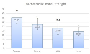
Figure 1 Mean µTBS (MPa) and Standard Deviation (SD) obtained in the tested groups. Different letters represent statistical differences among groups, according to Tukey test (p<0.05).
Table 2 shows the failure types of the dentin samples from the four disinfection groups. Generally, groups that showed low µTBS values had a higher tendency to adhesive failure. The CHX and Laser groups, which had the lowest µTBS values (95%), also predominantly showed adhesive failures. The control group, which had the highest bond strength value, showed the lowest number of adhesive and highest number of mixed failures.
Confocal laser scanning microscopy analysis
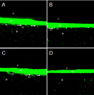
Figure 2 presents CLSM images showing the penetration of the adhesive material into the dentin in all groups. The control group exhibited a thick adhesive layer. Compared to other groups, thicker nano-layering, as well as longer and distinct resin tags, were observed across the entire surface (Figure 2.A). While thin nano-layering and short and thick resin tags were observed in the Ozone group, the adhesive layer was still evident (Figure 2.B). In the CHX group, a thicker adhesive layer and nano-layering were observed in some regions. Also, resin tags were evident and longer in some regions but shorter and irregular in some regions when compared to the control group (Figure 2.C).
The Laser group, which showed the lowest bond strength, had indistinct nano-layering and resin tags (Figure 2.D).
Discussion
After the mechanical removal of caries, residual bacterial species that cause secondary caries may multiply in dentin tubules, and the smear layer and bacterial proliferation may occur as irreversible pulpitis, apical periodontitis, and pulpal necrosis in the tooth (31). These undesirable outcomes increase the need to apply cavity disinfectant before the cavities are sealed with restorative materials (3). The ideal cavity disinfectant should have strong antimicrobial activity and avoid negative effects on the strength of the resin bond. Therefore, in this study, the effect of MDP-containing universal adhesive on µTBS was investigated by comparing the results gathered from groups of teeth treated with CHX, Ozone, and Er, Cr: YSGG laser irradiation. In the study, statistically significant differences were obtained between these cavity disinfectants. Thus, the null hypothesis was not accepted.
In dentistry, CHX has a strong antibacterial effect on oral bacteria, especially against S. mutans, and is one of the antimicrobial agents most commonly used to control dental plaque and tooth caries (25). CHX is expected to increase dentin bond strength as it has rewetting capacity, strong affinity, and high biocompatibility with the tooth structure (15,29). However, some researchers have reported that the pretreatment of CHX reduced the resin-dentin bond strength (25,29). In one study, using a self-etch adhesive has been associated with decreased bond strength when CHX was applied (25). Researchers stated that residues left on the dentin surface and tubules had caused a reduction in strength since the CHX is not washed off of the dentin surface (25). In a study by Ercan et al., the shear bond strength of the self- etch adhesive system to the dentin surface disinfected with NaOCl, H2O2, or CHX solutions was evaluated, and the CHX-treated material had significantly lower bond strength than the untreated control group (29). Similarly, in the current study, the µTBS value of dentin bonded to the universal adhesive by the self-etch method was significantly lower than the value of the control group as a result of the CHX application. In addition, Di Hipolito et al. have reported that CHX application showed a decreased number of calcium ions in the smear layer of dentin (32). CHX, which binds to both the organic components and minerals of dentin, may have a competitive approach by interacting chemically with the 10-MDP in the universal adhesive (33). This indicates that MDP may inhibit chemical bonding between the tooth and calcium (33). In this study, the application of CHX affected the bond strength values of the universal adhesive containing 10-MDP.
Ozone gas has been proposed as an antimicrobial agent that can reduce the number of microorganisms on the tooth surface (12,34). In an in vitro study, ozone application for 80 s was reported to reduce the number of microorganisms in an infected tooth cavity model to 2100ppm, which suggested that ozone has the potential to disinfect carious cavities (34). The disinfectant effect of ozone application for different durations has been studied, and the application of ozone for 20s or more was found to be effective in removing oral microorganisms (13,14).
Oxygen can react to free radicals during polymerization, affecting polymerization (35). Ozone is highly unstable and quickly converts to oxygen and can reduce the resin-dentin bonding by preventing the polymerization of the adhesive material because the reactivity of oxygen is much greater than that of the monomer (35,36). Similarly, after tooth whitening with hydrogen peroxide or carbamide peroxide, a decrease in bond strength has been seen due to the presence of residual oxygen (34,36). This study revealed a statistically significant decrease in µTBS values following ozone application compared to the control group. This might be because of the presence of residual oxygen after the use of ozone. Rodrigues et al. have stated that ozone application reduced the µTBS value at the resin-dentin interface (26). Dalkilic et al. (4), who assessed the µTBS of dentin after 60s of ozone application, have reported similar findings; ozone application decreased the bond strength.
Gurgan et al. (5) have shown that before restorative treatment, the application of ozone and Nd: YAG laser treatments to coronal and root dentin did not influence the shear bonding strength of self-etch adhesives. However, comparing the effects of three different cavity disinfectant agents on dentin in terms of shear bond strength of a self-etch and two universal adhesive systems, Akturk et al. (6) have recommended ozone as a cavity disinfectant. The inconsistency between these studies and the results mentioned above may be due to different times and doses, differences in adhesive types, and variability of the equipment supplying the ozone.
Erbium lasers, which have increased in popularity in recent years, are used for various applications such as cavity preparation, caries removal, sterilization, and improvement of dental hard tissues (15,18). In their studies, Turkun et al. have found that the antibacterial effect of an Er, Cr: YSGG laser against S. mutans, when used with 0.75W and 1W power output, was similar to that of CHX (37). Sufficient disinfection can be accomplished with an Er, Cr: YSGG laser, even at the low power setting of 0.75 W. However, attention should be paid to the irradiation settings in order to prevent thermal damage to the surrounding tissue while providing sufficient antibacterial effect (38). For these reasons, irradiation setting at a low dose (0.75 W) was used for cavity disinfection in our study.
There are conflicting reports of the bonding efficiency of adhesive systems applied to laserirradiated dentin surfaces in the literature. Some studies have reported decreased bond strength after laser irradiation, while others found that it increased (15, 39,40,41). In one study, an Er, Cr: YSGG laser was used at six different settings (3W/20 Hz, 3 W/35 Hz, 3 W/50 Hz, 1.5 W/20 Hz, 1.5 W/35 Hz, 1.5 W/50 Hz). The µTBS of self-etch adhesive on bovine teeth was compared with the control group. The results showed that different laser irradiation settings negatively affected the bonding strength of the adhesive systems at the resin-dentin interfaces (42). In another study, the µTBS of dentin surfaces irradiated with Er:YAG and Er, Cr: YSGG lasers were examined, and samples treated with laser irradiation had lower µTBS values than the control group (43). Deeb et al. (7) have evaluated the bond strength of surfaces treated with CHX, an Er, Cr: YSGG laser, and photodynamic therapy cavity disinfection protocols and found that the Er, Cr: YSGG laser-treated material showed lower bond strength than the untreated (control) group.
Similarly, in the current study, applying Er, Cr: YSGG laser irradiation as a cavity disinfectant reduced the µTBS. The decreased µTBS may be due to the complete melting and evaporation of the collagen in the organic matrix of the dentin surface from the increased temperature caused by the interaction between the laser radiation and the dentin. As a result, the denatured collagen fibrils fuse (44). Thus, diffusion of the adhesive resin into the interfibrillar collagen spaces is insufficient, and weaker bonding may occur in the dentin.
The variation within these results may be related to the composition of the adhesive systems. Recent universal adhesives are one-step self-etch adhesives designed under an ''all-in-one'' concept. In this concept, different etching modes allow clinically versatile use. In the current study, an MDP-containing universal adhesive was used. The chemical structure of 10-MDP contributes to the durability of the bond and improves the initial bonding performance of self-etch adhesives by intense and consistent adhesion to the calcium in hydroxyapatite (21).
When the CLSM images were examined, nano-layering was observed in all groups. More prominent nano-layering and resin tags were observed in the control group than the treated groups. In the CHX group, the resin tags are more prominent and longer in some regions, and the thickness of the nano-layering was close to the control group. This may be due to the MDP content of the universal adhesive. In addition, the nanolayering was the most indistinct in the Laser group, which showed the lowest µTBS value among all the groups.
Adhesive failure was predominant in all groups. But the CHX and Laser groups had more adhesive failures, which indicated a weaker resin-dentin bond and bond strength. Cohesive failure is associated with high adhesion strength (45). In this study, cohesive failure was observed in the Ozone and control groups. Mixed failure is more difficult to detect. If the bonding is strong, it is generally accepted that the fracture starts from the resin composite, propagates along the adhesive bond, and then spreads into the dentin (45). In the current study, the mixed failure rate of the control group, which demonstrated the highest bond strength, was higher than the other groups.
In this study, the effect of the different disinfectant methods applied to dentin surfaces on the initial (24h) µTBS values of the bonding of universal adhesives was studied; however, the long-term efficacy of the bond strength was not examined. Dental substrate type is another important factor that may affect the bonding strength. The age of the extracted teeth used in this study was not considered. Since the dentin tubule diameter changes with age, the dentin surface available for bonding may vary from tooth to tooth (46). Another factor contributing to the bond strength is the size of the dentin tubules. The size of the dentin tubules increases from the surface to the pulp chamber, and the dentin bond strength in the same tooth can vary depending on the location of the bonding (46). In this study, care was taken to remove all samples from the middle part of the tooth to eliminate this regional effect. The use of only one type of adhesive system is another constraint of this study.
Conclusion
According to the results of this study, the microtensile bond strength of G-Premio Bond to dentin decreases after disinfection with chlorhexidine, ozone, and Er, Cr: YSGG laser irradiation. The effect of these disinfection treatments on longterm bond strength should be investigated with long-term in vitro and in vivo studies.
DISCLOSURES
The authors have no conflict of interest to disclaim.
Author contribution statement
Conceptualization and design: D.B.C., A.D. and Ç.B.
Literature review: D.B.C. and A.D.
Methodology and validation: D.B.C. and A.D.
Formal analysis: D.B.C. and A.D.
Investigation and data collection: D.B.C. and A.D.
Data analysis and interpretation: Ç.B
Writing-original draft preparation: D.B.C.
Writing-review & editing: D.B.C. and A.D.
Supervision: Ç.B.

