


 English (pdf)
 Article in xml format
 Article references
 Send this article by e-mail
 Cited by SciELO
  Similars in
SciELO
Similars in
SciELO 
 Permalink
Introduction
Despite the advent of new methods for tooth preparation (such as lasers, air abrasion, and chemical dissolution), restorative dentistry still depends on the use of rotatory cutting instruments for enameloplasty, dentinoplasty, removal of previous restorations, caries removal, tooth reduction and preparation for fixed or removable prosthetics (1,2). High-speed handpieces using dental burs with diamond abrasives are the preferred cutting instruments used by dentists for tooth preparation over tungsten carbide and steel burs; mostly for their greater resistance to abrasion and wear, reduced heat generation during usage and longer service life (3,4,5).
A recent ADA ACE Panel Report reveals that 71% of participants in a national survey, use rotatory cutting instruments ''until worn out'' and the ''decrease of cutting efficiency'' was pointed as the primary indication to consider the bur as ''worn out'' (6). Although diamond burs can be cleaned, sterilized and re-used several times, there is a lack of information regarding clinical effects of using a worn bur. There is no consensus in the scientific literature about the maximum number of uses or number of procedures recommended for a clinician to use a dental bur; or even a standardized definition of what is considered a ''single use'', for example. Hence, it's not possible to be certain about the cutting efficiency of these instruments and usually one must rely on the bur's appearance to determine if, after multiple uses, it's still safe to use it avoiding unwanted side effects such as a greater heat generation, which could harm the dental pulp tissues, or an incomplete removal of infected or affected tissue.
As adhesion of restorative materials is a major influencing factor in the success of a prosthetic (or even direct) restoration nowadays, it is essential to understand the relationship between the type and condition of the applied bur during a tooth preparation and the resultant pattern of the substrate in terms of bonding performance. Different outcomes have been reported regarding the effect of different types and conditions (in terms of usage) of burs on dentin/resin bonding, depending also on the adhesive type employed (7,8,9,10). Currently, there are two main groups of commercially availa- ble adhesive systems, the etch-and-rinse and the self-etching. In the later, it can be included the so called ''universal adhesives'' or multi-mode adhesives which are essentially a modification of a self-etching adhesive which can be used applying both approaches, the etch-and-rinse and the self-etching. Due to its simpler and more permissive application technique (multi-mode), these single- component adhesives have become very popular recently (11). It has been proved that the various types of adhesive systems available, react diffe- rently to variations on dentin substrate roughness (and resultant smear layer after a tooth prepara- tion), produced by different types of burs (7,8,9,10). The etch-and-rinse adhesives are normally less affected by this situation, due to the employment of the phosphoric acid, which removes the surface pattern produced by any bur when the demineralization process of dental tissues occurs (11). Additionally, there is a growing tendency in restorative dentistry to avoid the etching step of the dentin with phosphoric acid, due to the possible higher risk of enzymatic and hydrolytic degradation that etch-and-rinse technique may induce, reason why self-etching strategy (carried out with self- etching adhesives or with universal adhesives in the self-etching mode) is preferred as it has shown similar bond strength and better interface longevity than etch-and-rinse adhesives (10). According to previous studies, bur type selection has a significant effect on the resin composite/dentin bond strength when using self-etching adhesives, as diamond burs produced significantly higher bond strength values than tungsten carbide burs when used to prepare a tooth (7,8). Also, softer or extra fine grid diamond burs tend to form thinner smear layers, which could eventually be removed easier or at least be better penetrated by a self-etching bonding agent, increasing the intimate contact between adhesive and dentin and producing stronger bonding, as reported previously (9,10). This may suggest that a bur, worn by multiple uses, may produce a smoother surface on dental tissues than a new one, and somehow it may affect future adhesion with resin-based materials by modifying the substrates' characteristics.
Considering this issue, it is still uncertain how a new or worn-by-use bur, may affect resin bonding to dentin when using a universal adhesive in a self-etching mode, as its performance depends between other factors, on the substrates' resultant roughness (after preparation process) and thick-ness and type of smear layer which may influence the intimate contact of the adhesive with the tissue. Thus, the aim of this study was to evaluate the effect of multiple uses of diamond burs on dentin superficial microroughness, burs' morphology and on resin-dentin bond strength when using a universal adhesive in a self-etching mode. The null hypothesis set was that multiple uses of diamond burs do not influence dentin surface roughness or resin-dentin bond strength.
Materials and methods
Burs and specimen preparation
The protocol for this in vitro study was reviewed and waivered by the Institutional Ethics Committee of the University of Costa Rica approval number CEC-180-2020. Komet parallel rounded tip diamond dental burs (Brasseler - Ref. No. 881 314 014) were selected. These burs are classified as a medium grain bur, have a cutting length of 8 mm and a head size of 1.4 mm, and are commonly used in dental preparations for fixed prosthetics. A previously calibrated single operator conducted all specimens' preparation procedures. Twenty- three extracted third molars, with no caries and no restorations, were anonymously donated on the six months before the experimental procedures were performed. All teeth were carefully pre-evaluated and randomly selected. The protocol for this study was reviewed and waivered by the Institutional Ethics Committee. Teeth were stored under controlled conditions of 100% humidity in saline solution and a temperature of 4ºC after cleansed and removal of the organic tissue with a dental curette. All procedures were performed using a high-speed handpiece (Kavo Unik) at a constant speed of 350,000rpm. The burs were randomly divided and tagged to be used in different molars until the corresponding number of uses was reached. Each use was defined as a complete preparation of a third molar for a fixed crown with parallel walls, contour's depth measuring the same as the bur's diameter and finished with a chamfer termination line. Once the number of uses was completed, the dental bur was cleaned using a soft toothbrush under running water and ultra- sonicated for 5 minutes. No thermal sterilization cycles were used to clean the burs to avoid the potential damage that extreme heat may cause to the bur's structure. Afterward, fresh clean molars were selected, and had its clinical crown removed at approximately one-half of the crown's length using a low-speed diamond precision saw (IsoMet 1000 Precision Sectioning Saw, Buehler) leaving a flat dentin surface. Each dentin surface was then divided into 4 areas randomly assigned to one experimental group depending on the number of previous dental preparations performed (0,1,5 and 10 uses respectively). After all the specimens were obtained, these were stored on saline solution under the same conditions previously detailed.
Scanning electron microscopy (SEM) analysis
Three dental burs from each experimental group were prepared and analyzed with a Scanning Electron Microscope (ASEM Microscopy JEOL JSM-6390LV) to describe the morphological changes suffered by burs' tip and body at 70X, 350X and 1000X. The most representative images were selected, and the defects observed were described according to the presence and severity of diamond crystals dislodgment and surface erosion.
SEM images were also prepared to illustrate and visually compare the effect on dentin's surface for the 0 uses bur and the 10 uses bur at 500X and 2000X. Finally, representative samples from the shear bond strength tests were analyzed with SEM to illustrate the failures observed.
Dentin microroughness analyses
Profilometry lectures were taken with a Dextak XT contact profilometer (Bruker) using a 2 µm radius stylus tip. The scan axis was taken with an applied force of 7mg and the resolution was set to 0.033µm/point corresponding to a stylus tip speed of 10µm/s. Triplicated measurements of a 1mm straight line of the prepared area were used to calculated mean roughness (Ra) for each experimental group. Representative 3D topographic images were randomly obtained selecting one specimen from each group, covering a total area of 500µm x 500µm. The image was constructed by stitching 500 scans with a separation of 1µm.
Shear bond strength analysis
For the shear bond strength analysis, the experiments were performed following the specifications from ''ISO 29022:213 Adhesion. Notchededge shear bond strength test''. Fifteen dental specimens from groups ''0 uses'' and ''10 uses'' were randomly selected. A customized device (Ultradent Products) was fixed to each specimen's occlusal flat surface with the aid of a two-face adhesive strip with a circular space matching the device's homolog space (2mm diameter x 2mm height). The isolated area was treated using a universal adhesive under the self-etching mode (Tetric N-Bond Universal, Ivoclar Vivadent) rubbing it for 20 seconds with a new micro brush. A thin coat was obtained after removing excesses and solvent was evaporated by air blowing the adhesive for 5 seconds. Adhesive layer was then photopolymerized for 10 seconds using a LED light curing unit at 1,200 mW/cm² (Bluephase N, Ivoclar Vivadent). Then, a 2mm resin composite (Tetric N-Ceram, Ivoclar Vivadent) increment was placed and photopolymerized for 10 seconds using the same intensity of the light curing unit, according to the manufacturer's indications, to build resin- composite cylinders (2x2mm). The plastic device was carefully removed, and all specimens were labeled and stored for 48 hours at 100% humidity at 37ºC temperature. The specimens were placed in a universal testing machine (Tinius Olsen HOIK-S) and a bond strength test was performed placing a customized chisel strictly parallel to the bonding area and applying a constant shear load at 1 mm/ min until failure. Data was collected to calculate bond strength values in MPa and specimens were preserved to be analyzed regarding the type of failure, with the aid of a digital optic microscope to determine if the failure was cohesive, adhesive, mixed or composite-cohesive.
Statistical analyses
Shapiro Wilks (W=0,97, p=0,03) and Levene tests (p=0,042) indicated that a non-parametric analysis was necessary to test the null hypothesis regarding surface roughness data. The Kruskall-Wallis test and the Siegel and Castellan post hoc analyses were used to determine statistical differences among groups. The Shapiro-Wilk test (W=0,897, p=0,007) and Levene test (p=0,88) also revealed that a non-parametric analysis was indicated to test the null hypothesis regarding bond strength data, hence the Wilcoxon test was used to determine statistical differences among groups. All tests were performed using a significance level of 95% (α=.05).
Results
SEM images at three different magnifications (70X, 350X, and 1000X) were obtained and analysed (Figure 1 and Figure 2). The morphological changes observed after increasing the number of uses of the diamond burs were evident. The burs' body and tip showed evidence of superficial deterioration, and dislodgement of the diamond particles is notorious as increasing the bur usage. Images of the new burs show a dense concentration of diamond crystals with sharp edges. After been used for 2 times, it was possible to observe flattened surfaces of the diamond crystals, and some areas where these were completely dislodged. After 5 uses and more evidently after the tenth use, the burs showed severely eroded surfaces, almost completely flattened and some visible pits where the diamond crystals were once adhered to the metal matrix of the bur. As the number of uses increased, the severity of the damage in the bur's morphology was more evident.
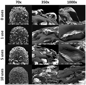
Figure 1 Representative SEM images of bur's tip at 70X, 350X, and 1000X by groups of 0, 1, 5, and 10 uses.
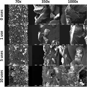
Figure 2 Representative SEM images of bur's body at 70X, 350X, and 1000X by groups of 0, 1, 5, and 10 uses.
Dentin microroughness analysis confirmed a tendency for an inverse relationship between the number of uses of a diamond bur and the Ra of the dentin's surface after cutting a flat dentin area with these burs (Figure 3). Statis- tical analysis showed significant differences between the 0 uses (Ra=6.01±1.16µm) and 10 uses (Ra=4.71±0.58µm) groups and between the 1 use (Ra=5.41±0.96µm) and the 10 uses groups. Topographic 3D images illustrate the dentin surface of the prepared specimens (Figure 4). In the 0 uses and 1 use groups, higher individual peaks and deeper valleys can be observed, while the 5 uses and 10 uses groups show a more flattened and a type of wavy topography. These 3D graphs confirmed the tendency for the inverse relationship between these two variables observed in the profilometry analysis.
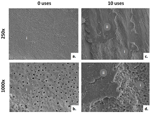
SEM images illustrate the surface of dentin at 500x and 2000x (Figure 5), of teeth prepared using a new bur and 10 uses bur before the bond strength analysis. Differences between groups are evident and support the microroughness previous findings. The results for shear bond strength between these two groups (Figure 6) showed no statistically significant differences (p=0.056) (median 14.86±8.08 MPa and 17.92±6.67 MPa respectively). When analysing the type of failure after the shear bond tests, the experimental groups showed a different distribution. In the new burs group, 53.3% of the samples had an adhesive failure while 40% had a mixed failure, and 6.7% showed dentin cohesive failure. In the 10 uses burs, 60% showed a mixed failure and 40% adhesive failure. Representative SEM images show the most prevalent type of failure for each group at 250X and 1000X (Figure 7). Figures 7 a. and b. present an adhesive failure were no resin composite or adhesive particles could be observed and the dentinal tubules appear open and empty (i). Figures 7 c. and d. show a mixed failure type where within the same specimen, open dentinal tubes (i) and adhesive or composite particles could be detected (ii).
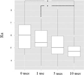
Figure 3 Box plot showing dentin microroughness median values (Ra) by experimental groups according to the number of previous uses of the bur (0, 1, 5, and 10 uses) (* statistical significative difference p<0,005)
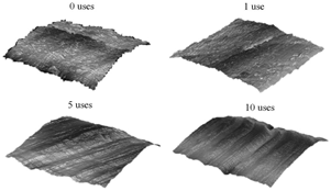
Figure 4 3D topographic images of dentin microroughness from representative samples of the experimental groups (0, 1, 5, and 10 uses) covering an area of 500µm x 500µm.
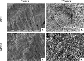
Figure 5 Representative SEM images of the dentin surface after preparation with 0 uses (a. and b.) and 10 uses (c. and d.).
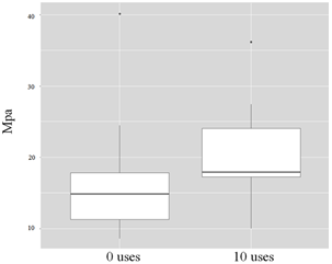
Figure 6 Box plot showing bond strength analysis between the 0 uses and 10 uses experimental groups in MPa.
Discussion
This study intended to clarify how multiple clinical uses of diamond burs affect their performance in terms of the damage they suffer, the roughness they may produce on dental tissues and how this affect bonding of resin materials to them when using self-etching adhesives. As the number of uses of the bur significantly affected the dentin surface's roughness, it didn't influence its bonding performance to a resin composite material when using a self-etch adhesive, the null hypothesis must be partly accepted.
Notorious morphology changes were observed on burs' anatomy when used several times (Figure 1 and Figure 2). SEM images of burs after its first use, already showed some signs of deterioration such as a loss of crystals' sharpness when compared to unused burs. Burs used for 5 and 10 procedures showed greater deterioration on this regard, as some diamond crystals were visibly flattened or even dislodged. These findings confirm that burs' morphology is clearly modified after usage, being this phenomenon greater when increasing the number of uses. The degree of worn suffered by the burs is not only related to the number of uses, but also to the hardness of the surface where the bur is working and of course its combined effect with the speed at which the bur is spinning, among other factors (11,12). This study was designed to analyse the effect on the burs integrity after using a bur multiple times to prepare sound tooth structures. The burs were cleaned between uses but no thermal sterilization was used to avoid the effect this procedure could have on bur's properties. It can be inferred that even after the first use of a bur to perform a complete tooth preparation for a fixed prosthetic, a significant worn on a diamond bur will be evident microscopically. These results are confirmed by previous studies (13,14).
Statistical analysis showed that new burs and those used after one procedure, produced rougher surfaces on dentin than burs used for 5 and 10 uses. This can be visually confirmed by the 3D profile for each group (Figure 4). According to these results, it can be concluded that as long as a diamond bur is being worn by multiple uses and its diamond crystals becoming flattened or even getting dislodged, the bur will produce less pronounced marks on the dentin surface, making a smoother or less rough pattern as confirmed previously (15,16. In the case that a bur has lost most of its diamond crystals, the burs' metallic matrix will become the cutting active site (instead of the sharpie diamond crystals in the new bur), reducing its cutting ability and producing a polishing effect or surface regularization in the substrate (16). For this reason, burs used multiple times produced less irregularities on the dentin surface, explaining the lower numerical roughness values obtained in the profilometry test. Using a bur in this condition (worn by usage) could probably also rise the temperature of the tissues where its being used as the metallic stem will be acting alone (with no aid of sharp-edged crystals), increasing the risk of affecting the pulp, however, this was not evaluated in this study reason why this specific issue should be confirmed in future investigations.
Regarding bond strength values, no significant differences were found between the two groups tested, indicating that no apparent relationship could be identified between dentin roughness generated by each bur group and the later bond strength. Based on data from a pilot study, the 0 uses group and the 10 uses group were selected to evaluate the bond strength values. Modern bonding strategies are divided into two major groups, the etch-and-rinse and the self-etch approaches (11,17,18). The etch-and-rinse technique includes the application of a phosphoric acid agent in order to remove part of the mineral matrix of the dental tissues with the intention of promoting micro irregularities at the surface and at certain deepness through the internal bulk of both enamel and dentin (both with different charac- teristics of course) which will serve as retentive areas when resin-based materials penetrate the tissue and due to its polymerization, a mechanical interlocking is established between both (17). This technique has been successful and validated for many years now in terms of in vitro bond strength and clinical performance, however, some issues of concern have been pointed regarding the usage of phosphoric acid in dentin, including the faster activation of enzymatic degradation, impossibility of total elimination of water from dentin after rinsing, high risk of incomplete infiltration of dental adhesives inside demineralized dentin (both rising the risk of hydrolytic degradation), and a high technical sensitivity. Due to those disadvantages, self-etching adhesives have recently gained more popularity among clinicians and researchers (11). The incorporation of acidic functional monomers to dental adhesives (not a new improvement) gave clinicians the possibility of bonding resin-based materials to tooth tissues by a chemical process instead of only by the micromechanical approach (18).
Burs' action become important in resin bonding specially when using self-etching bonding strategy because the chemical interaction between functional acidic monomers and calcium from hydroxyapatite depends highly on an intimate contact between both. This means that different thicknesses and compositions of the smear layer produced by widely used burs, may influence how well the adhesive can penetrate this layer and establish chemical bonds with the dentin tissue (18. This phenomenon is not very much signifi- cant when employing etch-and-rinse adhesives, because the phosphoric acid demineralizes the dentin around 8 µm deep (including smear layer), erasing any surface pattern previously produced by the burs' action (11,17,18). Also, as the self-etching strategy and simplified adhesives (universal for example) are currently preferred for the reasons exposed before, is that adhesive type was not a variable of interest to be evaluated here, and so it was used only a universal adhesive in the self-etching mode as an example of one of the most used type of available adhesive systems. In the current work, no significant differences in resin/dentin bond strength were found between 0 and 10 uses groups, although it was noted a numerical tendency of the median values of the 10 uses group to be higher than 0 uses group (Figure 6). Data did not fulfil parametric analysis assumptions, which means data were not normally distributed neither showed homogeneous varia- tions between groups so a high variation between values was a fact. Non-parametric analysis works with median values instead of averages and if data are not homogenous, as in this case, there is a higher probability to have not found significant differences between groups. This may explain why no statistical differences were detected between both groups even though numerical differences between averages were notorious. This great variability in data may be due to natural variability of dentin tissue, inherent factors of methodological procedures, or human error during the experimental data collection. However, the numerical difference reported in this work between bond strength produced by new burs and those used for 10 times seems logical as a worn bur may create not only a smoother surface on dentin for the reasons exposed before, but it also may have produced thinner a smear layer which favours intimate contact of self-etching adhesive to dentin, and thus improving chemical interaction between both, based on previous studies. (8,10,16,19) The results for the type of failure observed in the present study showed a slightly higher percentage of adhesive failures among the 0 uses bur group (53.3%) as compared to the 10 uses bur group (40%). Adhesive failure occurring at the transition zone between the smear layer and the intact dentin has been related to a weaker bond of the adhesive to dentin tissue, probably because the self-etch adhesive couldn't go completely through to establish a firm bond with the dentin (20).
The fact that a worn bur may improve bonding performance of a self-etch adhesive (as seen in previous studies and not supported in our results), should not be taken as the only effect of multiple uses of a bur, because this is a multifactorial issue. During tooth preparation, cutting efficiency is desired in order to prevent the dentist to increase the pressure to the tooth structure with the handpiece and bur, when feeling a lack of cutting action of the bur, because this may cause an undesirable rise in the temperature around the working area which may potentially cause an irreversible damage to the pulp tissues (not proved in the current work, as mentioned before). A worn bur will remove the tooth tissue mostly by friction produced by the bur's metal stem instead of the more efficient cutting from diamond crystals. (12,21) A previous study reported that new burs favoured a better bonding performance of an etch-and-rinse adhesive, suggesting that when using this kind of adhesives, a rougher dentin is beneficial because irregularities may allow better action of phosphoric acid and further improved mechanical retention (22). Consequently, it can be assumed that the possible effect of multiple uses of a diamond bur in dental adhesion of resin- based materials may be dependent on the type of adhesive system that will be used.
It is a fact that a new bur produces rougher surfaces compared to multiple used burs due to their improved cutting ability, allowing the clinician to reduce pressure and time when performing a dental preparation. So, from the results of this study, it is recommended to discard diamond burs after the first use, understanding ''one use'' as a complete tooth preparation for a crown, to guarantee the adequate cutting performance of these instruments. In terms of bonding, when using the self-etching approach, it is recommended to ''finish'' the preparation with low-grain size burs to produce thinner smear layer and smoother dentin surfaces. Further clinical research is needed to validate these observations.
Conclusions
After the first use, these diamond burs suffered morphological alterations, specifically flattened or dislodged diamond crystals, with an apparent relationship between the burs' deformation and the number of uses. Although, new or one-use burs produced rougher dentin surfaces than burs previously used 5 or 10 times, when using a self- etch adhesives or universal adhesives in the self-etching mode, no significantly difference in bond strength was found in vitro between resin composite and dentin when preparing the tooth with a new bur or a bur used for 10 previous times.
Author contribution statement
Conceptualization and design: D.C., A.P., M.M. Literature review, M.J., D.C., A.P., D.L., M.M.
Methodology and validation: D.C., F.M., J.V., D.L., M.M.
Formal analysis: F.M., A.P.
Investigation and data collection: M.J., D.C., F.M., D.L., M.M.
Resources: M.J., D.C., J.V., D.L.
Data analysis and interpretation: D.C., F.M., A.P., D.L, M.M.
Writing-original draft preparation: M.J., D.C., F.M., M.M.
Writing-review & editing: D.L., J.V., A.P. Supervision: D.C., M.M.
Project administration: M.M.
Funding acquisition: D.C., M.M.

