


 English (pdf)
 Article in xml format
 Article references
 Send this article by e-mail
 Cited by SciELO
  Similars in
SciELO
Similars in
SciELO 
 Permalink
Introduction
Cranial morphological parameters, such as maximum width or length, interorbital distance, biorbital width, nasion-prosthion height, bizygomatic breadth, mastoid height, bimastoid breadth, dimensions of the mastoid process, foramen magnum maximum width or length, and frontal or maxillary sinus measurements, have been studied in forensic science (1,2,3). It is important to be able to analyze bones even under conditions where body unity is damaged. The mastoid bone is suitable for forensic studies, including sex and age determination, because it is rather resistant to trauma due to its compact structure and anatomic position (4).
Studies have shown that the cranium is the next best contributor to sex assessment after the pelvis. Krogman and Iscan (5) have reported that sex can be determined from the pelvis with an accuracy of 95% and from the cranium with an accuracy of 92%. Cranium investigations are based on morphometric analyses, and accuracy rates have been investigated in different populations (6,7,8).
Osteometric measurements made using radiological methods have many advantages, such as being non-bone-destructuring methods, not requiring cleaning of the bones, and being more practical and applicable than many other methods (9-10).
To the best of our knowledge, there are no studies relating bimastoid diameter using conebeam computed tomography (CBCT). Therefore, the purpose of this study was to investigate the possibility of age and sex determination using bimastoid diameter with CBCT in a Turkish subpopulation.
Materials and methods
In this retrospective study, 200 patients aged 18-83 years were randomly selected and analyzed using CBCT. Based on the power analysis, the sample size was found to be at least 76. Ethical approval for this study was obtained from the Clinical Research Ethical Committee of Altınbaş University (approval number: 2020/25). Informed consent was routinely obtained from all patients prior to radiographic examinations. Equal number of male and female patients were selected to prevent possible effects of sex. Patients with radiologic evidence of cranial fracture, diseases affecting the cranial bones, or prior cranial surgery were excluded. Furthermore, patients aged <18 years were excluded due to the characteristics of skeletal development in childhood and adoles- cence (11-12).
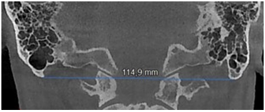
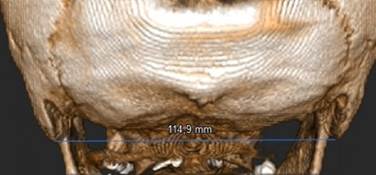
All CBCT examinations were performed using NewTom Vgi evo (CeflaGroup, Verona, Italy) at 110 kV and 1-32 mA, and NNT Viewer software (CeflaGroup, Verona, Italy) was used for measurements. The images were viewed in a quiet, dark room on a 22" high image quality Barco medical monitor (Barco, Kortrijk, Belgium) to provide an effective evaluation for CBCT. Bimastoid diameter is defined as the distance between the points of the mastoid process (1). To measure bimastoid diameter, an appropriate image was chosen from sagittal, coronal, and axial images in which measurement points could be best detected. Three-dimensional (3D) images were obtained by reconstruction. The maximum bimastoid diameter was measured on the coronal CBCT image (Figure 1), and then to verify the measurement, bimastoid diameter was measured on the coronal scan of 3D reconstruction (Figure 2).
Statistical analysis
The IBM SPSS Statistics 22 (IBM SPSS, Turkey) program was used for statistical analyses. The normal distribution of the parameters was evaluated using the Shapiro-Wilks test. A one-way analysis of variance (ANOVA) test was used to compare the normal distribution parameters with quantitative data. Students t-test was used to compare the normal distribution parameters between the two groups. Statistical significance was set at P<0.05.
Results
A total of 200 CBCT (100 female, 100 male) images were evaluated, and the mean bimastoid breadth was 106.12±6.22mm. The mean age of the patients was 45.55±16.28 years. A significant difference was detected between the sexes (p<0.05). The bimastoid diameter in male cases was higher than that in female cases (110.69±4.53mm vs. 101.65±4.00mm; Table 1). Additionally, participants were divided into age groups, and there was no significant difference in bimastoid breadth with advancing age (P>0.05; Table 1).
The sex distribution of each age group was assessed by one-way ANOVA analysis, and a statistically significant difference was not detected according to age groups for both sexes (P>0.05; Table 2).
Table 1 Mean values according to sex and age groups.
| - | - | Bimastoid Diameter (mm) |
|---|---|---|
| - | - | Mean±SD |
| Sex | Male | 110.69±4.53 |
| - | Female | 101.65±4.00 |
| - | p¹ | 0.000* |
| Age range | 18-29 | 104.83±6.33 |
| - | 30-39 | 106.18±6.63 |
| - | 40-49 | 108.14±6.24 |
| - | 50-59 | 107.27±5.75 |
| - | 60-69 | 104.88±5.77 |
| - | >70 | 105.09±6.24 |
| - | p² | 0.145 |
¹Student t Test ²Oneway Anova Test *p<0.05
Table 2 Analysis of bimastoid diameter by age groups in both sex.
| - | Bimastoid Diameter (mm) | - |
|---|---|---|
| - | Male | Female |
| Age range | Mean±SD | Mean±SD |
| 18-29 | 110.29±5.54 | 101.25±3.76 |
| 30-39 | 111.07±4.66 | 101.29±4.25 |
| 40-49 | 111.49±4.48 | 102.03±3.90 |
| 50-59 | 110.73±3.81 | 102.08±3.96 |
| 60-69 | 109.63±4.75 | 101.55±3.76 |
| > 70 | 109.95±4.47 | 102.66±5.69 |
| p² | 0.886 | 0.945 |
Oneway Anova Test.
Discussion
The cranium can be used to discriminate between the sexes, although different methods and areas were measured in studies of various populations in which cranial sex dimorphism was investigated. Data from living individuals are an important resource for identification studies. Advanced radiological methods, such as CBCT, multi-slice computed tomography (CT), and magnetic resonance imaging are important for osteometric measurements because they have some technical imaging advantages that prevent destructive procedures and maceration. These are time-consuming, contain ethical concerns, and have the opportunity to reach large populations. The specific standards of these radiologic methods may be obtained in anthropometric studies to provide clearer data on the skeleton (1). In this study, we aimed to obtain population-specific standards based on CBCT data of living individuals using bimastoid diameter measurements. In the literature, studies of sex discriminatory features in Turkish populations using cranial morphometric measures have reported that sex differences are clear in the dimensions of the frontal sinus, foramen magnum, and maxillary sinus of the cranium. In previous studies, the accuracy of discrimination was 80%-90% (13,14,15).
There was no significant difference in bimastoid breadth with advancing age in the present study. The mastoid area is usually maturated before puberty and is one of the slowest growing regions of the body in the postpubertal period (16). Therefore, bimastoid breadth is not available for age determination (17).
Since the male cranium is bigger than the female cranium, the mastoid process dimension is larger in men than in women due to the activity of longissimus capitis, splenius capitis, and sternocleidomastoid muscles (18). The bimastoid diameter in male patients was higher than that in female patients. The mean bimastoid diameter was 110.69±4.53mm in male patients and 101.65±4.00mm in female patients.
Marinescu et al. (19) reported that the accuracy rate for bimastoid diameter was 73%, and the measurements showed a significant difference in the Romanian population. In addition, Jain et al. (20) reported that the accuracy rate for bimastoid diameter was 75%, and its availability for sex determination was relatively high in an Indian population. A study conducted in a Turkish population by Buran et al. (17) found the accuracy rate of sex determination with 80.0% in men and 82.7% in women.
Apart from bimastoid diameter, different parameters, such as maximum cranial length, bizygomatic diameter, basion-bregma height, and cranial base length, can be used for sex determination from the cranium (1).
In a study conducted by Steyn et al. (21), 12 cranial parameters were measured in 47 female and 44 male skeletons in a South African population. The bizygomatic diameter with an accuracy rate of 80% was reported as the most dimorphic parameter. Similarly, Kranioti et al. (6) reported that the bizygomatic diameter with an 81.9% accuracy rate was reported as the most dimorphic dimension by evaluating 16 cranial parameters in 88 female and 90 male skeletons in a Crete population. In another study, Walker (22) reported that glabella had the best accuracy rate (82.6%) by measuring five cranial parameters in 140 female and 164 male skeletons in the United States. In a study conducted by Sakaue et al. (23), five cranial parameters were measured in 108 female patients and 205 male patients in a Japanese population. The supra-orbital ridge with an 80.5% accuracy rate was reported as the most dimorphic dimension.
Bimastoid diameter was measured for sex determination using cranial CT images by Buran et al. (17). The accuracy rate was 80.0% in men and 82.7% in women; therefore, they reported that the bimastoid diameter measurement can be used for sex determination in the Turkish population. In another study, 14 cranial parameters using multidetector CT were measured retrospectively in 200 female and 200 male patients in a Turkish population. The most remarkable measurements in dimorphism were the bizygomatic diameter, máximum cranial length, basion-bregma height, and cranial base length. Additionally, the bizygomatic diameter with an accuracy rate of 77% in men and 83% in women was the most dimorphic parameter (1). In a study by Cekdemir et al. (24), parameters, including maximum cranial length, minimum frontal breadth, bizygomatic breadth, parietal chord, maximum cranial breadth, bimastoid diameter, and length of the cranial base, were investigated. The variables with the most successful sex discrimination performance were bizygomatic breadth (89.2%), maximum cranial length (87.4%), and bimastoid diameter (84.8%). The important point here is to analyze existing bones, even in cases where body integrity is impaired. The mastoid bone is suitable for sex and age determination because it is resistant to trauma due to its compact structure and anatomic position (4).
Conclusion
There was no significant difference in bimastoid diameter with advancing age.
For sex determination, morphometric measurements of bimastoid diameter ensured a high rate of dimorphism in the Turkish subpopulation.
CBCT morphometric analysis may be reliable and convenient for evaluating sex and may be recommended to compare population data.

