


 Inglés (pdf)
 Articulo en XML
 Referencias del artículo
 Enviar articulo por email
 Citado por SciELO
  Similares en
SciELO
Similares en
SciELO 
 Permalink
INTRODUCTION
Prosthetic dental treatment has a wide spectrum of clinical applications, including fixed, removable, and implant-supported prostheses, with various materials used. The properties of materials are constantly being enhanced, and new products are being introduced. Polyether ether ketone (PEEK) is a material that has gained popularity in dentistry in recent years. It is a semi-crystalline thermoplastic biomaterial with the chemical formula (-C6H4-O-C6H4-O-C6H4-CO-)n that belongs to the family of poly(aryl ether ketone) polymers based on ultra-high molecular weight polyethylene (1). First produced in 1978 (2), this material has been used in orthopaedics (3), maxillofacial surgery (4), and dental treatments (5) as an alternative to metal substructure treatments. PEEK has found a wide range of applications in implants, orthodontic braces, temporary abutments, and fixed and removable prostheses as an esthetic alternative to metal systems in dental clinical practice (6,7,8,9,10). It owes its popularity to its excellent chemical, thermal, and mechanical properties and superior biocompatibility (11). PEEK is also a hard and durable material and exhibits less deformation than other thermoplastic materials at high temperatures (12).
In order for a material to be used in the mouth for many years, it must be minimally affected by the mouth fluids, and the plaque accumulation on it must be minimized. To that end, its surface must be finely polished. Surface roughness may cause coloration or discoloration, plaque accumulation, and abrasion of the opposing tooth (13,14). Surface topography, roughness, hardness, and abrasion properties are extremely important for optimum yield in a material (15). A restoration may also require additional processes (such as control of occlusion and proximal contacts) at the last rehearsal before it is inserted into the mouth. In such cases, the material is removed from the restoration surface, causing deterioration of the restoration’s surface finish. Conventional polishing of PEEK is performed in the laboratory. These procedures can sometimes be performed after cementation. In such cases, chairside polishing with polishing rubbers or kits can be applied to restore the lost surface finish of the restoration (16). For this purpose, various polishing kits have been introduced. Such kits include tungsten carbide finishing burs, diamond rotary instruments, silicone rubber discs, and silicon carbide or aluminum oxide-coated abrasive discs (17).
Surface roughness (profilometer) análisis (18) and Vickers hardness measurements (19) are tests used to examine a material’s surface properties. However, while these mechanical tests provide numerical values related to the surface structure of the material, they do not provide full information on its surface topography. Scanning electron microscopy (SEM), atomic force microscopy (AFM), three-dimensional (3D) optical profilometers, and confocal laser scanning microscope (CLSM) are used to clearly observe changes on the surface of the material and examine its topography. Environmental scanning electron microscopy (ESEM) is used to examine the natural conditions and microscopic properties of a material with no coating procedure on its surface (20).
In cases where there is not enough time, chairside polishing with polishing rubbers is applied to the restorative materials. For PEEK, knowing the success of chairside methods is necessary for clinical use. This study aimed to investigate the effects of different polishing procedures on the surface roughness and hardness of PEEK. The hypothesis was that the different surface finishing processes would not significantly affect the surface roughness and hardness of PEEK specimens.
MATERIALS AND METHODS
Power analysis performed using G*Power (v. 3.0.10) to obtain the highest power level with the smallest possible sample size showed that at least 22 specimens were required (power= 80, α= 0.05).
The specimens used in the study were prepared from PEEK blocks (CopraPeek; Whitepeaks Dental Solutions GmbH&Co, Essen, Germany) in the form of discs 10mm in diameter and 2mm in length. After checking the specimens’ suitability to the initial dimensions, their surfaces were ground with P600 and P800 grit silicon carbide paper (English Abrasives & Chemicals, London, UK) for 60s and polished with a fine pumice stone (Ernst Hinrichs Dental, Goslar, Germany) and goat hair brushes (Jiffy; Ultradent Products, South Jordan, UT, USA) for 60s in an automatic polishing device (Reco Dental, Wiesbaden, Germany) with a vertical force of 25 N to produce a standard surface. All specimens were then cleaned in an ultrasonic machine (CD-4800; Jeken, Dongguan, China) for 10min and stored in a dry place until surface roughening and polishing kit applications.
The obtained specimens were randomly divided into three groups (n=22), and each specimen was numbered. The three groups were as follows:
Control (Conventional Polishing) group (C). These specimens were subjected to mechanical tests with no prior surface treatment.
Meisinger polishing kit (Luster Intraoral Twist Kit; Hager & Meisinger, Neuss, Germany) group (M). The surfaces of these specimens were abraded with cylindrical diamond burs (837LF-FG-014; Meisinger, Neuss, Germany) under water cooling to remove 0.1mm of the material from the surface to simulate a finishing chairside procedure. A digital caliper was used to control the material thickness. The polishing rubbers included in the kit were then applied to the abraded surfaces in the order specified by the manufacturer (green-blue-red- yellow). All were applied by the same researcher in a circular motion for 60s each, with a maximum tip rotating speed of 10,000 rpm according to the manufacturer’s instructions, without water cooling, and with an average pressure of 2 N.
OptraFine polishing kit (OptraFine Assortment; Ivoclar Vivadent, Schaan, Liectenstein) group (O). The surfaces of these specimens were also abraded with cylindrical diamond burs (Meisinger) under water cooling to remove 0.1mm of the material from the surface to simulate a finishing chairside procedure. A digital caliper was used to control the material thickness. The polishing rubbers included in the kit were then applied to the abraded surfaces in the order specified by the manufacturer (light blue-dark blue). All were applied by the same researcher in a circular motion for 60s each, with a maximum tip rotating speed of 10,000rpm according to the manufacturer’s instructions, with water cooling, and with an average pressure of 2 N. Afterward, with the same tip speed and pressure, the polishing paste included in the kit (OptraFine HP Polishing Paste; Ivoclar Vivadent) was applied to the surfaces in a circular motion for 60s without water cooling.
One specimen from each group was randomly selected, and the specimens were subjected to ESEM examination. Surface imaging was performed by focused ion beam SEM transmission electron microscopy (Quanta 3D FEG; FEI, Hillsboro, OR, USA) with no coating on the specimen surfaces. Images were taken at 1500x magnification. The specimens’ surface topography was then analyzed with an AFM (Hitachi 5100N; Hitachi High-Technologies Corporation, Tokyo, Japan).
The polished surfaces of all specimens were analyzed with a 0.008-mm cutoff in a contact scanning profilometer (Tencor P-7 Stylus Profiler; KLA-Tencor, Milpitas, CA, USA) capable of 360° measurements. To ensure standardization, three separate measurements were obtained from each specimen, averaged, and recorded as its Ra value.
The Vickers hardness test was performed using a Vickers hardness tester (FM-800e Microhardness Tester, Future-Tech, Kawasaki, Japan) for 15s (dwell time), and the Vickers hardness values (HV) were recorded. To ensure standardization, three measurements were obtained from the different regions of each specimen, averaged, and recorded as its HV value.
Statistical analyses of both surface roughness and Vickers hardness tests were performed with the Kolmogorov-Smirnov homogeneity test and analysis of variance (ANOVA) using IBM SPSS Statistics v. 20 (IBM, Armonk, NY, USA) (p<0.05). The Tukey multiple comparison test was used for comparisons between the group means (α=0.05).
RESULTS
ANOVA showed that the differences between the three groups were not statistically significant (p>0.05) in terms of either surface roughness (p=0.23) or Vickers hardness (p=0.85). Mean and standard deviation values are shown in Table 1.
ESEM examination (Figure 1. A-C) revealed that although the surface of the group C specimen was smooth, it had hollows and micro cavities in some places. The M group specimen had micro cavities with small traces of burs on its surface and was smoother than the group C specimen. Although there were traces of burs in the O group specimen, its surface appeared to be smoother and denser. A comparison of the ESEM images of the group M and O specimens with the group C specimen showed that the polishing kits formed almost a layer on the surface of the material, filling the micro cavities in the material and making it appear more uniform.
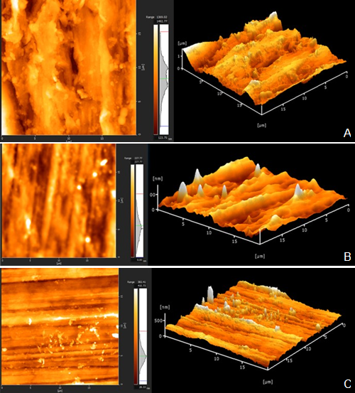
AFM examination (Figure 2. A-C) revealed more rough areas in the C group specimen, mainly in its centre. Although the surfaces of both the M and O group specimens showed less rough areas at the periphery than the C group, there was not much variation among the surface images of all groups.
Table 1 Least square means and Standard deviations for surface roughness and hardness.
| - | Surface Roughness (Ra) | - | - | Vickers Hardness (HV) | - | - |
|---|---|---|---|---|---|---|
| - | Mean | Std. Deviation | N | Mean | Std. Deviation | N |
| Control | 3.54 | 2.58 | 22 | 36.68 | 4.50 | 22 |
| Meisinger | 3.46 | 2.54 | 22 | 39.56 | 6.73 | 22 |
| OptraFine | 3.16 | 1.59 | 22 | 37.57 | 5.55 | 22 |
p>0.05.
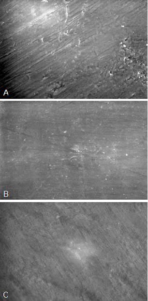
Figure 1 A. ESEM micrograph of the polished surfaces of control group (1500x). B. ESEM micrograph of the polished surfaces of Meisinger group (1500x). C. ESEM micrograph of the polished surfaces of OptraFine group (1500x).
DISCUSSION
This study aimed to investigate the effect of different polishing procedures on the surface roughness and hardness of PEEK specimens. As no significant differences were observed between the polishing procedures in terms of surface roughness and hardness, the study hypothesis was accepted.
Extra surface treatments applied to a material can change its surface properties. Since PEEK is a material recently introduced in clinical use, research on its surface properties is still being conducted. Studies have shown that treatments applied to the surface of PEEK specimens can change their surface roughness and morphology (21,22,23,24).
The hardness of a material is one of its most important mechanical properties. It plays a role in abrasion and is used as a criterion to determine the material’s wear resistance (25,26). For PEEK specimens, Nisa et al. (27) reported a surface hardness value of 22 HV, whereas Kumar et al. (28) reported values of 28-43 HV. The specimens of the control group in this study had a mean hardness of 36.68±4.5 HV, which is consistent with the literature.
Few studies have examined the effects of polishing procedures on the surface properties of PEEK. Heimer et al. (29) reported that chairside polishing yielded lower surface roughness than laboratory methods and that specimens polished using the two-body mode exhibited higher roughness than those polished using the three- body mode. Sturz et al. (30) found that chairside surface treatments affected the surface roughness of specimens and that air polishing resulted in the highest surface roughness of PEEK specimens.
ESEM is primarily used for biological materials or for materials that do not have a carbon coating, to monitor the changes in the material surface at the micron level and, the AFM is a powerful microscope that is used for high-resolution, sensitive examinations of surface roughness down to the atomic level (31). Both advanced imaging techniques provide clearer and detailed information about the surface changes of materials. In this study, the surface roughness values from lowest to highest were in the order of groups O, M, and C, although the difference was not statistically different. ESEM and AFM confirmed this order. We believe that the lowest roughness values observed in group O were due to the diamond paste included in the kit and applied last. In both polishing procedures, polishing is performed with progressively thicker rubber burs, which results in a smoother material surface. The fact that group C exhibited the lowest hardness values supports this hypothesis, despite the lack of statistical significance. We attribute the high mean standard deviation values for surface roughness values to the sensitivity of the test used. In the contact scanning profilometric test method used, mean roughness values of a disc-shaped specimen are obtained by scanning the entire surface. Since the measurement is made from the whole surface, not from a single point of the specimen surface, much more precise and accurate values are obtained. Consequently, extreme values that are far below and above the average may occur and increase the standard deviation.
One limitation of this study is the small number of polishing kits used. To confirm our findings, future studies should evaluate more kits. Another limitation is that bacterial retention analysis was not performed. Future research should investigate bacterial retention on PEEK surfaces, as this is important for clinical reliability.
CONCLUSIONS
The polishing kits used in this study did not result in statistically significant differences in the surface roughness and hardness of PEEK specimens compared to conventional polishing processes. The polishing procedures even smoothed the specimens, as revealed by ESEM and AFM examinations. Based on these results, we can conclude that polishing kits can be applied to PEEK and offer considerable convenience, especially in chairside procedures.

