-
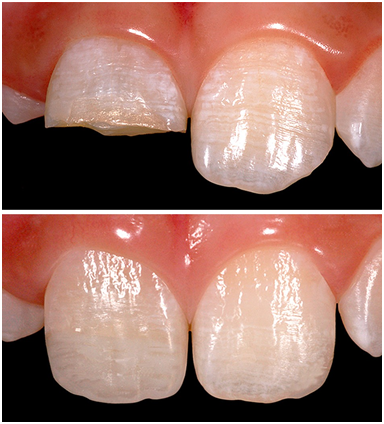
Restorative treatment of fractured anterior teeth represents a challenge for the dentists. Depending on the severity of the fracture, various materials and techniques may be used. Currently, one of the treatments of choice is the use of direct adhesive restorations with composite resins.
The esthetic result and longevity of such restorations are directly related to the quality of marginal adaptation. Improving the clinical performance and longevity of direct adhesive restorations has been the subject of several studies (1,2,3,4,5). Nevertheless, doubts still exist regarding how to prepare the cavosurface angle prior to adhesive reconstruction to optimize the clinical performance of the procedure.
Mechanical bevel preparation prior to restorative treatment was the most recommended technique in this regard (6,7,8). However, considering that anterior tooth fractures occur very often in young patients, it is important to estimate the actual need for bevel preparation in fractured anterior tooth restorations.
In addition to the lack of consensus in literature, there is little long-term clinical research evaluating the influence of unprepared cavosurface configuration on the clinical performance of direct adhesive restorations.
Clinical evaluation of anterior teeth restorations
One of the most elaborate studies on this topic was performed by Buonocore and Davila (1). They described an in vivo restorative technique for fractured anterior teeth. A total of 104 restorations with composite resin were performed. Initially, the fractured teeth were cleaned with no cavity preparation. Dental enamel was conditioned with phosphoric acid for 60 s after dentin protection with Dycal. A thin layer of adhesive was applied to the properly conditioned enamel a few millimeters beyond the fracture line (±2mm) for subsequent application of the resin material. The authors found that this technique requires some degree of overcontouring and that the larger and thicker the overcontour, the greater the retention and sealing. Data obtained after clinical and radiographic evaluation over a period of 8 to 24 months revealed that among 104 restorations, 102 were successful. Marginal integrity was maintained in all cases without evidence of marginal infiltration. The color compatibility was generally excellent. When slight marginal discoloration was observed, the defect could be corrected by polishing. Authors concluded that the proposed technique is conservative, fast, economical, and atraumatic. They also pointed out that in cases of restoration failure, the tooth would be in the same initial condition because no dental tissue was mechanically removed.
On the other hand, Crim (9) recommended beveling around the fracture line in order to allow a proper anatomical contour for restoration. According to the author, the bevel preparation improves marginal control, increases the surface area for adhesion and improves the transition of composite resin to dental structure in areas where esthetics is important. The author emphasized the efficiency of the procedure, since the entire preparation is confined to the enamel, without inducing damage or dental pulp injuries. He also stated that, without enamel removal, restoration may be unsatisfactory because of an overcontour and lesser resistance to displacement. Nowadays, this last statement is the most used one in justification to enamel bevel restoration. Some professionals recommended a 60° bevel in enamel to remove unsupported prisms as well as exposing them to acid conditioning, promoting better retention and sealing. They also stated that the bevel allows a gradual thickening of the composite resin, which makes it difficult to see the restorative interface. Moreover, a study stated that the execution of a bevel in the whole cavosurface angle, besides promoting a better cavity sealing, helps the esthetic harmony (10). Authors also pointed out that the bevel promotes greater retention as it increases the conditioned area, resulting in more space for the restorative material and thus improving the esthetic aspect of restoration.
Luiz N. Baratieri, one of the most recognized professionals in the field, reported that there are two alternatives regarding tooth preparation: beveled and unprepared (11). The author explained that, with the technique of total acid etching, current efficient adhesive systems, and the wide variety of composite resins, it is possible to satisfactorily restore fractured anterior teeth by a direct technique without any bevel preparation. For the authors, not carrying out a preparation was important because it helps in preservation of a healthy dental structure. The justifications for this type of approach are: a) the restorations will be considered reversible, since the tooth is not submitted to any kind of preparation; b) avoiding the wear of dental structure with the use of drills also avoids possible psychological trauma in children, in whom fractures of upper anterior teeth are more common; and c) when they fail and have to be replaced, there will be more healthy dental structure available for a new adhesive restorative procedure. However, the restoration of an unprepared fractured anterior tooth may have the following disadvantages: excesses may be lodged on the unconditioned surface causing marginal discoloration to this critical area due to microleakage as well as color alteration over time. As for the beveled alternative, whose demand stems from the need for optimum esthetics, the advantages are as follows: a) a defined marginal termination, allowing for adequate adaptation or marginal integrity of the composite resin; and b) ease of finishing with less risk of composite resin remaining lodged and the appearance of “white lines” on the restoration margins.
Araujo Jr. (12) evaluated the influence of cavosurface configuration (beveled and unprepared) on the esthetic outcome of direct composite resin restorations on fractured anterior teeth. Seventeen patients with at least one maxillary central incisor fracture or with any Class IV restoration with replacement indication were selected. Of the 34 selected incisors, 10 were healthy and 24 had deficient or coronal fracture restorations, which were performed by a single surgeon. The healthy and restored teeth were divided into 3 groups: group I=12 beveled teeth; group II=12 teeth restored without cavosurface preparation; and group III=10 healthy teeth. After restorative treatment, standardized photographs of the 34 specimens were collected. These were attached to evaluation questionnaires which were submitted to 120 evaluators assigned to 3 groups: group A=40 dentistry students; group B=40 specialists in esthetic dentistry; and group C=40 patients. The data obtained from the responses to the questionnaire was analyzed. According to the results, there was no difference observed between the beveled and unprepared groups in the esthetic appearance of restorations. Thus, the author concluded that it is possible to perform esthetically satisfactory restorations on fractured anterior teeth without promoting any type of dental wear.
Moreover, Araujo Jr. et al (13) in a clinical case report stated that the composite resins due to the significant evolution in their optical properties, allow restorations with translucency, texture and shape closer to the teeth, which improves the esthetics and functionality of restorations. With sufficient knowledge, determination and professional training, composite restorations are a safe treatment alternative with predictable and satisfactory results. The authors also pointed out that in direct adhesive restorations, any reduction in healthy dental structure should be avoided, particularly in young patients.
Since 2006, this theme was evaluated in my thesis for a master’s degree in Operative Dentistry. We performed an in vivo study to evaluate the influence of the cavosurface angle (beveled or non-prepared) on the clinical performance of direct adhesive composite resin restorations of the fractured anterior teeth. The restorations were performed by the same professional following a previously established, standardized protocol. Twenty-four upper central incisors with fracture or with indication for substitution were selected for the study. The teeth were divided into two groups: group 1 comprised of 12 Class IV composite resin restorations with bevel preparation of the cavosurface angle (bevel); and group 2 comprised of 12 Class IV composite resin restorations with no preparation of the cavosurface angle (non-preparation). The restorations were evaluated 7 days and 4 years after the treatment, according to the USPHS-modified criteria by two examiners. After 4 years, two restorations were excluded, and a final sample of 22 restorations (11 with bevel and 11 without preparation) was evaluated. The Fisher’s exact Test was performed in order to analyze the association between the two variables (bevel or non-preparation), and the results showed that there is no significant difference between groups (p>0.05). Therefore, we conclude that the configuration of the cavosurface angle does not influence the clinical performance of direct adhesive composite resin restorations in fractured anterior teeth and that the restoration of Class IV fractures can be accomplished without removing healthy tooth tissue.
Final considerations
Coronary fractures can occur at any age but generally affect children and adolescents. Owing to their high incidence and involvement of anterior teeth, they deserve special attention. Direct adhesive strategies are the most commonly used treatments for the conservative restoration of this type of defect. The combination of the esthetic expectation of the patient and desire for the development of a conservative treatment by the dentist resulted in the development of different clinical protocols. Moreover, contemporary restorative dentistry advocates minimally invasive conservative procedures to prevent unnecessary removal of healthy dental structure during the operative process. Evidence suggests that the functional and esthetic restoration of Class IV fractures can be accomplished without removing healthy tooth tissue.


