


Services on Demand
Journal
Article
 text in
  English (pdf)
 Article in xml format
 Article references
 Send this article by e-mail
Indicators
-
 Cited by SciELO
 -
 Access statistics
Related links
-
 Similars in
SciELO
Similars in
SciELO
Share

 Permalink
Odontología Vital
On-line version ISSN 1659-0775Print version ISSN 1659-0775
Odontología Vital n.38 San Pedro, Lourdes de Montes de Oca Jan./Jun. 2023
Article
Complications in the extraction of impacted, and retained third molars. Literature Review

1 Estudiante de Odontología, Universidad Hemisferios, Quito- Ecuador stevm1997@gmail.com ORCID: https://orcid.org/0000-0003-3549-9051
2 Docente de la Universidad Hemisferios, Especialista en Cirugía Buco- Maxilo Facial PhD en Patología Bucal, Quito- Ecuador cristinar@ uhemisferios.edu.ec ORCID: https://orcid.org/0000-0001-7945-2680
After the various articles compiled by different authors, is becomes clear that the third molars are very often what contemplate various complications at the time of the surgical procedure, due not only to their eruption but also to their different characteristics that occur such as anatomy, shape, position of its eruption, etc. For this reason, the comprehensive complementary study before proceeding to the surgical act is the first option that is made. For a correct post-surgical treatment to be effective both antibiotic-pharmacological, integral biomaterials, etc.
Purpose:
To establish through a review of the literatura which are the actions or surgical procedures being performed that can avoid the most prevalent complications in the extraction of included, retained and impacted mandibular third molars.
Materials and methods:
A descriptive and analytical study is proposed, respectively, with 2 types of electronic databases:
PubMed and SciELO, taking as support articles that include meta-analyses, systematic reviews, literary reviews, etc.
Results:
It was confirmed that the best procedure for perhaps a possible one: hemorrhage, fractures, lacerations, etc. It is good pharmacological surgical management during and after surgery.
Conclusion:
With this review of the literature, the idea is reached that a correct diagnosis, strict pharmacological management and knowledge of the complications that can arise during and after dental extractions are correct actions that are very commonly used during the surgical procedure. , which avoids their respective difficulties.
Keywords: impacted tooth; unerupted tooth; third molar
Introduction
The third molars often manifest a number of variations in their coronal and root morphology, this includes de possibility to be tri or tetra radicular at root level.
Frecuently they are smaller in size compared to other teeth such as second molars.(Palareti et al., 2016) They are the last teeth to erupt between 18 and 30 years of age, and are capable of causing dental or local anomalies at any period of their dental formation or eruption process.
However, it should be noted that according to the Pell and Gregory classification, it is useful to determine the degree of impaction, depth in relation to the occlusal plane of the lower second molar and the mesiodistal diameter in relation to the distance between the lower second molar and the anterior part of the mandibular branch identifying the degree of complication at the time of surgery. (Poblete et al., 2020)
As described in the literature, some of the main characteristics that increase the difficulty of surgery are the roots due to the chance of being fused, thin, without a conventional anatomical shape, making extraction more complex.(Loureiro et al., 2020)
At the level of the coronal diameter it seems to be similar to that of the lower first and second molars.
A key problem is that 60% do not occlude due to the loss of anatomical space, which induces to the adoption of inadequate positions, compromising the rest of the dental organs.(Moreno et al., 2019)
Different studies reveal that retained tooth are frequently associated with embryological conditions, since these teeth are formed from the epitelial cord going through a process of calcification and root formation.
In addition, this tooth must follow a known eruption path as a Capdepont curve so that it can erupt, but it is necessary to execute a concave straightening curve backwards and up.
Therefore most of these dental organs are not positioned in the correct way.(Rivera-Herrera et al., 2020)
Epidemiologically, retentions affect women more than men, 58.8% on the other hand, 9.70% impactions and 24.90% included.
The literature is consistent and finds similarities both in Mexico, Brazil, Colombia and Ecuador. It should be noted that in terms of people with African ancestry this condition has being reported in 2.2% of the population possibly in association with the size of the jaw. Nonetheless, the cause has yet to be confirmed.(Toledano-Serrabona et al., 2021)
The treatment for any retained tooth is the multidisciplinary one, establishing cost benefit and the difficulties that may occur before the extraction. (Cervino et al., 2019) A correct preoperative diagnosis considering radiological examinations, together with a clinical examination, complementary examinations and antibiotic prophylaxis also greatly reduces the risk of complications. Once the diagnostic process has been carried out, the surgical and pharmacological protocol will be the next action.(Staderini et al., 2019)
The most common complications in retained, included and impacted third molars can be classified as direct and indirect, that is, during and after surgery:
Direct: In a retained third molar (upper or lower) the most common complications vary from oroantral communication, mandibular scale fractures, and alveolar lesions to the inferior dental nerve and bleeding. In the case of an impacted molar it may implicate erroneous displacement of the instruments to anatomical spaces of great importance. In an included third molar the issues may be pericoronitis, the most frequent in severe cases, abscesses, tumors, cysts. (Gutiérrez Valdez & Pérez, 2016)Indirect: generally emphysema, inflammatory processes, hemorrhages, paresthesias,hyperesthesia, edema and others.
Thus, a review and analysis will be carried out regarding the essential maneuvers to avoid complications of the third molars during their removal, based on the PubMed, SciElo databases with selected articles between the years 2016 to 2021.
Objective:
To establish, through a literature review, which are the actions to be executed during surgical procedures that can avoid the most prevalent complications in the extraction of included, retained and impacted mandibular third molars.
Conflict of intrerest.
The authors declare that they have no conflict of interest, the literature review described is original and has not been previously published, nor has financial support been received prior to its preparation.
Materials and methods
A descriptive study of the literatura is presented, describing the análisis through 2 types of electronic databases:
Pubmed, SciELO, based on metaanalysis articles, review articles, brief reports, systematic reviews, etc.
Corresponding to the years 2016 to 2021 using the “PICO” strategy as support using search terms such as: Tooth, Impacted,Tooth, Unerupted, Molar, Third,Hemorrhage, Surgicaland their counterparts in Spanish respectively.
Articles were selected considering concordance between the title and objective in this way, examining whether they include meta-analyses, systematic reviews, literary articles, brief reports, etc.
Filtering exact years respectively corresponding to the complications of the third molars, resulting in the search of 137.
On the other hand, a total of 107 articles were excluded, being duplicate articles, comparative studies, clinical cases, etc. It results in a total of 30 articles for this literary review. All this information collected was reviewed in its entirety and exposed.
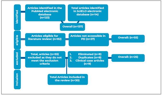
Fig.1 Flowchart for selecting articles from the literature review.
Table 1: Articles included
| (Borges et al., 2017) | literary article | soft tissue injuries | |||||
| (Calzavara & Lhano, 2019) | meta analysis | Radiographic study prior to surgery | |||||
| (Cammarata-Scalisi et al., 2018) | literary article | Complications of lower third molars before supernumerary teeth. | |||||
| (Cervino et al., 2019) | literary article | Pharmacotherapy after extraction. Post extraction antibiotic | |||||
| (Chugh et al., 2020) | Systematic review | therapy. Anxiety in the surgical act | |||||
| (Falci et al., 2017) | systematic article | Effect of dexamethasone at the time of third molar surgery. Ideal | |||||
| (Freitas, 2018) | meta analysis | option of kinesiology bandage to avoid post-extraction pain. | |||||
| (Glera-Suárez et al., 2020) | Systematic review | Correct maneuvers prior to dental extraction | |||||
| (Jaroń et al., 2020) | literary article | ||||||
| (Kim et al., 2018) | literary article | ||||||
| (Konkel et al., 2019) | literary review | Periodontal disease risk factor as a complication of mandibular | |||||
| (Loureiro et al., 2020) | systematic article | third molars | |||||
| (Mahardawi et al., 2020) | Systematic review | Form of eruption of impacted third molars | |||||
| (Marinkovic et al., 2020) | meta analysis | ||||||
| (Moreira Zevallos & Barona Terán, | literary article | Postoperative antibiotic therap y. Mandibular third molar | |||||
| 2021) | literary review | characteristics. Agenesis predisposing factor of a third molar | |||||
| (Moreno et al., 2019) | Review article | when erupting. third molar displacement | |||||
| (Di Nardo et al., 2019) | literary review | Third molar physiology at mandibular scale | |||||
| (Oda et al., 2021) | analytical study | Complications and generalities in oral surgery | |||||
| (Pacheco-vergara & Cartes- | Research work | Cardiovascular disease factor as a complication | |||||
| velásquez, 2016) | literary review | Higher incidence of post oral surgery complication | |||||
| (Palareti et al., 2016) | literary review | Classification of third molars | |||||
| (Poblete et al., 2020) | Systematic review | Consequences of bad practice during surgery | |||||
| (Rivera-Herrera et al., 2020) | Post extraction hyaluronic acid therapy | ||||||
| (Saber et al., 2018) | meta analysis | Surgical management of mandibular third molars | |||||
| (Shuborna et al., 2019) | literary review | Post-extraction therapy with chlorhexidine gel to prevent alveolar | |||||
| (Staderini et al., 2019) (Teshome, | meta analysis | osteitis | |||||
| 2017)z(Xiang et al., 2019) (Yu et al., | meta analysis | Treatment for mandibular fracture complication | |||||
| 2017) | Platelet-rich fiber therapy (healing) | ||||||
| Anesthetic methods for mandibular third molars | |||||||
Results
The complications that occur during the extraction of third molars are very frequent and to prevent them from occurring it is essential to consider various factors such as: genetics, environment, development or any pathology in order to be able to carry out the respective surgery.(Borges et al., 2017)Difficulties not only occur in lower teeth but also in upper teeth due to important anatomical repairs such as the floor of the maxillary sinus or pterygomaxillary or cortical bone fossa. (Poblete et al., 2020)
Subsequent complications of extraction result in alveolitis, bleeding or injury to the alveolar nerve, neurosensory damage.(Calzavara & Lhano, 2019)The absence of precaution or the incorrect surgical technique will increase soft tissue inflammation, risks of emphysema(Nardo et al., 2019)and other direct and indirect local complications, considering that these may manifest
Themselves after 24 hours of the execution of the procedure. (Mahardawi et al., 2020).
Complications
are unpredictable (Cammarata-Scalisi et al., 2018)and they occur mainly due to the difficulty of the extraction. (Oda et al., 2021)
The affectation of neighboring teeth is very common (Marinkovic et al., 2020), ( Pacheco-vergara & Cartes-Velásquez, 2016)with the second molars being the most affected .(Saber et al., 2018) Y (Yu et al., 2017)
Discussion
After a complete literary compilation, the most frequent complications for retained, included and impacted third molars are: involvement of soft bone tissues, mandibular fractures, and rupture. They is also an agreement in the fact that non-cooperative patients experience the largest number of complications due to the lack of selfcare.(Kim et al., 2018) (Jaroń et al., 2020)
Xiang et al., (2019) agreed with the restof the reviewed literature in the fact that these complications are common.
Also, they recommend that in cases of laceration to soft tissue, the use of platelet-rich fibrin is optimal, since it heals and favors epithelial regeneration.
Kim et al. (2018) disagree, since they considere that there is not enough scientific evidence to support this protocol. Shuborna et al. (2019) propose the use of hyaluronic acid in the event of bleeding and Teshome (2017) also considers that the use of these biomaterials in addition to 50ml chlorhexidine gel is a preventive or solution to possible alveolar osteitis in the area of the extracted tooth, thus promoting healing.
Other published articles such as that of Chugh et al. (2020)agree that the correct use and pharmacologicalantibiotic administration after third molar extraction are essential to avoid any infection or adverse reaction(Tg, 2020) Y (Konkel et al., 2019) emphasized that the correct diagnosis with its respective radiographic and complementary examinations are essential for correct surgical management.
The research carried out presented limitations related to the fact that most of the articles were clinical cases, repeated articles and in-vitro studies with respect to third molars, comparative studies.
Conclusion
The different methods that may be carried out for the clinical management of included, retained and impacted third molars must be deeply studied in order to reduce the risk of complications such as bleeding, hemorrhage, edema, fractures and lacerations.
We found that, as an innovative and interesting measure, the Kinesiotape bandage may provide important benefits. For example it has shown to relieve pain, control post-surgical inflammation, the mobility of response after an injury, improving the neuromechanical response, reducing bruising, accelerating drainage in the affected area, as well as blood circulation and fluid removal. Muscle sti mulation with the ability to relieve pain before contractions and finally postural problems.
Referencias bibliográficas
Borges, T. S., Vargas-Ferreira, F., Kramer, P. F., & Feldens, C. A. (2017). Impact of traumatic dental injuries on oral health-related quality of life of preschool children: A systematic review and metaanalysis. PLoS ONE, 12(2), 1-13. https://doi.org/10.1371/journal. pone.0172235 [ Links ]
Calzavara, N., & Lhano, D. (2019). AS. [ Links ]
Cammarata-Scalisi, F., Avendaño, A., & Callea, M. (2018). Main genetic entities associated with supernumerary teeth. Archivos Argentinos de Pediatria, 116(6), 437-444. https://doi.org/10.5546/aap.2018.eng.437 [ Links ]
Cervino, G., Cicciù, M., Biondi, A., Bocchieri, S., Herford, A. S., Laino, L., & Fiorillo, L. (2019). Antibiotic prophylaxis on third molar extraction: Systematic review of recent data. Antibiotics, 8(2), 1-14. https://doi.org/10.3390/antibiotics8020053 [ Links ]
Chugh, A., Patnana, A. K., Kumar, P., Chugh, V. K., Khera, D., & Singh, S. (2020). Critical analysis of methodological quality of systematic reviews and meta-analysis of antibiotics in third molar surgeries using AMSTAR 2. Journal of Oral Biology and Craniofacial Research, 10(4), 441-449. https://doi.org/10.1016/j.jobcr.2020.07.011 [ Links ]
Falci, S. M., Lima, T., & Martins, C. C. (2017). Efecto preventivo de la dexametasona en la cirugía del tercer molar: un metaanálisis. Anesthesia Progress, 64(3), 136-143. [ Links ]
Glera-Suárez, P., Soto-Peñaloza, D., Peñarrocha-Oltra, D., & Peñarrocha-Diago, M. (2020). e233 Med Oral Patol Oral Cir Bucal. 25(2), 233-242. https://doi.org/10.4317/medoral [ Links ]
Gutiérrez Valdez, D. H., & Pérez, D. (2016). Incidencia de infecciones postquirúrgicas de terceros molares en pacientes atendidos en clínica de enseñanza odontológica. Avances En Odontoestomatologia, 32(5), 259-264. https://www.fisiofocus.com/es/articulo/que-beneficios-tiene-el-kinesiotaping [ Links ]
Jaroń, A., Jedliński, M., Grzywacz, E., Mazur, M., & Trybek, G. (2020). Kinesiology taping as an innovative measure against post-operative complications after third molar extraction-systematic review. Journal of Clinical Medicine, 9(12), 1-13. https:// doi.org/10.3390/jcm9123988 [ Links ]
Kim, C., Hwang, K.-G., & Park, C.-J. (2018). Local anesthesia for mandibular third molar extraction. Journal of Dental Anesthesia and Pain Medicine, 18(5), 287. https://doi.org/10.17245/jdapm.2018.18.5.287 [ Links ]
Konkel, J. E., O’Boyle, C., & Krishnan, S. (2019). Distal consequences of oral inflammation. Frontiers in Immunology, 10(JUN). https://doi.org/10.3389/fimmu.2019.01403 [ Links ]
Loureiro, R. M., Sumi, D. V, Tames, H., Ribeiro, S. P. P., Soares, C. R., Gomes, R. L. E., & Abstracto, M. M. D. (2020). Imágenes de corte transversal de tercer [ Links ]
Mahardawi, B., Kumar, K. C., Arunakul, K., Chaiyasamut, T., & Wongsirichat, N. (2020). Judgement in artificial eruption of embedded teeth from an oral surgery perspective: Review article. Journal of the Korean Association of Oral and Maxillofacial Surgeons, 46(1), 12-18. https://doi.org/10.5125/jkaoms.2020.46.1.12 [ Links ]
Marinkovic, D., Azócar, D., & Romo, L. (2020). Terapia antibiótica postoperatoria en pacientes sanos sometidos a cirugía de terceros molares impactados. International Journal of Interdisciplinary Dentistry, 13(3), 186-190. https://doi.org/10.4067/s2452- 55882020000300186 [ Links ]
Moreira Zevallos, P., & Barona Terán, J. (2021). Características De Los Terceros Molares Inferiores Impactados Observados Por Medios Radiográficos. Revista Científica Especialidades Odontológicas UG, 1(2). https://doi.org/10.53591/eoug.v1i2.16 [ Links ]
Moreno, M. T., Díaz, A., González, A., Manríquez Soto, G., & Toro-Ibacache, V. (2019). Is third molar agenesis an anomaly or just a sign of variation? Prevalence and manner of presentation of this condition in a sample from the metropolitan region of Chile. International Journal of Morphology, 37(4), 1382-1386. https://doi.org/10.4067/S0717-95022019000401382 [ Links ]
Nardo, D. Di, Mazzucchi, G., Lollobrigida, M., Passariello, C., Guarnieri, R., Galli, M., De Biase, A., & Testarelli, L. (2019). Immediate or delayed retrieval of the displaced third molar: A review. J Clin Exp Dent, 11(1), 55-61. https://doi.org/10.4317/jced.55379 [ Links ]
Oda, M., Nishida, I., Habu, M., Takahashi, O., Tsurushima, H., Otani, T., Yoshiga, D., Saeki, K., Tanaka, T., Wakasugi-Sato, N., Matsumoto-Takeda, S., Nagasaki, Y., Miyamoto, I., Kito, S., Sasaguri, M., & Morimoto, Y. (2021). Overview of radiological studies on visualization of gubernaculum tracts of permanent teeth. Journal of Clinical Medicine, 10(14). https://doi.org/10.3390/ jcm10143051 [ Links ]
Pacheco-vergara, M. J., & Cartes-velásquez, R. A. (2016). de cirugía bucal . Revisión de la literatura. 20, 13-21. [ Links ]
Palareti, G., Legnani, C., Cosmi, B., Antonucci, E., Erba, N., Poli, D., Testa, S., & Tosetto, A. (2016). Comparison between different D-Dimer cutoff values to assess the individual risk of recurrent venous thromboembolism: Analysis of results obtained in the DULCIS study. International Journal of Laboratory Hematology, 38(1), 42-49. https://doi.org/10.1111/ijlh.12426 [ Links ]
Poblete, F., Dallaserra, M., Yanine, N., Araya, I., Cortés, R., Vergara, C., & Villanueva, J. (2020). Incidencia de complicaciones post quirúrgicas en cirugía bucal. International Journal of Interdisciplinary Dentistry, 13(1), 13-16. https://doi.org/10.4067/s2452- 55882020000100013 [ Links ]
Rivera-Herrera, R. S., Esparza-Villalpando, V., Bermeo-Escalona, J. R., Martínez-Rider, R., & Pozos-Guillén, A. (2020). Análisis de concordancia de tres clasificaciones de terceros molares mandibulares retenidos. Gaceta Medica de Mexico, 156(1), 22-26. https://doi.org/10.24875/GMM.19005113 [ Links ]
Saber, A. M., Altoukhi, D. H., Horaib, M. F., El-Housseiny, A. A., Alamoudi, N. M., & Sabbagh, H. J. (2018). Consequences of early extraction of compromised first permanent molar: A systematic review. BMC Oral Health, 18(1), 1-15. https://doi.org/10.1186/ s12903-018-0516-4 [ Links ]
Shuborna, N. S., Chaiyasamut, T., Sakdajeyont, W., Vorakulpipat, C., Rojvanakarn, M., & Wongsirichat, N. (2019). Generation of novel hyaluronic acid biomaterials for study of pain in third molar intervention: a review. Journal of Dental Anesthesia and Pain Medicine, 19(1), 11. https://doi.org/10.17245/jdapm.2019.19.1.11 [ Links ]
Staderini, E., Patini, R., Guglielmi, F., Camodeca, A., & Gallenzi, P. (2019). How to manage impacted third molars: Germectomy or delayed removal? A systematic literature review. Medicina (Lithuania), 55(3), 1-14. https://doi.org/10.3390/medicina55030079 [ Links ]
Teshome, A. (2017). The efficacy of chlorhexidine gel in the prevention of alveolar osteitis after mandibular third molar extraction: A systematic review and meta-analysis. BMC Oral Health, 17(1). https://doi.org/10.1186/s12903-017-0376-3 [ Links ]
Tg, M. (2020). Ghaeminia H, Nienhuijs MEL, Toedtling V, Perry J, Tummers M, Hoppenreijs TJM, Van der Sanden WJM, Mettes TG. https://doi.org/10.1002/14651858.CD003879.pub5.www.cochranelibrary.com [ Links ]
Vista de Control inflamatorio postquirúrgico mediante Kinesiotape en exodoncia de terceros molares. Retrieved June 2, 2022, from https://reciamuc.com/~recimund/index.php/es/article/view/1351/1868 [ Links ]
Xiang, X., Shi, P., Zhang, P., Shen, J., & Kang, J. (2019). Impact of platelet-rich fibrin on mandibular third molar surgery recovery: A systematic review and meta-analysis. BMC Oral Health, 19(1), 1-10. https://doi.org/10.1186/s12903-019-0824-3 [ Links ]
Received: May 2022; Accepted: July 2022
 Este es un artículo publicado en acceso abierto bajo una licencia Creative Commons
