


Services on Demand
Journal
Article
 English (pdf)
 Article in xml format
 Article references
 Send this article by e-mail
Indicators
-
 Cited by SciELO
 -
 Access statistics
Related links
-
 Similars in
SciELO
Similars in
SciELO  uBio
uBio
Share

 Permalink
Revista de Biología Tropical
On-line version ISSN 0034-7744Print version ISSN 0034-7744
Rev. biol. trop vol.61 n.1 San José Mar. 2013
Phytochemistry, anti-inflammatory and analgesic activities of the aqueous leaf extract of Lagenaria breviflora (Cucurbitaceae) in laboratory animals
*Dirección para correspondencia:
Abstract
The plant, and especially the fruit of Lagenaria breviflora is widely used in folklore medicine in West Africa as a herbal remedy for the treatment of human measles, digestive disorders, and as wound antiseptics (e.g. umbilical incision wound), while livestock farmers use it for Newcastle disease and coccidiosis treatment in various animal species, especially poultry. The purpose of this study was to contribute with new information on this plant leaves extract effect, as few studies have considered their effects. We collected fresh leaves of Lagenaria breviflora from the school farm of the University of Ibadan, Nigeria in May 2011. Dried leaves were ground and a 200g sample was used to prepare the extract. The grounded leaves material was allowed to shake in 1 000mL distilled water for 48h, in an orbital shaker at room temperature of 24°C. The obtained extract was filtered and concentrated to dryness under reduced pressure at 40ºC, and the thick solution was lyophilized, for a final extract yield of 12.6%. Standard phytochemical methods were used to test the presence of saponins, alkaloids, tannins, anthraquinones, cardiac glycosides, cyanogenetic glycosides and flavonoids. The anti-inflammatory activity of the aqueous leaf extract of the plant was assessed using carrageenan-induced paw edema and histamine-induced paw edema in rats. The analgesic effect was determined using the acetic acid writhing method as well as formalin test in mice. Our results showed that the extract at 100 and 200mg/ kg body weight significantly reduced the formation of the oedema induced by carrageenan and histamine. In the acetic acid-induced writhing model, the extract showed a good analgesic effect characterized by reduction in the number of writhes when compared to the control. The extract caused dose-dependent decrease of licking time and licking frequency in rats injected with 2.5% formalin, signifying its analgesic effect. These results were however less than those of indomethacin, the reference drug used in this study. Since the plant extract reduced significantly the formation of oedema induced by carrageenan and histamine, as well as reduced the number of writhes in acetic acid-induced writhing models and dose-dependent decrease of licking frequency in rats injected with 2.5% formalin, the results have validated the basis for the traditional use of Lagenaria breviflora against inflamed purulent wounds, swellings, and bruises seen in some infectious diseases such as New Castle disease.
Key words: Lagenaria breviflora, anti-inflammation, analgesic, histamine, carrageenan, rats, mice.
Resumen
La planta, y sobre todo el fruto de Lagenaria breviflora es ampliamente utilizada en medicina tradicional en África occidental como un remedio herbal para el tratamiento del sarampión humano, trastornos digestivos y como antiséptico de la herida umbilical (por ejemplo, herida de incisión), mientras que los ganaderos la utilizan para tratar la enfermedad de Newcastle y la coccidiosis en varias especies animales, especialmente aves de corral. El propósito de este estudio fue analizar el efecto del extracto de esta planta, ya que hay pocos estudios sobre la temática. Se recolectaron hojas frescas de Lagenaria breviflora en la finca demostrativa de enseñanza de la Universidad de Iba- dan, Nigeria, en mayo 2011. Las hojas secas se trituraron y una muestra de 200g fue utilizada para preparar el extracto. El material se mezcló en 1 000ml de agua destilada durante 48 horas, en un agitador orbital a temperatura ambiente de 24°C. El extracto obtenido se filtró y se concentró hasta sequedad a una presión baja y a 40°C, la solución espesa se liofilizó, para un rendimiento de extracto final de 12.6. Para probar la presencia de saponinas, alcaloides, taninos, antraquinonas, glucósidos cardíacos, glucósidos cianogénicos y flavonoides se utilizaron los métodos fitoquímicos estándares. La actividad anti-inflamatoria del extracto acuoso de hojas de la planta se evaluó mediante la inducción de un edema por carragenina e histamina en la pata de las ratas. El efecto analgésico se determinó utilizando el método de contorsiones inducidas por ácido acético y la prueba de formalina en ratones. Nuestros resultados mostraron que el extracto de 100 y 200mg/kg de peso corporal redujo significativamente la formación de edema inducido por la carragenina e histamina. En el modelo de contorsiones inducidas por ácido acético, el extracto mostró un buen efecto analgésico caracterizado por una reducción en el número de retortijones en comparación con el control. El extracto causó una disminución dependiente de la dosis en el tiempo y frecuencia de lameo en ratas inyectadas con 2.5% de formalina, demostrando su efecto analgésico. Estos resultados sin embargo fueron menores que los de la indometacina, fármaco de referencia utilizado en este estudio. El extracto de la planta redujo significativamente la formación de edema inducido por carragenina e histamina, así como la baja en el número de retortijones por ácido acético y una disminución de la dosis-dependiente de la frecuencia de lameo en ratas inyectadas con formalina al 2.5%, los resultados validan el uso tradicional de Lagenaria breviflora contra la inflamación de las heridas purulentas, inflamaciones y contusiones que se dan en algunas enfermedades infecciosas como la enfermedad de New Castle.
Palabras clave: Lagenaria breviflora, anti-inflamatorio, analgésico, histamina, carragenina, ratas.
The inflammation process is a pathophysiological response of mammalian tissues to a variety of hostile agents including infectious organisms, toxic chemical substances, physical injury or tumor growth leading to local accumulation of plasmic fluid and blood cells (Sobota et al. 2000). Although a defense mechanism, the complex events and mediators involved in the inflammatory reaction can induce, maintain and aggravate many disorders. The use of nonsteroidal anti-inflammatory drugs (NSAIDs) in the treatment of diseases associated with inflammatory reactions has adverse effects which pose a major problem in their clinical use (Vasudevan et al. 2007). Hence, new anti- inflammatory and analgesic drugs lacking such effects are being searched as alternatives to NSAIDs. According to Kumara (2001), plant based drugs in traditional medicine are being paid much attention because of their minimal side effects, cheapness and also the fact that 80% of the world population still rely on them.
Several plants have been used in folkloric medicine for the treatment and prevention of infectious and non-infectious diseases in man and his animals, and this has led to renewed scientific interest in the use of plants for these purposes (Oridupa et al. 2011). Besides, there is global resurgence in the use of herbal preparations and in some developing countries like Nigeria; it is being gradually integrated into the primary and secondary health care systems. Nearly all societies have used herbal materials as sources of medicines and the development of these herbal medicines have depended on the local botanical flora (Adedapo et al. 2009).
Lagenaria breviflora, a perennial climber of the family Cucurbitaceae was used in this study. It occurs from Senegal to West Cameroons, and is generally widespread in tropical Africa (Oridupa et al. 2011). The whole fruit of Lagenaria breviflora Roberts is used for the prevention and treatment of Newcastle disease in poultry and measles in humans (Yasuyuki et al. 2005, Hanno et al. 2009, Adedapo & Bankole 2011). The plant has also been reported for its antibacterial (Tomori et al. 2007), anti- implantation (Elujoba et al. 1985), miracicidal and cercaricidal activities (Ajayi et al. 2002), and haematinic and immuno-stimulatory activities (Saba et al. 2009a, Saba et al. 2009b, Onasanwo et al. 2011a, Onasanwo et al. 2011b).
For this species, many studies have been carried out on the various parts of the plant but no significant work has been done on leaves, which are in even more quantity and easily available, as compared to the fruit of the plant. Not much is also said concerning the phytochemical constituents of the leaves of this plant. The present study was therefore undertaken to investigate the phytochemical constituents, anti- inflammatory, analgesic and safety potentials of the aqueous leaf extract of Lagenaria breviflora Roberts and their effect in experimental animals, especially that virtually, all infectious diseases involves inflammation and pain.
Materials and methods
Plant material and preparation of extracts
Fresh leaves of Lagenaria breviflora Roberts were collected from the school farm of University of Ibadan, Nigeria in May 2011, the rainy season period in the country. The leaves from adult plants were collected in the field and were identified by botanists; a voucher specimen (UIH ADE/001/2011) was deposited at the herbarium of the Department of Botany, University of Ibadan. The leaves were dried under shade for one week, grounded and 200g of plant material were shaken in 1 000mL of distilled water in an orbital shaker at 24°C, for 48h. The obtained extract was then filtered using a Buckner funnel and Whatman No. 1 filter paper. Each filtrate was concentrated to dryness under reduced pressure at 40ºC using a rotary evaporator. The thick solution was lyophilized using freeze drying system for biological investigations, for a final extract yield of 12.6%.
Animals
The experimental animals used in this study were male Wistar rats weighing between 100 and 150g as well as mice weighing between 20 and 25g. They were maintained at the Experimental Animal House of the Faculty of Veterinary Medicine, University of Ibadan in rat cages and fed on commercial rabbit cubes (Ladokun and Son Livestock Feeds, Nigeria Ltd). The animals were allowed to clean fresh water ad libitum in bottles. All experimental protocols were in compliance with University of Ibadan Ethics Committee on Research in Animals, as well as internationally accepted principles for laboratory animal use and care.
Chemicals
Carrageenan and acetic acid were obtained from Sigma-Aldrich (Chemie Gmbh, Steinheim, Denmark). The standard drugs used in the various experiments were indomethacin and histamine obtained from Sigma-Aldrich.
All the chemicals and drugs used were of analytical grade.
Phytochemical screening
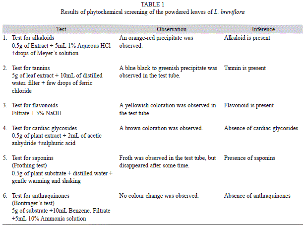
Standard phytochemical methods were used to test for the presence of saponins, alkaloids, tannins, anthraquinones, cardiac glycosides, cyanogenetic glycosides and flavonoids, following standard methods, as described below:
Saponins: About one gram of the powdered extract sample was boiled with 10mL of distilled water for ten minutes (Abate 1989). The samples were filtered using a Buckner funnel and Whatman No 1 filter paper while hot, cooled and the following tests were carried out:
a. Frothing: 2.5mL of the filtrate was diluted to 10mL of water and was shaken vigorously for two minutes. Frothing observed indicates a positive test.
b. Emulsification: 2.5mL of the filtrate was shaken vigorously for two minutes with a few drops of olive oil. An emulsified layer indicated a positive test.
Alkaloids: About one gram of the powdered sample was extracted with 10mL of 10% hydrochloric acid by boiling for five minutes on a water bath (Shale et al. 1999). The extract was filtered using a Buckner funnel and Whatman No. 1 filter paper and the pH of the filtrate was adjusted to about six by adding a few drops of a diluted ammonia solution and tested with litmus paper after which few drops of Dragendorff’s, Mayer’s and Wagner’s reagent were added separately to aliquots of the filtrate in different test tubes. A reddish brown, cream and reddish brown precipitate, respectively, indicated a positive test.
Tannins: About one gram of the powdered sample was boiled with 10mL of distilled water for five minutes, filtered while hot and a few drops of ferric chloride reagent was added to the filtrate (Gupta et al. 2005). A red colouration indicates a positive test.
Anthraquinones: One gram of the powdered sample was extracted with 10mL of 10% sulphuric acid, containing traces of ferric chloride solution for 15 minutes (Moody et al. 2006). It was filtered while hot and the cooled filtrate was extracted while hot with two equal volumes of 97% chloroform. The presence of a rose pink colour in the aqueous layer indicated a positive test.
Cardiac glycosides: One gram of the powdered sample was extracted with 10mL of 80% alcohol for five minutes on a water bath; the extract was filtered and diluted with an equal volume of distilled water. A few drops of lead acetate solution was added, shaken, and filtered after standing for a few minutes. The filtrate was extracted with aliquots of chloroform. The combined chloroform extracts were divided into two portions and Keller Killiani and Kedde tests were carried out on them (Sawadogo et al. 2006).
Keller Killiani test: The extract was evaporated to dryness and 3mL of ferric chloride reagent was added to the cooled residue in a clean test tube. Two ml of concentrated sulphuric acid was gently poured down the side of the test tube. A purple or reddish brown ring at the interface and green colour in acetic acid layer indicated a positive test for 2-de-oxy sugar.
Kedde test: The dry residue was mixed with 1mL of 2% 3,5-dinitrobenzoic acid in ethanol. The solution was made alkaline with five percent sodium hydroxide. A brown purple colour indicated a positive test for the presence of unsaturated lactone ring.
Cyanogenetic glycosides: Half a gram of the powdered sample was placed in three different test tubes A, B, C. Test tubes A and B were mixed with water with a suspended moist sodium picrate paper in the neck of the tube, trapped by means of cork. Test tubes B and C were kept at room temperature while test tube A was placed in boiling water bath (Sawadogo et al. 2006). At the end of about half an hour, a change in colour of the sodium picrate paper indicated a positive test.
Flavonoids: A small quantity of the extract was dissolved in diluted sodium hydroxide (4%) and hydrochloric acid was added to the mixture (Sawadogo et al. 2006). A yellow solution that turns colorless on addition of hydrochloric acid indicated the presence of flavonoids.
Acute toxicity test
The acute toxicity of L. breviflora aqueous extract was determined in rats according to the method of Hilaly et al. (2004) with slight modifications. Rats fasted for 16h were randomly divided into five groups of six rats each. Graded doses of the plant’s extract (200, 400, 800, 1 600 and 3 200mg/kg p.o.) were separately administered to the rats in each of the groups by means of bulbed steel needle. All the rats in the groups were then allowed free access to food and water, and were observed over a period of 48h for acute toxicity signs. The number of deaths within this period of time was recorded.
Anti-inflammatory activity tests
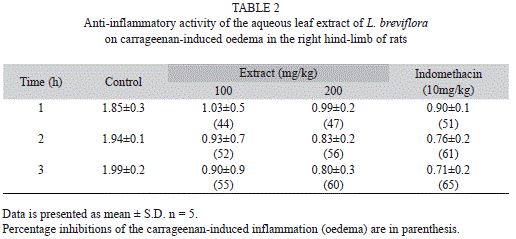
Carrageenan-induced paw edema: Twenty animals used in this study were divided into four groups of five animals each. The first group served as the control, the second, third and fourth group received, respectively, indomethacin (10mg/kg body weight), and the L. breviflora extract at two doses of 100 and 200mg/kg. The plant extract and indomethacin were dissolved in distilled water. Carrageenan solution (0.1ml of 1% carrageenan solution) was injected into the sub-plantar region of the right hind paw of the rats 1h after intraperitoneal administration of distilled water, indomethacin and extract (Silva et al. 2005). The paw volume was measured after administration of drug and extract using a micrometer screw gauge at: 0hr, 1hr, 2hr and 3hr.
The anti-inflammatory effect of the extract was calculated with the use of the following equation: anti-inflammatory activity (%)=(1- D/C) X 100, where D represented the average paw volume after the extract was administered to the rats and C was the paw volume in the control groups. The percentage inhibition of the inflammation was calculated from the formula: % inhibition = D0-Dt/D0 X 100 where D0 was the average inflammation (hind paw oedema) of the control group at a given time; and Dt was the average inflammation of the drug treated (i.e. extracts or reference indomethacin) rats at the same time (Gupta et al. 2005, Sawadogo et al. 2006).
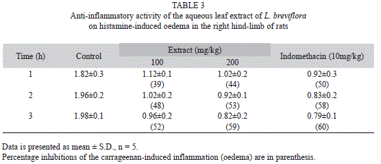
Histamine induced paw edema: For this test we used the method of Perianayagam et al. (2006), the paw edema was produced by subplantar administration of 0.1% freshly prepared solution of histamine into the right hind paw of the rats. Sixteen rats divided into four groups of four rats per group were used. The paw volume was recorded before 0h and 1h after the histamine injection. The four groups of the rats were pre-treated with the plant extract (100, 200mg/ kg), 2mL/kg of distilled water (vehicle control) or 10mg/kg indomethacin (reference drug). These were administered intraperitoneally 1hr before eliciting paw edema. The anti-inflammatory activity was calculated as described for carrageenan-induced edema.
Analgesic activity tests
Acetic acid-induced writhing in rats: The acetic acid-induced writhing test measures abdominal constrictions together with stretching of the hind limbs resulting from intraperitoneal (i.p.) injection of 0.7% acetic acid (10mL/ kg). This was carried out according to the procedures described by Sawadogo et al. (2006). Four groups of five animals per group were used in this study comprising the control (2mL/ kg distilled water), indomethacin (10mg/kg), or plant extract (100, 200mg/kg). After 30min, acetic acid was administered intraperitoneally.
The number of writhing movements was counted for 30min.
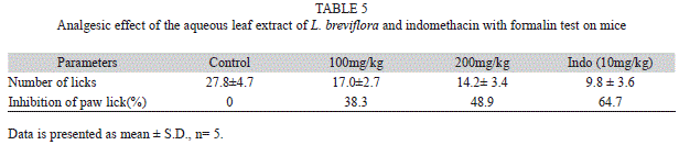
Formalin test in rats: The procedure was essentially similar to that described by Correa & Calixto (1993). Formalin solution (0.05mL of 2.5% formalin) was injected into the sub- plantar of the right hind paw. The number of times and the time spent licking the injected paw was recorded and was considered as indicative of pain. The animals were pretreated with indomethacin and plant extract (100 and 200mg/kg) 30min before being administered with formalin, and the responses were observed for 30 min.
The data were expressed as mean ±S.D. Where applicable the difference in response to test drugs was determined by student’s t-test. p<0.05 was considered significant.
Results
Phytochemical screening
Phytochemical screening of L. breviflora leaves showed the presence of alkaloids, tannin, flavonoids and saponins. Anthraquinones and cardiac glycosides were however absent (Table 1).
Acute toxicity test
No deaths were recorded in the control, 200 and 400mg/kg dose groups. All animals that received 800 and 1 600mg/kg doses died. Behavioral changes exhibited by the animals included dullness, lethargy and anorexia, with severity depending on the dose. Dull animals that did not die later recovered and became active.
Anti-inflammatory activity test
Carrageenan-induced paw edema: When compared to the control, the extract and indomethacin significantly reduced the paw edema hours after carrageenan injection. For instance, the 100 mg/kg extract produced its highest effect at 3h (55%) after carrageenan injection, while the 200mg/kg extract was more effective 3h (60%) after injection. The reference drug, indomethacin produced time-dependent reduction as the effect was more pronounced at 3h (65%) of carrageenan administration (Table 2).
Histamine-induced paw edema: Inhibition of histamine-induced edema was observed in this study. The anti-histaminic activity of the 100mg/kg extract was most pronounced at 3h (52%) while that of the 200mg/kg (59%), and indomethacin (60%) was also at 3h (Table 3).
Anti-nociceptive activity tests
Acetic acid-induced writhing: The effect of the aqueous leaf extract of L. breviflora on writhing response in mice showed that the extract at 100 and 200mg/kg caused 48 and 61% inhibition, respectively, on the writhing response induced by acetic acid. An 82% inhibition was observed with indomethacin, the reference drug used in the study (Table 4).
Formalin test: In this study, the extract caused a dose-dependent decrease in licking time by the mice injected with formalin. The indomethacin group showed better analgesic effect than the two doses. The analgesic effects of these groups were significantly different from that of control at p<0.05 (Table 5).
Discussion
Phytochemical analysis of the leaves of Lagenaria breviflora showed the presence of tannins, saponins, alkaloids and flavonoids. The presence of flavonoids and tannins in the leaves of this plant is likely to be responsible for the anti-inflammatory and analgesic effects observed. Flavonoids and tannins are phenolic compounds and plant phenolics are also a major group of compounds that act as primary antioxidants or free radical scavengers (Adedapo et al. 2008a, 2008b, Ayoola et al. 2008). Tannins and saponins are also found to be effective antioxidants, antimicrobial, and anti-carcinogenic agents (Lai et al. 2010). Flavonoids are known to target prostaglandins which are involved in the late phase of acute inflammation and pain perception (Rajnarayana et al. 2001, Chakraborty et al. 2004). Hence, the presence of flavonoids may be contributory to the anti-inflammatory and analgesic activities of aqueous leaf extract of the plant.
The results of the acute toxicity study however showed that from 800mg/kg dose, all the mice used in this study died. This might suggest that at a dose of 800mg/kg and above, the leaves from this plant are toxic, signifying that caution must be exercised in its use for medicinal purpose.
The results of this investigation revealed that the aqueous extract of the leaves of L. breviflora possessed a dose dependent activity against carrageenan- and histamine-induced paw oedema in rats. The activity of the 200mg/ kg of the extract was slightly less effective than that of indomethacin. Carrageenan-induced hind paw edema is the standard experimental model of acute inflammation; it is the phlogistic agent of choice for testing anti-inflammatory drugs as it is not known to be antigenic and is devoid of apparent systemic effects. Moreover, the experimental model exhibits a high degree of reproducibility (Winter et al. 1962). Carrageenan-induced edema is a biphasic response. The first phase is mediated through the release of histamine, serotonin and kinins whereas the second phase is related to the release of prostaglandin and slow reacting sub- stances which peak at 3h (Vinegar et al. 1969). The carrageenan-induced inflammation model which is a predictive test for anti-inflammatory agent acts by inhibiting the mediators of acute inflammation (Ozaki 1990, Mossai et al. 1995, Silva et al. 2005), therefore, the result is an indication that L. breviflora can be effective in acute inflammatory disorders.
In the histamine-induced paw edema, 200mg/kg of the extract exhibited inhibitory effect than the reference drug at 2h. It should be noted that the early phase (1-2h) in the induced paw edema model is mainly mediated by histamine, serotonin and the increase of prostaglandin (PG) synthesis in the surroundings of the damaged tissues. The late phase on the other hand, is mainly mediated by bradykinin, leukotrienes, polymorphonuclear cells and PGs produced in tissue microphages (Antonio & Souza-Brito 1998, Cuman et al. 2001, Linardi et al. 2002, Vasudevan et al. 2007). The results of the present study suggest that the suppression of inflammation at the early phase was as a result of the antihistamine activity of the plant extract.
The abdominal constriction response induced by acetic acid is a sensitive procedure to establish peripherally acting analgesics. This response is thought to involve local peritoneal receptors (Chakraborty et al. 2004). The acetic acid induced writhing test, a non specific but nevertheless sensitive method widely used for analgesic screening (Le Bars et al. 2001). The number of writhing movements during a 30min observation in the control group was 61.4 ± 2.3. In this study, the reference drug gave an 82% inhibition of writhing in the animals while the 200mg/kg dose gave 61% inhibition showing that it is not as effective as the reference drug. Acetic acid has been found to cause an increase in peritoneal fluid levels of prostaglandins (PGE2 and PGF2), hence causing inflammatory pain by inducing capillary permeability (Amico-Roxas et al. 1984). The observed effects in the present study suggest that nonetheless L. breviflora had an inhibitory effect on prostaglandins synthesis.
The formalin test has been described as a convenient method for producing and quantifying pain in rats (Dubuisson & Dennis 1977, Tjolsen et al. 1992). The test employs an adequate painful stimulus to which the animals show a spontaneous response and it is sensitive to commonly used analgesics. The pain stimulus, a continuous rather than a transient one, may have resemblance to some kind of clinical pain, and observations are made on animals which are restrained only lightly or not at all (Hunskaar et al. 1985, Ghannadi et al. 2005). Intraplantar injection of 2.5% formalin evoked a characteristic licking response in the Wistar rats. In this study, the extract caused a dose-dependent decrease in licking frequency on rats injected with formalin, showing the analgesic effect of the extract. However, the active doses of the plant extract showed lower potency in bringing about analgesia than that of the reference drug.
Since the plant extract reduced significantly the formation of oedema induced by carrageenan and histamine, as well as reduced the number of writhes in acetic acid-induced writhing models and dose-dependent decrease of licking frequency in rats injected with 2.5% formalin, the results have validated the basis for the traditional use of L. breviflora against infectious diseases such as New Castle disease which is characterized by inflammation and pain.
Acknowledgments
We acknowledge the assistance of the technical staff of Pharmacognosy laboratory of the University of Lagos with respect to phytochemical screening of the leaves of the plant used in this study.
References
Abate, G. 1989. In S. Demissew (ed.). Ethiopian Traditional Medicine, p. 3-5. Este Debdabe. Department of Biology, Science Faculty, Addis Ababa University, Ethiopia. [ Links ]
Adedapo, A.A., S. Koduru, F.O. Jimoh, P.J. Masika & A.J. Afolayan. 2008a. Evaluation of the medicinal potentials of the methanol extracts of the leaves and stem of Halleria lucida. Bioresource. Tech. 99: 4158-4163. [ Links ]
Adedapo, A.A., F.O. Jimoh, A.J. Afolayan & P.J. Masika. 2008b. Antioxidant activities and phenolic contents of the methanol extracts of the stems of Acokanthera oppositifolia and Adenia gummifera. BMC Complement. Alternat. Med. 8:54. [ Links ]
Adedapo, A.A., F.O. Jimoh, A.J. Afolayan & P.J. Masika. 2009. Antioxidant activities of the methanol extracts of the leaves and stems of Celtis africana. Rec. Nat. Prod. 3: 23-31. [ Links ]
Adedapo, A.A. & B.O. Bankole. 2011. Ethnoveterinary survey of poultry management in some selected farms in Ibadan, Nigeria. Recent Prog. Med. Plant 31: 305-316. [ Links ]
Ajayi, G.O., N.C. Awujo & L.E. Abulu. 2002. The miracicidal and cercaricidal activity of the methanolic extracts of Lagenaria breviflora Robert Family Cucurbitaceae fruit on Schistosoma mansoni. Nig. Qtr. J. Hosp. Med. 12: 57-59. [ Links ]
Amico-Roxas, M., A. Caruso, S. Trombadore, R. Scifo & U. Scapagnini. 1984. Gangliosides antinociceptive effects in rodents. Arch. Intl. Pharmacodyn. Ther. 272: 103-117. [ Links ]
Antonio, M.A. & A.R.M. Souza-Brito. 1998. Oral antiinflammatory and anti ulcerogenic activities of a hydroalcoholic extract and partitioned fractions of Turnea ulmifolia (Turneaceae). J. Ethnopharmacol. 61: 215-228. [ Links ]
Ayoola, G.A., H.A.B. Coker, S.A. Adesegun, A.A. Adepoju-Bello, K. Obaweya, E.C. Ezennia & T.O. Atangbayila. 2008. Phytochemical Screening and Antioxidant Activities of Some Selected Medicinal Plants Used for Malaria Therapy in Southwestern Nigeria. Trop. J. Pharm. Res. 7: 1019-1024. [ Links ]
Chakraborty, A., R.K.B. Devi, S. Rita, K. Sharatchandra & T.I. Singh. 2004. Preliminary studies on anti-inflammatory and analgesic activities of Spilanthes acmella in experimental animal models. Indian J. Pharmacol. 36 (3): 148-150. [ Links ]
Correa, C.R. & J.B. Calixto. 1993. Evidence of participation of B1 and B2 kinin receptors in formalin-induced nociceptive response in mouse. Br. J. Pharmacol. 110: 193-198. [ Links ]
Cuman, R.K.N., C.A. Bersani-Amadio & Z.B. Fortes. 2001. Influence of type 2 diabetes on the inflammatory response in rat. Inflammation Res. 50: 460-465. [ Links ]
Dubuisson, D. & S.G. Dennis. 1977. The formalin test: a quantitative study of the analgesic effects of morphine, meperidine and brain stem stimulation in rats and cats. Pain 4: 161-174. [ Links ]
Elujoba, A.A., S.O. Olagbende & S.K. Adesina. 1985. Anti-implantation activity of the fruit of Lagenaria breviflora Robert. J. Ethnopharmacol. 13: 281-288. [ Links ]
Ghannadi, A., V. Hajhashemi & H. Jafarabadi. 2005. An investigation of the analgesic and anti-inflammatory effects of Nigella sativa seed polyphenols. J. Med. Food. 8: 488-493. [ Links ]
Gupta, M., U.K. Mazunder, R. Sambath Kumbar, P. Gomath, Y. Rajeshwar, B.B. Kakoti & V. Tamil Selven. 2005. Anti-inflammatory, analgesic and antipyretic effects of methanol extract from Bauhina racemosa stem bark in animal models. J. Ethnopharmacol. 98: 267-273. [ Links ]
Hanno, S., H. Christoph & S.R. Susanne. 2009. Gourds afloat: adapted phytogeny reveals an Asian origin of the gourd family (Cucurbitaceae) and numerous oversea dispersal events. Proc. N. Soc. 276: 843-845. [ Links ]
Hilaly, J.E., Z.H. Israili & B. Lyoussi. 2004. Acute and chronic toxicological studies of Ajuga iva in experimental animals. J. Ethnopharmacol. 91: 43-30. [ Links ]
Hunskaar, S., O.B. Fasmer & K. Hole. 1985. Formalin test in mice: a useful technique for evaluating wild analgesics. J. Neurosci. Meth. 4: 69-76. [ Links ]
Kumara, N.K.V.M.R. 2001. Identification of strategies to improve research on medicinal plants used in Sri Lanka. In WHO Symposium. University of Ruhuna, Galle, Sri Lanka. [ Links ]
Lai, F.R., Q.B. Wen, L. Li, H. Wu & X.F. Li. 2010. Antioxidant activities of water-soluble polysaccharide extracted from mung bean (Vigna radiata L.) hull with ultrasonic assisted treatment. Carbohyd. Polym. 81: 323-329. [ Links ]
Le Bars, D., M. Gozariu & S. Cadden. 2001. Animal models of nociception. Pharmacol. Rev. 53: 597-652. [ Links ]
Linardi, A., S.K.P. Costa, G.R. DeSilva & E. Antunes. 2002. Involvement of kinins, mast cells, and sensory neurons in the plasma exudation and paw edema induced by staphylococcal entrotoxin B in the mouse. Eur. J. Pharmacol. 399: 235-242. [ Links ]
Moody, J.O., V.A. Robert, J.D. Connolly & P.J. Houghton. 2006. Anti-inflammatory activities of the methanol extracts and an isolated furanoditerpene constituent of Sphenocentrum jollyanum Pierre (Menispermaceae). J. Ethnopharmacol. 104: 87-91. [ Links ]
Mossai, J.S., S. Rafatullah, A.M. Galal & M.A. Al-Yahya. 1995. Pharmacological studies of Rhus retinorrhea. Intl. J. Pharmacol. 33: 242-246. [ Links ]
Onasanwo, S.A., A.B. Saba, O.A. Oridupa, A.A. Oyagbemi & B.V. Owoyele. 2011a. Anti-nociceptive and anti-inflammatory properties of the ethanolic extract of Lagenaria breviflora whole fruit in rat and mice. Niger. J. Physiol. Sci. 26:71-6. [ Links ]
Onasanwo, S.A., N. Singh, A.B. Saba, A.A. Oyagbemi, O.A. Oridupa & G. Palit. 2011b. Anti-ulcerogenic and in vitro antioxidant activities of Lagenaria breviflora (LB) whole fruit ethanolic extract in laboratory animals. Pharmacognosy. Res. 3:2-8. [ Links ]
Oridupa, O.A., A.B. Saba & L.K. Sulaiman. 2011. Preliminary report on the antiviral activity of the ethanolic fruit extract of Lagenaria breviflora Roberts on New- castle Disease virus. Trop. Vet. 29 (1): 22-33. [ Links ]
Ozaki, Y. 1990. Anti-inflammatory effects of Curcuma xanthorrhiza Roxb, and its active principle. Chem. Pharm. Bull. 38: 1045-1048. [ Links ]
Perianayagam, J.B., S.K. Sharma & K.K. Pillai. 2006. Anti-inflammatory activity of Trichodesma indicum root extract in experimental animals. J. Ethnopharmacol. 104: 410-414. [ Links ]
Rajnarayana, K., M.S. Reddy, M.R. Chaluvadi & D.R. Krishna. 2001. Bioflavonoids classification, pharmacological, biochemical effects and therapeutic potential. Indian J. Pharmacol. 33:2-16. [ Links ]
Saba, A.B., O.A. Oridupa & S.O. Ofuegbe. 2009a. Evaluation of haematological and serum electrolyte changes in Wistar rats administered with ethanolic extract of whole fruit of Lagenaria breviflora Roberts. J. Med. Plants Res. 3: 758-762. [ Links ]
Saba, A.B., O.A. Oridupa, M.O. Oyeyemi & O.D. Osanyigbe. 2009b. Spermatozoa morphology and cha- racteristics of male Wistar rats administered with ethanolic extract of Lagenaria breviflora Roberts. Afr. J. Biotech. 8: 1170-1175. [ Links ]
Sawadogo, W.R., R. Boly, M. Lompo & N. Some. 2006. Anti-inflammatory, analgesic and antipyretic activities of Dicliptera verticillata. Intl. J. Pharmacol. 2: 435-438. [ Links ]
Shale, T.L., W.A. Stirk & J. Van Staden. 1999. Screening of medicinal plants used in Lesotho for antibacterial and anti-inflammatory activity. J. Ethnopharmacol. 67: 347-354. [ Links ]
Silva, G.N., F.R. Martins & M.E. Matheus. 2005. Investigation of anti-inflammatory and antinociceptive activities of Lantana trifolia. J. Ethnopharmacol. 100: 254-259. [ Links ]
Sobota, R., M. Szwed, A. Kasza, M. Bugno & T. Kordula. 2000. Parthenolide inhibits activation of signal transducers and activators of transcription (STATs) induced by cytokines of the IL-6 family. Bioch. Biophys. Res. Comm. 267: 329-333. [ Links ]
Tjolsen, A., D.G. Berge, S. Hunskaar, J.H. Rosland & K. Hole. 1992. The formalin test: an evaluation of the method. Pain 51: 5-17. [ Links ]
Tomori, O.A., A.B. Saba & H.O. Dada-Adegbola. 2007. Antibacterial activity of ethanolic extract of whole fruit of Lagenaria breviflora Robert. J. Anim. Vet. Adv. 6: 752-757. [ Links ]
Vasudevan, M., K.K. Gunman & M. Parle. 2007. Antinociceptive and anti-inflammatory effects of Thespesia populnea bark extract. J. Ethnopharmacol. 109: 264-270. [ Links ]
Vinegar, R., W. Schreiber & R. Hugo. 1969. Biphasic development of carrageenin oedema in rats. J. Pharmacol. Exp. Ther. 166: 96-103. [ Links ]
Winter, C.A., E.A. Risley & G.W. Nuss. 1962. Carrageenan induced edema in hind paw of the rat as an assay for anti-inflammatory drugs. Proc. Soc. Exp. Biol. Med. 111: 544-547. [ Links ]
Yasuyuki, M., M. Patrick, F. Hiroshi & M. Hiroko. 2005. Diversity of landraces of the white-flowered Gourd (Lagenaria siceraria) and its wild relatives in Kenya. Genet. Res. Crop. Evol. 52: 737-747. [ Links ]
*Correspondencia:
Adeolu Adedapo: Department of Veterinary Physiology, Biochemistry and Pharmacology, University of Ibadan, Nigeria. adedapo3a@ yahoo.co.uk
Temitayo Adewuyi: Department of Veterinary Physiology, Biochemistry and Pharmacology, University of Ibadan, Nigeria. temitayoemmanuel12@yahoo.com
Margaret Sofidiya: Department of Pharmacognosy, University of Lagos, Nigeria. toyin_sofidiya@yahoo.co.uk
1. Department of Veterinary Physiology, Biochemistry and Pharmacology, University of Ibadan, Nigeria; adedapo3a@ yahoo.co.uk, temitayoemmanuel12@yahoo.com
2. Department of Pharmacognosy, University of Lagos, Nigeria; toyin_sofidiya@yahoo.co.uk
Received 10-I-2012. Corrected 20-V-2012. Accepted 21-VI-2012.

