


Services on Demand
Journal
Article
 English (pdf)
 Article in xml format
 Article references
 Send this article by e-mail
Indicators
-
 Cited by SciELO
 -
 Access statistics
Related links
-
 Similars in
SciELO
Similars in
SciELO  uBio
uBio
Share

 Permalink
Revista de Biología Tropical
On-line version ISSN 0034-7744Print version ISSN 0034-7744
Rev. biol. trop vol.60 suppl.1 San José Mar. 2012
*Dirección para correspondencia
Abstract
The study of coral diseases, coral pathogens, and the effects of diseases on tropical and subtropical coral reefs are all current, high-profile research areas. This interest has grown steadily since the first report of a coral disease in 1973. The author of this report was Arnfried Antonius and the publication was an abstract in the proceedings of a scientific meeting of the Association of Marine Laboratories of the Caribbean, or AMLC (then known as the Association of Island Marine Laboratories of the Caribbean). Since Antonius’ pioneering communication he continued working on coral diseases on reefs throughout the world, often documenting the first observation of a novel pathology in a novel location. Each of the coral diseases Antonius first described, in particular black band disease, is the subject of current and ongoing investigations addressing pathogens, etiology, and their effects on coral reefs. Many of the points and observations he made in his early papers are highly relevant to research today. This paper reviews aspects of Antonius’ early work, highlighting contributions he made that include the first in situ experimental studies aimed at discerning coral epizootiology and the first quantitative assessments of the role of environmental factors in coral disease. Antonius’ early findings are discussed in terms of relevant current controversies in this research area.
Key words: Caribbean coral diseases, black band disease, white band disease, Arnfried Antonius.
Resumen
El estudio de las enfermedades de los corales, los patogenos de los corales y los efectos de estas enfermedades sobre los arrecifes tropicales y subtropicales son actualmente areas importantes de investigacion. El interés en este tema ha crecido continuamente desde el primer informe sobre una enfermedad de coral que se publico en 1973. El autor de este informe fue Arnfried Antonius y la publicacion fue un resumen en el Libro de Programa y Resumenes de la Decima Reunion de la Asociacion de Laboratorios Marinos Islenos del Caribe (conocida ahora como la Asociacion de Laboratorios Marinos del Caribe). Desde esta comunicacion pionera, Antonius siguio trabajando sobre las enfermedades de los corales en arrecifes alrededor del mundo, a menudo documentando la primera observacion de una nueva enfermedad en un nuevo lugar. Cada enfermedad de coral descrita por primera vez por Antonius es actualmente el objeto de investigaciones actuales en lo que se refiere a patogenos, ecologia de las enfermedades y los efectos sobre los arrecifes de coral. Muchas de las observaciones en sus trabajos tempranos siguen siendo relevantes en la investigacion actual. Este trabajo examinara ciertos aspectos de los estudios tempranos de Antonius sobre las enfermedades de los corales, poniendo de relieve sus contribuciones novedosas que incluyen los primeros experimentos in situ que tenian como objetivo el estudio de la etiologia de las enfermedades de los corales y los primeros analisis cuantitativos de la incidencia de las enfermedades de corales y de los patrones de distribución en funcion de los factores ambientales. Las contribuciones iniciales de Antonius se discuten en terminos de las controversias actuales sobre el tema.
Palabras clave: Enfermedades de corales, enfermedad de banda negra, enfermedad de banda blanca, Arnfried Antonius
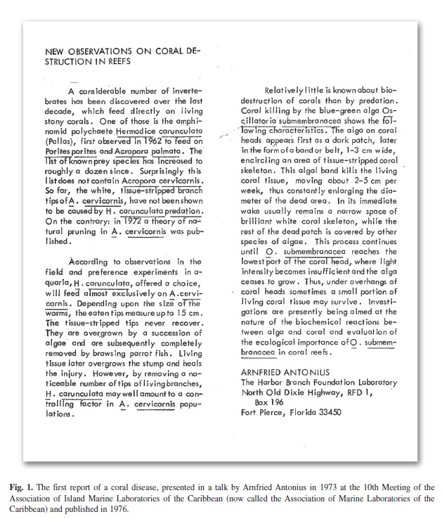
In 1973, at the 10th meeting of the Association of Island Marine Laboratories of the Caribbean, Arnfried Antonius gave the first report of a coral disease. Abstracts from this meeting were published in 1976, and Antonius’ report, titled “New observations on coral destruction in reefs”, is now widely cited as the first published documentation of coral disease. This abstract is reproduced in Fig. 1. Although the publication was produced in 1976, this report has been consistently cited as Antonius (1973) by Antonius himself in his subsequent publications and by subsequent investigators in the field. The actual first publication of a coral disease was by Garrett and Ducklow in 1975.
In the four decades since this report the study of coral diseases has increased steadily and dramatically, with the number of published papers focusing on this topic increasing exponentially since the early 1990s (Sokolow 2009). The increase in both coral disease research and the number of associated papers appearing in the literature has unfortunately been matched by an increase in coral disease incidence and prevalence on tropical and subtropical reefs worldwide, along with an increase in the appearance and spread of novel coral diseases
(Sutherland et al. 2004, Weil 2004).
Although widely recognized as the “father of coral disease” for his first reports in this research area, Antonius’ early work also is highly notable in that he was the first to combine field observations with experimental work in the laboratory using controlled conditions; the first to document the relationships between coral disease incidence and environmental factors (temperature, nutrient enrichment, and pollution); the first to point out that some coral diseases may have similar disease signs but different potential pathogens; and the first to extrapolate coral disease activity on reefs to overall reef ecology. Examples of his contributions in each of these areas are summarized and discussed below.
Recognition and Description of the First Coral Diseases
Black Band Disease: The subject of Antonius’ first coral disease report (Antonius 1976) was black band disease (BBD). Although not referred to as a disease in his report (Fig. 1), he conferred this name in the literature in a later publication (Antonius 1981a). In addition to describing the pattern of coral tissue death that results from BBD he identified a bluegreen algal (cyanobacterial) pathogen which he identified as Oscillatoria submembranacea (Fig. 1) based on a personal communication with Drouet (Antonius 1985a). At that time Drouet was a recognized expert in cyanobacterial taxonomy, using morphological characteristics as the defining criteria. Antonius described the BBD cyanobacterial pathogen as an unbranched filamentous type with “differing end cells, one tapering, the other rounded” (Antonius 1985a). Since this early work this cyanobacterium was redescribed as Phormidium corallyticum in three back-to-back papers (Rutzler & Santavy 1983, Rutzler et al. 1983, Taylor 1983) on which Antonius was co-author of one (Rutzler et al. 1983). In the 2000s the new application of molecular genetics (16SrRNA gene sequencing) to the study of BBD led to much controversy in the literature as to the identification of the dominant BBD cyanobacterium, with various genera proposed that included Oscillatoria, Geitlerinema, and Leptolyngbya (Cooney et al. 2002, Frias-Lopez et al. 2003, Myers et al. 2007) in the Caribbean and Oscillatoria and Pseudoscillatoria in the Indo-Pacific and Red Sea (Sussman et al. 2006, Rasoulouniriana et al. 2009, Sato et al. 2009). Only very recently has this cyanobacterium been formally described under the International Code of Botanical Nomenclature as the new cyanobacterial genus and species Roseofilum reptotaenium (Casamata et al. 2012), which translates as “creeping band of red thread”. The formal characterization includes description of an identifying morphology, that of one tapering and one rounded end cell, as noted and reported by Antonius (1985a).
As seen in Antonius’ first report (Fig. 1), BBD was attributed to the “coral killing” cyanobacterium he described. This contention also became a subject of investigation and controversy, and for many years the BBD pathogen was proposed to be, in addition, to the cyanobacterium, a sulfide-oxidizing bacterium (Ducklow & Mitchell 1979), various heterotrophic bacteria (Cooney et al. 2002, Frias-Lopez et al. 2004), and a marine fungus (Ramos-Flores 1983). BBD was also proposed as a polymicrobial disease, i.e. caused by a community of bacteria with no primary pathogen (Richardson et al. 1997). A recent meta-analysis of clone libraries constructed from 87 BBD samples collected between 2002 and 2010 (Miller & Richardson 2011) revealed that by far the most common 16S rRNA gene sequence was for R. reptotaenium, detected in 78% of the clone libraries examined. The three next most abundant sequences, each detected in 13% of clone libraries, were three heterotrophic bacteria, each of which was detected previously in BBD samples (Miller & Richardson 2011). These were an alphaproteobacterium, Roseovarius crassostreae, known to be the pathogen of Juvenile Oyster Disease, an uncultured alphaproteobacterium associated with Juvenile Oyster Disease, and a Cytophaga sp. (Cooney et al. 2002, Frias-Lopez et al. 2002, Sekar et al. 2008). Recently, a unialgal laboratory culture of R. reptotaenium was shown to infect coral in the laboratory and produce BBD (Casamata et al. 2012). While the culture has bacterial contaminants, none of these were found in the BBD-derived clone libraries (Miller & Richardson 2011). Since the culture cannot live without associated bacteria (a pure culture could not maintain viability) Koch’s Postulates cannot be fulfilled and thus the “proof” of R. reptotaenium as the BBD primary pathogen is currently not feasible. For a discussion of this issue in general see Fredricks & Relman (1996). In 1981 (Antonius 1981a) Antonius modified his identification of a single (cyanobacterial) BBD pathogen to a cyanobacterial pathogen “in association with bacteria”. It appears that he has been right all along.
White Band Disease: White band disease (WBD), first described by Antonius in 1981 (Antonius 1981a, 1981b), was described as “a band of brilliant white skeleton always visible in the wake of a moving front of tissue destruction” (Antonius 1981b). As is the case with BBD, WBD has been, since Antonius’ first report, the subject of ongoing and controversial research and discussion. In particular, a bacterial pathogen of WBD has been elusive and confusing to the point that it has been suggested that various disease signs given names such white plague and white death, in addition to white band, should be referred to as “white syndrome” (reviewed in Pantos et al. 2003). This same point was made by Antonius in his first report of the disease (1981a) in which he states that WBD is “variously called White Death, Plague, etc.” Antonius experimentally demonstrated that WBD could not be transmitted between coral hosts, either by direct contact or by injection with fresh disease material (Antonius 1981b), and could not be cured using antibiotics (Antonius 1985b). In contrast, in his 1985 study he successfully demonstrated that BBD could be easily transmitted and was curable with antibiotics. Antonius’ overall conclusions about WBD etiology, that it is a “physiological syndrome that needs only a trigger” (Antonius 1981b), remains the most viable working hypothesis today.
Shut Down Reaction: In 1977 Antonius first described the coral disease he termed shut down reaction (SDR). He noted that this disease only occurred in corals maintained in aquaria (Antonius 1977), an observation that holds true today (Sutherland et al. 2004). The name is based on the extremely rapid rate of tissue destruction, 10 cm per hour along a moving front, that can be transmitted using infected tissue or contact with an infected colony, but only when the recipient colony is under stress (Antonius 1981b). No definitive work has been carried out to define the etiology of SDR.
Skeleton Eroding Band: The final coral disease that Antonius was the first to describe is skeleton eroding band (SEB) (Antonius & Lipscomb 2000). This disease is caused by a protozoan, Halofolliculina corallasia, which appears as a dark band that moves across corals while lysing tissue. The fact that Antonius recognized this as separate from BBD attests to his exceptional observational skills, for it is only by close visual examination that one can tell the difference between the two. Antonius reported SEB on corals in the Indo-Pacific. A similar protozoan infection on Caribbean corals has only relatively recently been noted (Croquer et al. 2006)
Combining Field Observations and Experimental Work
While it is well-known that Antonius was the first to report and name several coral diseases, less well know is the fact that he was the first to conduct the combination of field and laboratory experimental work required to understand coral disease etiology. One example will be provided.
In Antonius’ ongoing work to determine the pathogens associated with BBD and WBD, summarized above, he made the observations that: i) BBD could be easily transmitted between corals, whereas WBD could not; ii) inoculation of a healthy coral with BBD would produce an infection on a healthy coral whereas inoculation with WBD would not; iii) exposure of infected coral colonies in situ to antibiotics would cure a BBD infected coral but not one infected with WBD; and iv) new BBD infections appeared to occur only on corals infected with WBD, specifically at the site of WBD infection.
The last observation (reported in Antonius 1981a, 1981b, and 1985b) was the basis for a remarkable seven year field/laboratory experiment carried out to determine how BBD infections initiate on the reef (Antonius 1985a). The time required to complete the experiment was due to the fact that, since WBD could not be artificially transmitted, experiments relied on finding naturally occurring WBD-infected colonies in situ on the reef for experimental use. Furthermore, indicative of Antonius’ detailed and exhaustive approach to research, the experiment included corals on reefs of both the Caribbean and the Indo-Pacific. In each region four species of WBD and BBD susceptible corals were studied, a total of eight coral species investigated. Finally, all experiments were replicated 12 times at three different temperatures (26, 28 and 30˚C, selected based on his observations of temperature thresholds for BBD - see below).
Antonius’ experimental design (Antonius 1985a) consisted of field experiments in which a BBD-infected coral was placed near a WBDinfected coral, and laboratory experiments using aquaria in which he manipulated WBD and BBD-infected corals. BBD-infected corals were easy to obtain since he could artificially and easily infect healthy corals, but WBD-infected corals for laboratory experiments had to be collected on the reef. Field experiments consisted of monitoring WBD-infected corals for which BBD-infected corals were placed 0.5-1 m upstream. Control WBD-infected colonies were in areas of the reef that were free of BBD, or had BBD-infected corals placed a minimum of 10 m downstream. Laboratory experiments consisted of placing BBD-infected corals into aquaria with WBD-infected corals, controls being aquaria with WBD-infected corals together with healthy or injured corals (no BBD). All experiments consisted of observing whether or not the WBDinfected corals developed BBD.
Antonius’ results were, in his word, “unequivocal” proof of the positive relationship between BBD and WBD. Analysis of the results of the 12 experiments showed that at 30˚C the appearance of a BBD infection on a WBD lesion ranged, for Caribbean corals, from 58-83% on the reef and from 69-92% in aquaria. For Indo-Pacific corals the values were 62-86% on the reef and 71-93% in aquaria. At 28˚C Caribbean corals exhibited 54-78% BBD infection on the reef and 67-85% in aquaria, and Indo-Pacific corals 42-80% and 50-92% respectively. At 26˚C (below the field temperature threshold of BBD) there were zero cases of BBD infection on the reef, and a maximum of 8% infection in aquaria, where stress may have been a factor. Half of the species tested (both Caribbean and Indo-Pacific corals) did not become infected in aquaria with BBD at 26˚C. None of the controls on the reef or in aquaria became infected with BBD.
As remarkable as these experiments is the fact that in the 27 years since this paper was published none of the many investigators in coral disease research has further investigated these fascinating and compelling findings.
Coral Disease Incidence and Environmental Factors
In the last few decades, as coral disease incidence and prevalence have increased on reefs world-wide, there has been a major focus on the relationship between coral disease and environmental factors, in particular those associated with human activity. Virtually all of the studies that found a positive relationship were preceded by similar work by Antonius.
Antonius was the first to report a temperature threshold for coral disease, specifically BBD and WBD. His first mention of this (Antonius 1981a) was the observation that both BBD and WBD were seasonal on reefs of high latitude (Bermuda and Florida), with diseased colonies present in the summer but not the winter and with the BBD season longer than that for WBD on the same reefs. Based on extensive field surveys on reefs in the Red Sea combined with temperature data, he demonstrated that BBD activity was strongest at 30˚C but did not occur at or below 26˚C (Antonius 1985b). In this same study he noted that WBD was not affected by temperature except for one period when it fell to an unusual low of 22˚C. At this time WBD frequency decreased, but was still present.
Antonius was also the first to report a correlation of a disease (BBD) with nutrients and sewage outflow. His first observation occurred when he was conducting BBD infection experiments to determine coral host species susceptibility and an aquarium water source became “contaminated” (Antonius 1981a). He observed that two normally non-susceptible coral species were infected and killed by BBD and concluded that the newly observed susceptibility was due to a 400x increase in nitrate and 10x increase in phosphate in the water (which he called “artificial hypernutrition”). He also observed that WBD progression was not affected by exposure to this water. His correlation of the disease with sewage was noted during extensive surveys of disease at 33 sites along the Red Sea (Antonius 1988a). In this study he documented the highest BBD frequency near Jeddah, which he attributed to “sea water pollution, especially eutrophication”. Since this work other investigators have shown a positive correlation between BBD and elevated nutrients (Bruckner & Bruckner 1997, Kuta & Richardson 2002, Kaczmarsky et al. 2005).
Coral Diseases and Coral Reef Ecology
Antonius’ body of work also illustrates his insight into the effect of coral diseases on coral biology and coral reef ecology. Three examples are summarized. The first, part of his ongoing study on BBD infection and transmission, involved experimentally testing the hypothesis that wounds on corals caused by lysing of tissue by mesenterial filaments of aggressive corals of other species could be infected by BBD (Antonius 1985a). He did document successful infection, which he recorded as lower than BBD infections on WBD lesions (40-50% on the reef, 30-40% in aquaria) a difference he attributed to the fact that wounds due to aggression were stationary and temporary, whereas WBD induced lesions on infected colonies were present over days to weeks due to the steadily advancing disease front. Of note is his interpretation of his observation in the context of coral biology. He concluded that BBD susceptibility was an “unexpected bonus for the aggressor” in that BBD infection would likely completely remove the encroaching coral from the reef. This outcome confers a much greater benefit when compared to the limited ability of the aggressor to inflict tissue damage on a small area of an immediately adjacent competing coral colony via extrusion of mesenterial filaments (Antonius 1985a).
The second example also arose from BBD infection studies. In this case Antonius noted that BBD often began at the site of a clump of green algae growing near or onto a coral colony. His interpretation was that the pathogenic BBD cyanobacterium resided in the green algal turf, which served as a reservoir, and that as the turf moved against the coral via wave action it abraded the coral surface, resulting in an opening into which the BBD cyanobacterium could invade (Antonius 1985a). This was the first consideration of a reservoir for a coral pathogen on the reef.
Finally, Antonius showed that susceptibility to coral disease could be used as a coral taxonomic tool. In Antonius (1988b) he noted that there was confusion as to whether Platygyra lamellina and P. daedalea were one or two species. The controversy was resolved when he determined that P. daedalea could not be infected with BBD, whereas P. lamellina was susceptible. In addition to these three examples there are many more cases of extrapolation of his work to coral reef ecology in his papers.
Coral Diseases and the AMLC
Since Antonius’ first report of a coral disease at the AMLC meeting in 1973 there has been a steady stream of papers presented at AMLC scientific meetings that focused on, or were related to, coral diseases (see proceedings on the AMLC website, http://www.amlc-carib.org/). And, beginning in 1997 and at every AMLC scientific meeting since, there were sessions dedicated to studies on coral diseases. As research in this field continues AMLC marine laboratories and individual AMLC members continue to contribute to the advancement of knowledge in this critical area of research.
Acknowledgments
Arnfried Antonius passed away in 2010. The coral disease session at the 35th Scientific Meeting of the AMLC, held at the Universidad de Costa Rica, San Pedro, Costa Rica, was dedicated to his memory as is this paper, presented at the meeting.
References
Antonius, A. 1976. New observations on coral destruction in reefs. 10th Mtg. Assoc. Isl. Mar. Lab. Carib., Mayaguez, Puerto Rico, 10: 3. [ Links ]
Antonius, A. 1977. Coral mortality in reefs: a problem for science and management. Proc. 3rd Intl. Coral Reef Symp., Miami 2: 618-623. [ Links ]
Antonius, A. 1981a. The “band” diseases in coral reefs. Proc. 4th Intl. Coral Reef Symp., Manila 2: 7-14. [ Links ]
Antonius, A. 1981b. Coral reef pathology: a review. Proc. 4th Intl. Coral Reef Symp., Manila 2: 3-6. [ Links ]
Antonius, A. 1985a. Black band disease infection experiments on hexacorals and octocorals. Proc. 5th Intl. Coral Reef Symp. Tahiti 6: 155-160. [ Links ]
Antonius, A. 1985b. Coral diseases in the Indo-Pacific: a first record. P.S.Z.N.I: Mar. Ecol. 6: 197-218. [ Links ]
Antonius, A. 1988a. Distribution and dynamics of coral diseases in the Eastern Red Sea. Proc. 6th Intl. Coral Reef Symp., Townsville 2: 293-298. [ Links ]
Antonius, A. 1988b. Black band disease behavior on Red Sea reef corals. Proc. 6th Intl. Coral Reef Symp., Townsville 3: 145-150. [ Links ]
Antonius, A.A. & D. Lipscomb. 2000. First protozoan coral-killer identified in the Indo-Pacific. Atoll Res. Bull. 48: 1-21. [ Links ]
Bruckner, A. & R. Bruckner. 1997. The persistence of black band disease in Jamaica: impact on community structure. Proc. 8th Intl. Coral Reef Symp., Panama 1: 601-606. [ Links ]
Casamata, D.A., D. Stanić, M. Gantar & L.L. Richardson. 2012. Characterization of Roseofilum reptotaenium (Cyanobacteria, Oscillatoriales) gen. et sp. nov. Isolated from Caribbean black band disease. Phycologia (in press). [ Links ]
Cooney, R., O. Pantos, M. Le Tissier, M. Barer, A. O’Donnell & J. Bythell. 2002. Characterization of the bacterial consortium associated with black band disease in coral using molecular microbiological techniques. Env. Microbiol. 4: 401-413. [ Links ]
Croquer, A.C. Bastidas & D. Lipscomb. 2006. Folliculinid ciliates: a new threat to Caribbean corals? Dis. Aqua. Org. 69: 75-78. [ Links ]
Ducklow, H. & R. Mitchell. 1979. Observations on naturally and artificially diseased tropical corals: a scanning electron microscope study. Microb. Ecol. 5: 215-223. [ Links ]
Fredricks, D. & D. Relman. 1996. Sequence-based identification of microbial pathogens: a reconsideration of Koch’s postulates. Clin. Microbiol. Rev. 9: 18-33. [ Links ]
Frias-Lopez, J., A. Zerkle, G.T. Bonheyo & B.W. Fouke. 2002. Partitioning of bacterial communities between seawater and healthy, black band diseased, and dead coral surfaces. Appl. Env. Microbiol. 68: 2214-2228. [ Links ]
Frias-Lopez, J., G.T. Bonheyo, J. Qusheng & B.W. Fouke. 2003. Cyanobacteria associated with coral black band disease in Caribbean and Indo-Pacific reefs. Appl. Env. Microbiol. 69: 2409-2413. [ Links ]
Frias-Lopez, J., J.S. Klaus, G.T. Bonhey & B.W. Fouke. 2004. Bacterial community associated with black band disease in corals. Appl. Env. Microbiol. 70: 5955-5962. [ Links ]
Garrett, P. & H. Ducklow. 1975. Coral disease in Bermuda. Nature 253: 349-350. [ Links ]
Kaczmarsky, L.T., M. Draud & E.H. Williams. 2005. Is there a relationship between proximity to sewage effluent and the prevalence of coral disease? Car. J. Sci. 41: 124–137. [ Links ]
Kuta, K.G. & L.L. Richardson. 2002. Ecological aspects of black band disease of corals: relationships between disease incidence and environmental factors. Coral Reefs 21: 393-398. [ Links ]
Miller, A.W. & L.L. Richardson. 2011. A meta-analysis of 16S rRNA gene clone libraries from the polymicrobial black band disease of corals. FEMS Microbiol. Ecol. 75: 231-241. [ Links ]
Myers, J.L., R. Sekar & L.L. Richardson. 2007. Molecular detection and ecological significance of the cyanobacteria Geitlerinema and Leptolyngbya in black band disease of corals. Appl. Env. Microbiol. 73: 5173-5182. [ Links ]
Pantos, O., R. Cooney, M. Le Tissier, M. Barer, A. O’Donnell & J. Bythell. 2003. The bacterial ecology of a plague-like disease affecting the Caribbean coral Montastraea annularis. Env. Microbiol. 5: 370-382. [ Links ]
Ramos-Flores, T. 1983. Lower marine fungus associated with black line disease in star corals (Montastraea annularis). Biol. Bull. 165: 429-435. [ Links ]
Rasoulouniriana, D., N. Siboni, E. Ben-Dov, E. Kramarsky- Winter, Y. Loya & A. Kushmaro. 2009. Pseudoscillatoria coralii gen. nov., sp. nov., a cyanobacterium associated with coral black band disease (BBD). Dis. Aqua. Org. 87: 91-96. [ Links ]
Richardson, L.L., K.G. Kuta, S. Schnell & R.G. Carlton. 1997. Ecology of the black band disease microbial consortium. Proc. 8th Intl. Coral Reef Symp., Panama 1: 597-600. [ Links ]
Rutzler, K. & D.L. Santavy. 1983. The black band disease of Atlantic reef corals. I. Description of the cyanophyte pathogen. P.S.Z.N.I: Mar. Ecol. 4: 301-319. [ Links ]
Rutzler, K., D.L. Santavy & A. Antonius. 1983. The black band disease of Atlantic reef corals. III. Distribution, ecology and development. P.S.Z.N.I: Mar. Ecol. 4: 329-358. [ Links ]
Sato, Y., B.L. Willis & D.G. Bourne. 2009. Successional changes in bacterial communities during the development of black band disease on the reef coral, Montipora hispida. ISME J. 1-12. [ Links ]
Sekar, R., L.T. Kaczmarsky & L.L. Richardson. 2008. Microbial community composition of black band disease on the coral host Siderastrea siderea from three regions of the wider Caribbean. Mar. Ecol. Prog. Ser. 362: 85-98. [ Links ]
Sokolow. S.H. 2009. Effects of a changing climate on the dynamics of coral infectious disease: a review of the evidence. Dis. Aqua. Org. 87: 5-18. [ Links ]
Sussman, M., D. Bourne & B. Willis. 2006. A single cyanobacterial ribotype is associated with both red and black bands on diseased corals from Palau. Dis. Aqua. Org. 69: 111-118. [ Links ]
Sutherland, K.P., J.W. Porter & C. Torres. 2004. Disease and immunity in Caribbean and Indo-Pacific zooxanthellate corals. Mar. Ecol. Prog. Ser. 266: 273-302. [ Links ]
Taylor, D. 1983. The black band disease of Atlantic reef corals. II. Isolation, cultivation, and growth of Phormidium corallyticum. P.S.Z.N.I: Mar. Ecol. 4: 320-328. [ Links ]
Weil, E. 2004. Coral reef diseases in the Wider Caribbean, p. 35-68. In: Y. Loya & E. Rosenberg (eds). Coral Health and Disease. Springer-Verlag, Berlin. [ Links ]
*Correspondencia:
Laurie L. Richardson: Department of Biological Sciences, Florida International University, Miami, Florida 33199 USA; richardl@fiu.edu
1 Department of Biological Sciences, Florida International University, Miami, Florida 33199 USA; richardl@fiu.edu
Received 22-VII-2011. Corrected 17-XI-2011. Accepted 20-XII-2011.

