


Services on Demand
Journal
Article
 Article in xml format
 Article references
 Send this article by e-mail
Indicators
-
 Cited by SciELO
 -
 Access statistics
Related links
-
 Similars in
SciELO
Similars in
SciELO  uBio
uBio
Share

 Permalink
Revista de Biología Tropical
On-line version ISSN 0034-7744Print version ISSN 0034-7744
Rev. biol. trop vol.48 n.4 San José Dec. 2000
Carmen Rivera 1 2 and Lisela Moreira Carmona1
Recibido: 1-III-00 Corregido: 16-VI-00 Aceptado: 16-VI-00
Abstract
Two types of inclusion bodies were consistently observed under light microscopy in bean (Phaseolus vulgaris) leaf tissue infected with bean rugose mosaic virus (BRMV), a species of the genus Comovirus, family Comoviridae. One type consisted of vacuolated inclusions found mainly in the cytoplasm of epidermal cells. The other type consisted of abundant crystalloid inclusions of different sizes and shapes found consistently in glandular hairs, guard cells, phloem tissue, xylem elements and occasionally in epidermal and mesophyll tissues. The two types of inclusion bodies stained with Azure A and Luxol Brilliant Green Bl-Calcomine Orange 2RS (O-G), and were similar to those seen to be caused by other species of comoviruses.
Keywords
Comovirus, cytopathology, light microscopy, diagnosis, bean.
Bean rugose mosaic virus (BRMV) belongs to the genus Comovirus and to the family Comoviridae of plant viruses. It was first described infecting beans (Phaseolus vulgaris, L) in Costa Rica (Gámez 1972, 1982); it also occurs in Guatemala (Cartín 1973), El Salvador (Gálvez et al.1977), Brazil (Lin et al. 1981), Colombia (Acosta et al. 1986), Honduras and Nicaragua (Gámez 1977). In bean, it causes a severe mosaic, rugosity and deformation of leaves and pods, and in some cases severe stunting and blistering of leaves. Its host range is restricted to the species Phaseolus vulgaris and Vicia faba, and Chenopodium amaranticolor of the Leguminosae and Chenopodiaceae families respectively, and it is transmitted by leaf-feeding beetles (Gámez 1972, 1982).
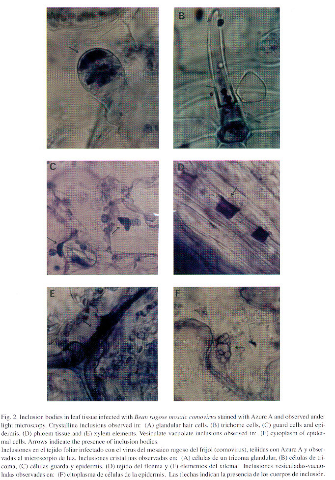
Fifteen species have been identified for the Comovirus genus (Murphy et al. 1995), eight of which have been studied cytologically. All comoviruses studied thus far induce the formation of two different types of cytoplasmic inclusion bodies: a) crystalline inclusions of various and unusual shapes composed of viral particles, which stain violet in Azure A and green in the OG combination (Luxol Brilliant Green BL-Calcomine Orange 2 RS), and are frequently observed in glandular hair cells, guard cells and phloem tissue and occasionally in the epidermis and mesophyll; and b) vesiculate-vacuolate inclusions observed in the cytoplasm of epidermal cells which stain green in OG and red to magenta in Azure A (Gibbs and Paul 1970, Kim and Fulton 1972, Kitajima et al. 1974, Langenberg and Schroeder 1975, Bruening 1978, Edwardson and Christie 1991). Both types of inclusion bodies and their location are considered to be main diagnostic characteristics of the Comovirus genus (Edwardson and Christie 1978). Xylem elements blocked with masses of virus particles have also been observed in squash mosaic virus and cowpea mosaic virus infections (Christie and Edwardson 1977). In this paper, inclusion bodies observed by light microscopy in BRMV-infected bean leaf tissue are described and compared with those induced by other members of the genus Comovirus.
Materials and Methods
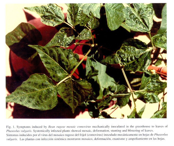
Virus isolate: the isolate of BRMV was initially obtained from plants of P. vulgaris infected in the field and later maintained in the greenhouse by mechanical inoculation on bean plants. Leaves from plants showing severe mosaic and leaf deformation, and stunting and blistering of leaves and pods (Fig. 1) were selected for examination by light microscopy. Healthy leaves from the same bean variety were also examined.
Virus identification: the identity of the virus in all plants selected for light microscopy was confirmed by DAS-ELISA using rabbit polyclonal antibodies produced by C. Rivera, (1990).
Light microscopy: strips from the lower epidermis of the leaves of inoculated and non- inoculated bean plants were removed and stained in 0.1% Azure A and OG (Christie and Edwardson 1977). After removal of the epidermis, the mesophyll was soaked in 2-methoxyethanol for 30 min to remove the chlorophyll and stained with Azure A. The excess stain was removed in 95% ethanol. The tissue was mounted directly in Euparal and observed with a light microscope and photographed.
Results
Two types of inclusion bodies were consistently observed by light microscopy in bean leaf tissue infected with bean rugose mosaic comovirus. Crystalline cytoplasmic inclusions of different size and shape were frequently found in glandular hair cells (Fig. 2 A), trichome cells (Fig. 2 B), epidermis and guard cells (Fig. 2 C) and phloem tissue (Fig. 2 D). The xylem elements observed were blocked with masses of crystalline inclusions (Fig. 2 E). Vesiculate-vacuolate inclusions were also frequently found in the cytoplasm of epidermal cells (Fig. 2 F). Both types of inclusions stained with Azure A and OG combination.
Discussion
All comoviruses studied to date are reported to induce vacuolate-vesiculate inclusions and crystalline inclusions of virus particles. Both types of inclusion bodies are now considered to be main characteristics of the Comovirus genus ( Edwardson and Christie 1991) . Vacuolate-vesiculate inclusions in epidermal cells and crystalline inclusions of virus particles have been described in thrichome cells, spongy parenchyma, phloem, mesophyll cells and xylem elements by electron microscopy of BRMV infected leaves (Kitajima et al. 1974, Gálvez et al. 1977, Gámez 1982, Muñoz & Kitajima 1990). Both types of inclusion bodies were easily identified in this research by light microscopy of BRMV-infected tissue stained with Azure A and with O-G. Thus light microscopy should be useful in the diagnosis of BRMV comovirus infections.
Acknowledgements
We thank Melanie Hord for review manuscript and William Villalobos for technical assistance. This research was supported by the PCDV-CIBCM (801-94-905).
Resumen
Se observaron dos tipos de inclusiones virales, mediante microscopia de luz, en hojas de plantas de frijol (Phaseolus vulgaris) previamente infectadas con el virus del mosaico rugoso del frijol ("bean rugose mosaic comovirus", BRMV), especie del género Comovirus, familia Comoviridae. Se hallaron inclusiones vesiculadas, principalmente en el citoplasma de células de la epidermis, y abundantes inclusiones cristalinas de diferentes formas y tamaños siempre en células guarda, tricomas glandulares, floema, elementos del xilema y ocasionalmente en células epidérmicas y del mesófilo. Ambos tipos de inclusiones tiñeron con Azure A y con la tinción, verde naranja (Luxol Brilliant Green BL-Calcomine Orange 2 RS) conocida como OG, y son similares a las inclusiones inducidas por otras especies del género Comovirus.
References
Acosta O., Alegría A. & R Lastra. 1986. Some biological and physicochemical properties of bean rugose mosaic virus. Phytopathology 76: 1182-1184. [ Links ]
Bruening G. 1978. Bean pod mottle comovirus. CMI/AAB Descriptions of Plant Viruses No. 199, 6 pp. [ Links ]
Cartín F. 1973. Caracterización de dos nuevas razas del virus del mosaico rugoso del frijol. Tesis de Licenciatura, Facultad de Agronomía, Universidad de Costa Rica, San José, Costa Rica. [ Links ]
Christie R.G. & J.R. Edwardson. 1977. Light and electron microscopy of plant virus inclusions. Fla. Agric. Exp. Stn. Monogr. No. 9, 150 p. [ Links ]
Edwardson J.R. & R.G. Christie. 1978. Use of virus-induced inclusion bodies in classification and diagnosis. Ann. Rev. Phytopathol. 16: 31-55. [ Links ]
Edwardson J.R. & R.G. Christie. 1991.Comoviruses. In: Edwardson & Christie eds., Handbook of viruses infecting legumes, CRC, Boca Raton, Florida, 504 p. [ Links ]
Gálvez G.E., Cardenas M., Kitajima E.W., Díaz A.J. & M.P. Nieto. 1977. Purification, serology, electron microscopy, and properties of the Ampollado strain of bean rugose mosaic virus. Turrialba 27: 343-350. [ Links ]
Gámez R. 1972. Los virus del frijol en Centro América. II. Algunas propiedades y transmisión por crisomélidos del virus del mosaico rugoso del frijol. Turrialba 22: 249-257. [ Links ]
Gámez R. 1977. Las enfermedades virales como factores limitantes en la producción de frijol (Phaseolus vulgaris) en América Látina. Fitopatología 12: 24-27. [ Links ]
Gámez R. 1982. Bean rugose mosaic virus. CMI/AAB Descriptions of Plant Viruses No. 246, 4 pp. [ Links ]
Gibbs A.J. & H.L. Paul. 1970. Broad bean true mosaic comovirus. CMI/AAB Descriptions of Plant Viruses No. 20, 4 pp. [ Links ]
Kim K.S. & J.P. Fulton. 1972. Fine structure of plant cells infected with bean pod mottle virus. Virology 49: 112-121. [ Links ]
Kitajima E.W., Tascon A., Gámez R. & G.E. Gálvez. 1974. Ultrastructural studies on bean leaf tissues infected with two strains of bean rugose mosaic virus. Turrialba 24: 393-397. [ Links ]
Langenberg W.G. & H.F. Schroeder. 1975.The ultrastructural appearance of cowpea mosaic virus in cowpea. J. Ultrastruct. Res. 51: 166-175. [ Links ]
Lin M.T., Gámez R. & E.W. Kitajima. 1981. Bean "mosaico-em-desenho"virus is a member of the bean rugose mosaic serogroup. Fitopatol. Brasil. 6: 293-296. [ Links ]
Muñoz J. & E.W. Kitajima. 1990. Estudio comparativo da citopatologia induzida por algunos virus do feijoeiro (Phaseolus vulgaris) em infeccoes simples. Fitopatol. Brasil. 15: 276-284. [ Links ]
Murphy F.A., Fauquet C., Bishop D.H.L., Ghabrial, Jarvis A.W., Martelli G.P., Mayo M.A. & M.D. Summers. 1995. Virus Taxonomy. Sixth Report of the International Committee on Taxonomy of Viruses. Springer, Viena, Austria. 586p. [ Links ]
1 Centro de Investigación en Biología Celular y Molecular (CIBCM)
2 Facultad de Microbiología, Universidad de Costa Rica, San José, Costa Rica, fax (506) 207 3190, e-mail: crivera@sol.racsa.co.cr

