


 Articulo en XML
 Referencias del artículo
 Enviar articulo por email
 Citado por SciELO
  Similares en
SciELO
Similares en
SciELO  uBio
uBio 
 Permalink
Revista de Biología Tropical
versión On-line ISSN 0034-7744versión impresa ISSN 0034-7744
Rev. biol. trop vol.48 no.1 San José mar. 2000
capuchin monkey, Cebus apella (Primates: Cebidae)
Juan T. Borda1 Vanessa Nunes-Bastos 2 Silvia Pérez-Escalá 3 and Marcial Sánchez-Negrette4
Received 12-I-1999. Corrected 27-IX-1999. Accepted 29-IX-1999.
Abstract
The renal lesions are of special importance in the captive primates. The most commonly pathologies are: pyelonephritis, nephrocalcinosis, glomerulonephritis, congenital malformations, hydronephrosis and functional diseases. We report the histopathological study of renal lesions of five cases of deaths in Cebus apella (Primates) of the Argentinean Primate Center. The ages of the monkeys were from 4 months to 15 years old. Microscopically, we have observed principally acute diffuse proliferative glomerulonephritis, hilar mesangio proliferative glomerulonephritis, extracapilar glomerulonephritis with crescents, chronic interstitial nephritis and chronic pyelophritis.
Key words
Cebus apella, primate, kidney disease, glomerulonephritis, nephritis.
Non-human primates are utilized as experimental models in biomedical research and in pharmacological tests. In order to achieve this, a healthy state is an essential condition as well as a fundamental ethic premise (Bennett 1982).
Some renal lesions have been reported in primates in the wild as well in captivity. Among the species in the Old World, the Rhesus monkey (Macaca mulatta), proliferative arteriopathies characterized by the eccentric nodular thickening and disordered cellular proliferation accompanied by abundant mononuclear cellular infiltrate have been reported. Slight / mild isolated lesions to extensive glomerular change were mentioned among the glomerular change. Sclerosed and hyalinized glomeruli were observed in young animals (Skelton-Stroud and Glaister 1987). Mesangioproliferative glomerulonephritis of light to moderate severity was observed in apparently normal pigtailed macaques (Macaca nemestrina), and the tubular nephrosis was the lesion most frequently found (Boyce 1981, Giddens 1981). A case of malignant nephroblastoma was described in a 4 month-old individual of cynomolgus monkey (Macaca fascicularis) (Bennett 1982).
In baboons (Papio Cynocephalus) the presence of interstitial lymphoreticular infiltrates, occasional hyalinization of glomeruli and embolic pyelonephritis associated with septicemia in younger animals was described (Brack 1981). Renal infarcts and mesangioproliferative glomerulonephritis immunologically mediated were observed in others studies (Heidel 1981).
Primates in the New World also suffer several renal lesions. Among the owl monkeys (Aotus nancymae, Aotus vociferans and Aotus trivirgatus) the most common detected lesion was the immunomediated glomerulonephritis associated with hemolytic anemia in Aotus trivirgatus (Chalifoux 1981). Aotus vociferans and Aotus nancymae showed chronic nephropathy in about 10 % of the animals dead at the Peruvian Primatological Project breeding center (Gozalo and Montoya 1990).
Spontaneous Ig M mesangioproliferative nephropathies were described in callithricids. Morphologically, it was a mesangial hyperplasia accompanied by sub-acute to chronic interstitial inflammation (Brack 1988). Mesangial sclerosis, proliferation and sclerosis of Bowman capsule and glomerular sclerosis were also described (Potkay 1992). Some cases of renal lithiasis, pyelonephritis and glomerulonephritis are frequent in the squirrel monkey (Saimiri boliviensis) (Strickland and Clarkson 1985). The glomerulonephritis were classified into four types in this research: focal sclerosing glomerulonephritis, membranoproliferative glomerulonephritis, membranous glomerulonephritis and a combination of the last two-type (Stills and Bullock 1981).
Chronic glomerulonephritis was associated with microfilariasis in the brown capuchin monkey (Cebus apella) (Sanchez Negrette 1983) and this was the only report about this specie in spite of being utilized in biomedical and behavioral research (Nagle 1989, Ruiz and Arzuaga 1990).
In this report we describe renal lesions detected in necropsies of Cebus apella at the Argentinean Primate Center (Centro Argentina de Primates).
Materials and Methods
The Argentinian Primate Center (CAPRIM) has a colony of 70 Cebus apella housed in social group in outdoor cages.
The diet consisted of a commercially balanced diet that provides the animals with a minimum 25 % of protein, a minimum 3 % of fat, a maximum 5 % of fiber, 0.7 % of calcium, 0.6 % of phosphorus, 13 % of maximum humidity, 8.5 % of maximum ashes and a daily 9 % of each animal corporal weight plus seasonal fruit and water ad libitum.
The animals were clinically examined at least six times a year and laboratory tests are carried out twice a year.
The necropsies were made to all the dead animals using conventional methods followed by a systematic study of all the organs. The results of the histopathologic study were on five differently aged animals (four and nine months old; one, nine and fifteen years old). The samples for this study were recollected during four months period. In spite of this other animals died for different causes during this time.
The pieces obtained were fixed in 10 % neutral buffered formalin, included in paraffin cut at 5 µm and colored with the Hematoxylin-Eosin, PAS, Masson thrichromic and Argentic impregnation techniques.
Results
Necropsy and histopathology findings in five animals are reported in this work.
Case 1: A 15 year-old male clinically presented a paranasal abscess in relation to the upper molar. It did not respond to the treatment with wide spectrum antibiotics and died 24 hr after the treatment.
It showed a large purulent collect at the maxilla in the necropsy.
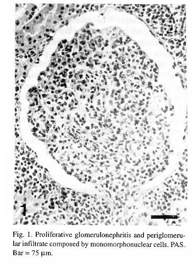
An interstitial leukocytory infiltrate composed by lymphocytes, monocytes and plasmocytes could be observed microscopically in both kidneys at the cortical area. Interstitial fibrosis and glomerular atrophy were shown at some sections. Necrosis and the presence of protein material in the lumen were detected at the tubules. The glomeruli were bloodless and showed high cellularity presenting an average of 300 cells per glomerulus. The hypercellurarity was caused by proliferation of endothelial and mesangial cells and leukocyte infiltration composed by neuthrophils and monocytes (Fig. 1).
The medullar zone showed congestion and calcium deposits.
Diagnosis: Acute proliferative diffuse glomerulonephritis and chronic interstitial nephritis.
Case 2: A 9 year-old male did not present clinical symptoms before it died. It did not show relevant data in the necropsy.
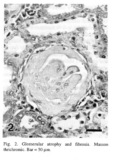
Both kidneys histopathologically showed abundant interstitial lymphoplasmocyte infiltrate, isolated glomerular atrophy and interstitial fibrosis in the cortical area. The rest of the glomeruli presented an increase of the intra and extra glomerular mesangium principally at the hilium and thickening of the parietal wall of the Bowman capsule. The glomerular atrophy and fibrosis were corroborated through the Goldner thrichromic technique (Fig. 2). The increase of the mesangial matrix as well as the thickening of the Bowman capsule was seen through the toluidin blue and PAS technique.
Diagnosis: Chronic interstitial nephritis with fibrosis and glomerular atrophy and hilar mesangial hyperplasia.
Case 3: A 1 year-old male presented loss of appetite and adynamia. The hematocrit was 18.56 %, hemoglobin 6.4 g/dl, glucose 0.034 g/l and total proteins 3.53 g/dl. It died a few hours later.
The microscopic changes involved predominantly tubules and interstitium. The tubules showed atrophy in some areas and hypertrophy in others, or dilatation. Dilated tubules were filled with colloid casts showing pronounced thyroid-like.
Diffuse interstitial lymphoplasmocyte infiltrate at the cortical as well as the medullar area and renal pelvis was observed. At a cortical area the tubules were dilated with eosinophilic material showing pronounced thyroid-like. The Bowman capsule thickening and the intra glomerular fibrosis of some of the glomeruli were evident. In this case the hyaline obliteration of glomerulus, transforming them into acellular eosinophilic PAS-positive masses. The arterioles were thickened at the expense of the hyperplasia and hyalinization of the media tunica.
Diagnosis: Chronic pyelonephritis.
Case 4: A 9 month-old male presented a symptomatology characterized by intense adynamia and therefore clinical blood tests were made showing a 6% hematocrit and 2.9 g/l hemoglobin.
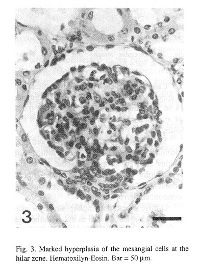
In the microscopic study of both kidneys, isolated focuses of lymphoplasmocyte interstitial infiltrate were detected at a cortical zone; the glomeruli showed marked increase of the mesangial matrix in the hilar zone, characterized by proliferation of mesangial cells and increase of mesangial matrix (Fig. 3).
Diagnosis: Hilar mesangioproliferative glomerulonephritis with interstitial nephritis.
Case 5: Before its death, a lactating 4 month-old male presented asthenia and was unable to hold onto his mother.
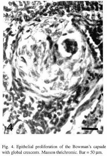
In both kidneys, microscopic observation showed isolated focuses of interstitial nephritis and degeneration of the tubular epithelium. Lymphocytes, monocytes and plasmocytes composed the interstitial infiltrate. The tubular degeneration was characterized by vacuolar change. The glomeruli presented epithelial proliferation of the Bowmans capsule forming; in this way, epithelial global crescent. Crescent was formed by proliferation of parietal cells. The crescents eventually obliterate Bowmans space and compress the glomerular tuft, sometimes the glomerular capillary loops were completely collapsed (Fig. 4). Minimal intraglomerular fibrosis was detected through the Masson thricromic technique.
Diagnosis: Extracapillary glomerulonephritis with global crescents.
Discussion
There are a variety of antecedents about renal disease in non-human primates and certain diseases are more likely to occur in some species in particular. For instance the glomerulonephritis is frequently reported in owl monkeys (Aotus sp) (Gozalo and Montoya 1990), Squirrel monkeys (Saimiri sp) (Stills and Bullock 1981), in the Callithricids (Brack and Rothe 1981, Potkay 1992) and pigtailed macaques (Macaca nemestrina) (Boyce 1981, Giddens 1981).
Cebus apella is one of primates species of the New World that is utilized in biomedical research (Nagle 1989, Ruiz and Arzuaga 1990). However, the bibliography describing renal alteration is scarce (Sánchez Negrette 1983).
Histopathological studies, from the material obtained through necropsy of five primates, showed that all the kidneys studied presented morphological characteristic of severe renal disease and in some cases it was correlated with anemia, this situation may be caused by damage kidneys, it can not produce erythropoietin and this would cause a fall in red blood cell production and the consequent anemia (Guyton 1976). The hypoproteinemia principally hipoalbuminemia would be the result of an increase in the glomerular membrane permeability and the protein loss through the urine (Iotti and García Dadone 1979). But in other cases, they presented neither alteration in the normal biochemical values nor clinical manifestations indicating renal disease.
The extracapillary glomerulonephritis with global crescents is also known with the names of glomerulonephritis rapidly progressive and malignant sub-acute and its generally fatal depending on the percentage of affected glomeruli with crescents and their type (focal or diffuse). More than 70 % of the human case are almost always fatal (Iotti and García Dadone 1979). As it was shown in our study, this kind of glomerulonephritis affected the youngest animal (4 months old) and the microscopic finding corresponded to a rapidly fatal course. In the human being this glomerulopaty have a fatal course and it is more frequent in childhood. In our casuistic a similar case was present in a four month old monkey.
The mesangioproliferative glomerulonephritis is characterized by an increase of the mesangial matrix and mesangial cells, accompanied by a thickening of the capillary wall. This kind of glomerulonephritis was observed in Macaca nemestrina (Boyce 1981, Giddens 1981), Saimiri sciureus (Stills and Bullock 1981), and in Callithrichidae family (Brack 1988) in these monkeys, there were Ig M deposits inside the mesangial cells and matrix. According to another study carried out on baboons (Papio cynocephalus), 15 of them suffered mesangioproliferative glomerulonephritis, in which deposits of Ig G were found in 6, of Ig M in 5, of C3 in 4 and of Ig A and C4 in 2 (Heidel 1981). As it was shown by our study, both kidneys presented glomeruli with a proliferation of mesangio, mainly in the hilar region accompanied by isolated focuses of interstitial nephritis corresponding to the cases of Callithricids (Brack 1988).
In the acute diffuse proliferative glomerulonephritis, all glomeruli were thoroughly affected in both kidneys. The Bowmans space disappeared because of the increase in the number of cells (mainly endothelial but also mesangial and leukocytes) and there was not thickening of the capillary wall. In humans, this kind of glomerulonephritis is related to the streptococcus B hemolytic, Lancelfield group A, of which the most common strains are 12, 4 and 1; these strains cause upper respiratory tract infection or skin lesions (streptodermitis-strain 49) from 1 to 3 weeks before the nephropathy (Iotti and García Dadone 1979). In our case the glomerulonephritis probably occurred in relation to the purulent collect of the maxilla, however, both kidneys presented a chronic interstitial nephritis characterized by lymphoplasmocyte infiltrate, glomerular atrophy and interstitial fibrosis.
In animals with pyelonephritis chronic there used to be antecedents of repeated infection in the urinary tract or acute pyelonephritis which leads to the chronic form with renal insufficiency (Maxie 1980). In many cases, there are no previous antecedents and the diagnosis of chronic infection in the kidney can only be achieved in necropsy. Severe acute pyelonephritis with purulent exudate was described as the possible cause of death in Macaca nemestrina (Giddens 1981) and pyelonephritis was of the chronic type in a 1 year-old individual without any manifestation of renal insufficiency in our study.
The interstitial nephritis corresponds to inflammatory lesions in the renal interstice. This kind of nephritis is not very frequent in most of the animals species or in humans but it is common in dogs (Maxie 1980) and it was reported in Callithrichids (Brack 1988). We found chronic interstitial nephritis characterized by isolated focuses of lymphoplasmocyte infiltrate at the cortical level in all the animals examined.
Due to the fact that the aspects referred to as the etiology, pathology or clinical features of the nephritis are still unknown, specific research becomes necessary to know normal histologic parameters as well as renal function tests that determine how the morphological changes are related to the health conditions of individuals of different ages, sex, origin and life conditions.
Acknowledgements
The authors thank the technicians Francisca Morales, Ramón Romero and Miguel Blanco.
References
Bennett, B.T., F.Z. Belunhan & T.J. Welsh. 1982. Malignant nephroblastoma in Macaca fascicularis. Lab. Anim. Sci. 32: 403-404. [ Links ]
Boyce, J.T., W.E. Giddens Jr & R. Seifert. 1981. Spontaneous mesangioproliferative glomerulonephritis in pigtailed macaques Macaca nemestrina. Vet. Pathol. 18: 82-88. [ Links ]
Brack, M. 1981. Renal pathology in captive baboons Papio cynocephalus. Vet. Pathol. 18: 55-58. [ Links ]
Brack, M. 1988. Ig M-mesangial nephropathy in Callithricids. Vet. Pathol. 25: 270-276. [ Links ]
Brack, M & H. Rothe. 1981. Chronic tubulointerstitial nephritis and wasting disease in marmoset Callithrix jacchus Vet. Pathol. 18: 45-54. [ Links ]
Chalifoux, L.V., R.T. Bronson, P. Sehgal, B.J. Blake, & N.W. King. 1981. Nephritis and hemolytic anemia in owl monkeys Aotus trivirgatus. Vet. Phatol. 18: 23-37. [ Links ]
Iotti, R.M & L. García Dadone. 1979. Enfermedades del riñón y de las vías urinarias, p. 523-591. In B. Elsner., R.M. Iotti., C.E. Parisi., M. Caputti (eds.). Lecciones de Patología. Prensa Médica Argentina, Buenos Aires. [ Links ]
Giddens, W.E. Jr, J.T. Boyce, G.A. Blakley & W.R. Morton. 1981. Renal disease in the pigtailed macaque Macaca nemestrina. Vet. Pathol. 18: 70-81. [ Links ]
Gozalo, A & E. Montoya. 1990. Mortality causes of owl monkeys Aotus nancymae and Aotus vociferans in captivity. J. Med. Primatol. 19: 69-72. [ Links ]
Guyton A.,C. 1976. Micción, enfermedades renales y diuresis, p.500-513. In A.C. Guyton (ed.). Tratado de Fisiología Médica. Interamericana, México. [ Links ]
Heidel, J.R. 1981. Renal pathology of catheterized Papio cynocephalus. Vet. Pathol. 18: 59-69. [ Links ]
Maxie, M.G. 1980. El sistema urinario, p. 393-469. In K.V.F. Jubb., P.C. Kennedy & N. Palmer (eds.). Patología de los animales domésticos. Hemisferio Sur, Montevideo, Uruguay. [ Links ]
Nagle, C.A., N. Paul, I. Mazzoni, S. Quiroga, M.L. Torres, A.F. Mendizabal & Z. Farinati. 1989. Interovarian relationship in the secretion of progesterone during the luteal phase of the capuchin monkey Cebus apella. J. Reprod. Fert. 85: 389-396. [ Links ]
Potkay, S. 1992. Disease of the Callitricidae: a review. J. Med. Primatol. 21: 189-236. [ Links ]
Ruiz, J.C & E. Arzuaga. 1990. Valores hematológicos y de química sanguínea de monos capuchinos Cebus apella en cautiverio. Bol. Primatol. Lat. 29: 14-22. [ Links ]
Sánchez Negrette, M. 1983. Microfilariasis en monos platirrinos Cebus apella. Gaz. Vet. 381: 614-625. [ Links ]
Skelton-Stroud, P.N & J.R. Glaister. 1987. Naturally ocurring renal disease in non-human primates, p. 189-210. In P.H. Bach & E.A. Lock (eds.). Nephrotoxicity in the experimental and clinical situation. Martinus Nijhoff, New York. [ Links ]
Stills, H.F.Jr & B.C. Bullock. 1981. Renal disease in squirrel monkeys Saimiri sciureus. Vet. Path. 18: 38-44. [ Links ]
Strickland, H.T & Clarkson. 1985. Use of the squirrel monkey in cardiovascular research, p.309-310. In L.A. Rosemblum & C.L. Coe (eds.). Handbook of squirrel monkey research. Plenum, New York. [ Links ]
1 Consejo Nacional de Investigaciones Científicas y Técnicas -CONICET. Facultad de Ciencias Veterinarias de la Universidad Nacional del Nordeste, Corrientes, Argentina. Medrano 2250 G2 M1 Pb1, Corrientes. C.P. 3400. Corrientes. Argentina. Telfax: 54 3783 451268 e-mail: borda@compunort.com.ar
2Universidade Federal do Río do Janeiro, Brasil.
3Facultad der Ciencias Veterinarias de la Universidad Nacional de La Plata, Buenos Aires, Argentina.
4 Facultad de Ciencias Veterinarias de la Universidad Nacional del Nordeste, Corrientes, Argentina.

